Dielectrophoretic separation and immunoassay methods on active electronic matrix devices

an electronic matrix and immunoassay technology, applied in electrostatic separators, diaphragms, electrolysis, etc., can solve the problems of insufficient detection limited application of eps for cell manipulation, and inability to detect eps, so as to reduce the amount of non-specific background binding, improve the efficiency of immunochemical labeling and capture functions, and achieve rapid and efficient

Inactive Publication Date: 2005-05-03
GAMIDA FOR LIFE +1
View PDF93 Cites 50 Cited by
  • Summary
  • Abstract
  • Description
  • Claims
  • Application Information

AI Technical Summary

Benefits of technology

[0013]The present invention represents a substantial improvement in previous active electronic matrix chip constructs and dielectrophoretic methods by utilizing immunochemical components to perform specific capture and / or reporting functions in methods combining dielectrophoresis (DEP) and electrophoresis (EP). By utilizing active electronic matrix chip devices, the speed of traditional immunochemical labeling and capture functions can be dramatically increased, while reducing the amount of non-specific background binding. When combined with DEP methods on the active electronic matrix chip devices, sample preparation and immunochemical analysis can be done quickly and efficiently in the same instrument volume.

Problems solved by technology

Because most biological cells have similar electrophoretic mobilities, EP for manipulation of cells has limited applications.
These prior devices have several problems, including the following.
A first problem is that both separated and unseparated cells bind nonspecifically to the exposed glass surface of the slide and to the exposed electrode surfaces.
A second problem is that the volume of the flow chamber (several hundred μl) is so large that thermal convection disturbs and pushes off cells initially retained by the electrodes.
A third problem is that washing off any undesired cells is not easily accomplished without disturbing the cells that are desirably retained on the electrodes, as the desired cells and electrodes stand in the way of fluidic flow and, hence, block the wash flow containing any undesired cells.
These conventional electronic lysis / electroporation techniques have several problems.
A first problem is that the electronic lysis conditions specified by commercial macro-devices do not release medium to large proteins, organelles, and DNA molecules of high molecular weight (larger than 20 Kb) because they do not fit through the pores created in the cell membrane by the prior lysing methods.
A second problem is that some molecules of interest originally released in the lysis chamber are lost due to their non-specific binding to the surface of the lysis chamber.
A third problem is that the conventional electronic lysis macro-device works as a stand-alone unit such that both dielectrophoretic cell separation and electronic lysis cannot be performed on the same module.
These conventional preparation and processing techniques have several problems, including the following.
A first problem is that the steps of sample preparation and processing are typically performed separately and apart from the other main steps of the analysis.
In addition, most of these techniques involve carrying out numerous operations (e.g., pipetting, centrifugations, and electrophoresis) on a large number of samples.
They are often complex and time consuming, and generally require a high degree of skill.
Many a technique is limited in its application by a lack of sensitivity, specificity, or reproducibility.
Traditional immunoassay methods utilizing microtiter-plate formats, dipsticks, etc., are labor and time extensive.
Multiple steps requiring human intervention either during the process or between processes are sub-optimal in that there is a possibility of contamination and operator error.
Further, the use of multiple machines or complicated robotic systems for performing the individual processes is often prohibitive except for the largest laboratories, both in terms of the expense and physical space requirements.
However, for the reasons stated above, these traditional techniques involve the disadvantages of multiple sample / analyte transfer steps, and often require large sample volumes to obtain the desired sensitivity and specificity for the assay.

Method used


Image

  • Dielectrophoretic separation and immunoassay methods on active electronic matrix devices
  • Dielectrophoretic separation and immunoassay methods on active electronic matrix devices
  • Dielectrophoretic separation and immunoassay methods on active electronic matrix devices

Examples


example 1

Active Electronic Matrix Chip Devices Used in the Experiments

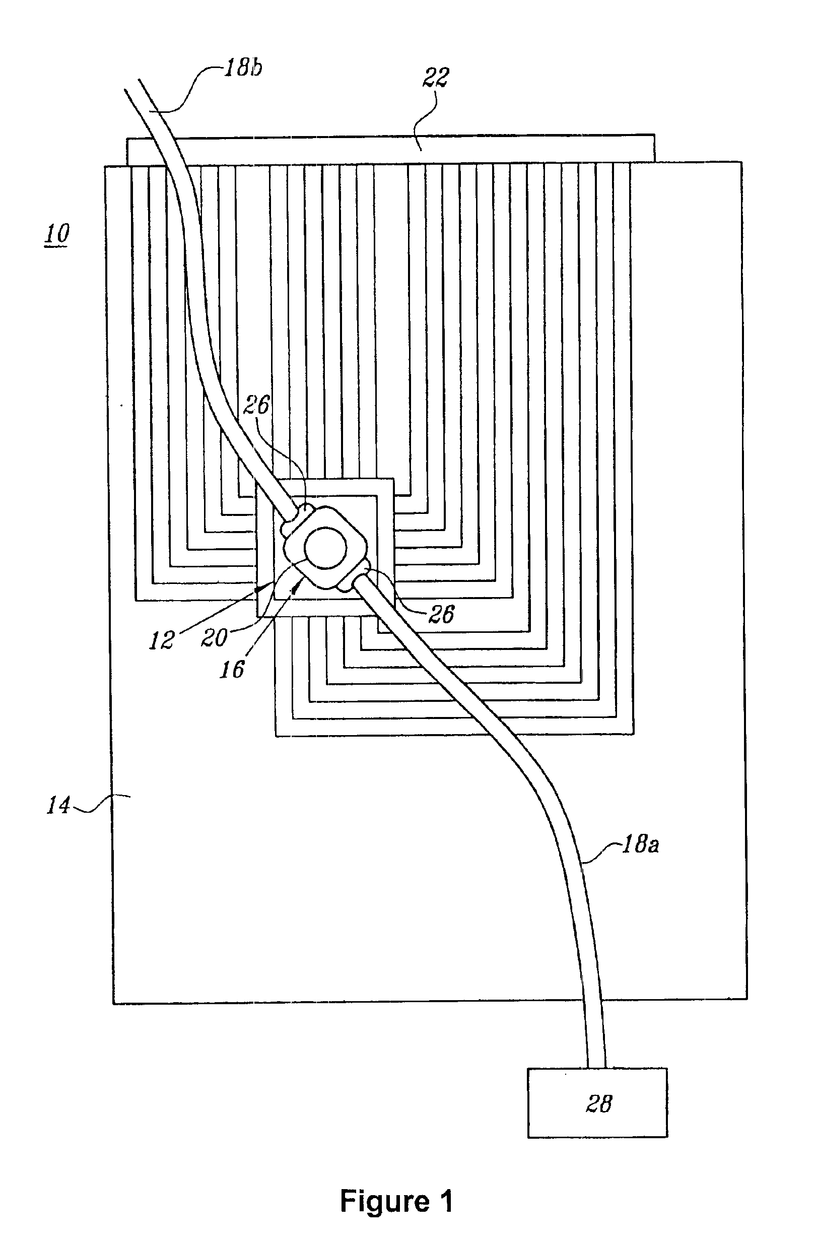
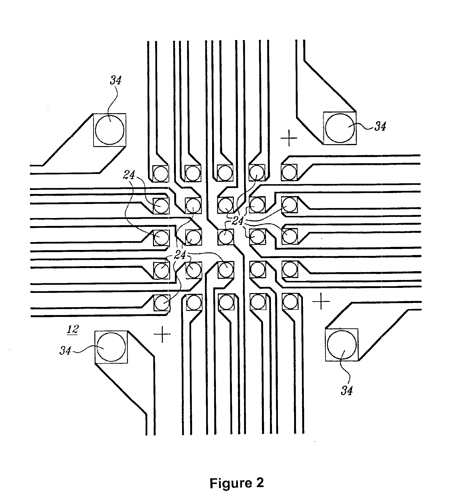
[0098]Active electronic matrix chips (like those pictured in FIG. 6), designated as the ‘5×5 array’, were fabricated on silicon wafer using semiconductor processing technique. The 5×5 array chip consisted of 25 circular, platinum electrodes that were 80 μm in diameter on a 200 μm center-to-center spacing and covered an area of 0.88×0.88 mm2, with reference electrodes surrounding the center array. Other areas on the chip were used for the connection pads to external signal sources and for electric wires between 5×5 array to these pads. An approximately 1 μm agarose permeation layer that contained streptavidin was prepared on the 5×5 array chip using a spin-coating technique.

[0099]The chip cartridge was formed by wire binding of the chip to a printed circuit board and was assembled with a flow cell (similar to that pictured in FIG. 1). A polycarbonate molded flow cell and a cover slip were attached to the chip with a UV-cure...

example 2

Immunoassays on Active Electronic Devices for Bacterial Toxin Proteins

2A: Modification and Determination of Isoelectric Points for Antigens to be Assayed

[0101]Staphylococcal enterotoxin B (SEB) and cholera toxin B (CTB), as well as fluorescein labeled CTB, were obtained from Sigma (St. Louis, Mo.). SEB toxin was modified with fluorescein isothiocyanate. The fluorescein labeling of the toxins allowed for direct visualization in a capture immunoassay without adding a secondary detection step. Analyte derivatization (e.g., with a non-specific label such as fluorescein isothiocyanate) has proven practical in other assay contexts, and could be incorporated into a single-device format by utilizing a reaction chamber in between fluidly connected DEP preparation and immunoassay areas of a closed active electronic matrix chip device. Isoelectric points for the modified and unmodified toxins were determined by isoelectric focusing using pH 4-10 NOVEX IEF gels (Invitorgen, Carlsbad, Calif.).

[0...

example 3

Immunochemical Detection of Bacillus globigii Spores

[0113]A single-antibody direct immunoassay was used for B. globigii spores, which consisted of two steps of DC current application: collecting B. globigii spores on the electrodes, and addressing the detecting antibodies to the electrodes. The assay was performed on 5×5 array chips. Bacillus globigii spores (the Biological Defense Research Directorate, Bethesda, Md.) were stocked in Phosphate Buffered Saline (PBS) (pH 7.2) (Life Technologies, Grand Island, N.Y.) at a concentration of 1.4×10⁹ spores / ml. in 50 mM histidine buffer. For the B. globigii assay, polyclonal goat anti-B. globigii antibody (Biological Defense Research Directorate) and polyclonal rabbit anti-Malathion antibody (Fitzgerald Industries International, Concord, Mass.) was labeled in house with Texas Red (TR)-x-succinimidyl ester (Molecular Probes, Eugene, Oreg.), and used as the detecting antibodies.

3A: Electronic Addressing of Spores to Selected Microlocations

[01...


PUM

PropertyMeasurementUnit
volumeaaaaaaaaaa
volumeaaaaaaaaaa
AC voltageaaaaaaaaaa

Abstract

This invention relates to devices and methods for performing active, multi-step molecular and biological sample preparation and diagnostic analyses employing immunochemical techniques. It relates generally to bioparticle separation, bioparticle enrichment, and electric field-mediated immunochemical detection on active electronic matrix devices utilizing AC and DC electric fields. More specifically, the invention relates to devices and methods for sample preparation/manipulation, immunoimmobilization, and immunoassays, all of which can be conducted on one or more active electronic chip devices within a single system. These manipulations are useful in a variety of applications, including, for example, detection of pathogenic bacteria and biological warfare agents, point-of-care diagnostics, food or medical product quality control assays, and other biological assays.

Description

FIELD OF THE INVENTION[0001]This invention relates to devices and methods for performing active, multi-step molecular and biological sample preparation and diagnostic analyses employing immunochemical techniques. It relates generally to bioparticle separation, bioparticle enrichment, and electric field-mediated immunochemical detection on active electronic matrix devices utilizing AC and DC electric fields. More specifically, the invention relates to devices and methods for sample preparation / manipulation, immunoimmobilization, and immunoassays, all of which can be conducted on one or more active electronic chip devices within a single system. These manipulations are useful in a variety of applications, including, for example, detection of pathogenic bacteria and biological warfare agents, point-of-care diagnostics, food or medical product quality control assays, and other biological assays.BACKGROUND OF THE INVENTION[0002]The basis for many molecular-biological and immunoassays, di...

Claims


Application Information

Patent Timeline
Patent Type & Authority Patents(United States)
IPC IPC(8): B01L3/00B03C5/02B03C5/00
CPCB01L3/502761B03C5/026B01J2219/00274B01L2200/0668B01L2300/0877B01L2400/0415B01L2200/10B03C5/005B03C2201/26B03C5/00G01N33/5438
Inventor HUANG, YINGEWALT, KARLAHAIGIS, ROBERTFORSTER, ANITAKRIHAK, MICHAEL K.
Owner GAMIDA FOR LIFE
Why Eureka
Try Eureka