Single pass attenuated total reflection fourier transform infrared microscopy apparatus and method for identifying protein secondary structure, surface charge and binding affinity

Inactive Publication Date: 2007-08-14
NORTH CAROLINA STATE UNIV
View PDF19 Cites 30 Cited by
  • Summary
  • Abstract
  • Description
  • Claims
  • Application Information

AI Technical Summary

Benefits of technology

[0018]A single-pass FTIR-ATR method of identifying secondary structure present in one or more solublized samples having or suspected to have secondary structure is also disclosed. The method comprises: (a) providing one or more solublized samples; (b) providing an internal reflecting element (IRE) comprising a functionalized tip; (c) contacting an IRE with one of the solublized samples to form a sample-IRE interface; (d) directing a beam of IR radiation through the IRE under conditions such that the IR radiation interacts with the aqueous solution-IRE interface once, thereby generating a reflectance profile using single-pass FTIR-ATR; (e) recording the reflectance profile over a preselected frequency range, the reflectance profile comprising observed frequencies; (f) repeating steps (c) through (e) for each of the solublized samples; and (g) correlating one or more observed frequencies with frequencies indicative of secondary structure composition, whereby secondary structure present in one or more solublized samples having or suspected to have secondary structure is ident

Problems solved by technology

Indeed, the prediction of secondary structure based on primary sequence is one of the most important problems in biology.
This effort, however, is far from complete and currently available technology is expected to rely heavily on the field of proteomics to determine the structure and function of the vast number of proteins now known only by their primary amino acid sequence.
The characterization of protein samples almost exclusively by the amide I and amide II bands of the protein's IR spectrum, is due to limitations imposed by the presence of large solvent bands of both water (H2O) and deuterium oxide (D2O) that obscure regions of the infrared spectrum where additional informative bands can be observed.
Often, however, conventional IR techniques cannot identify this information due to overlap of solvent signals with protein signals.
However, when used in transmission FTIR

Method used


Image

  • Single pass attenuated total reflection fourier transform infrared microscopy apparatus and method for identifying protein secondary structure, surface charge and binding affinity
  • Single pass attenuated total reflection fourier transform infrared microscopy apparatus and method for identifying protein secondary structure, surface charge and binding affinity
  • Single pass attenuated total reflection fourier transform infrared microscopy apparatus and method for identifying protein secondary structure, surface charge and binding affinity

Examples


example 1

IR Spectrum Acquisition

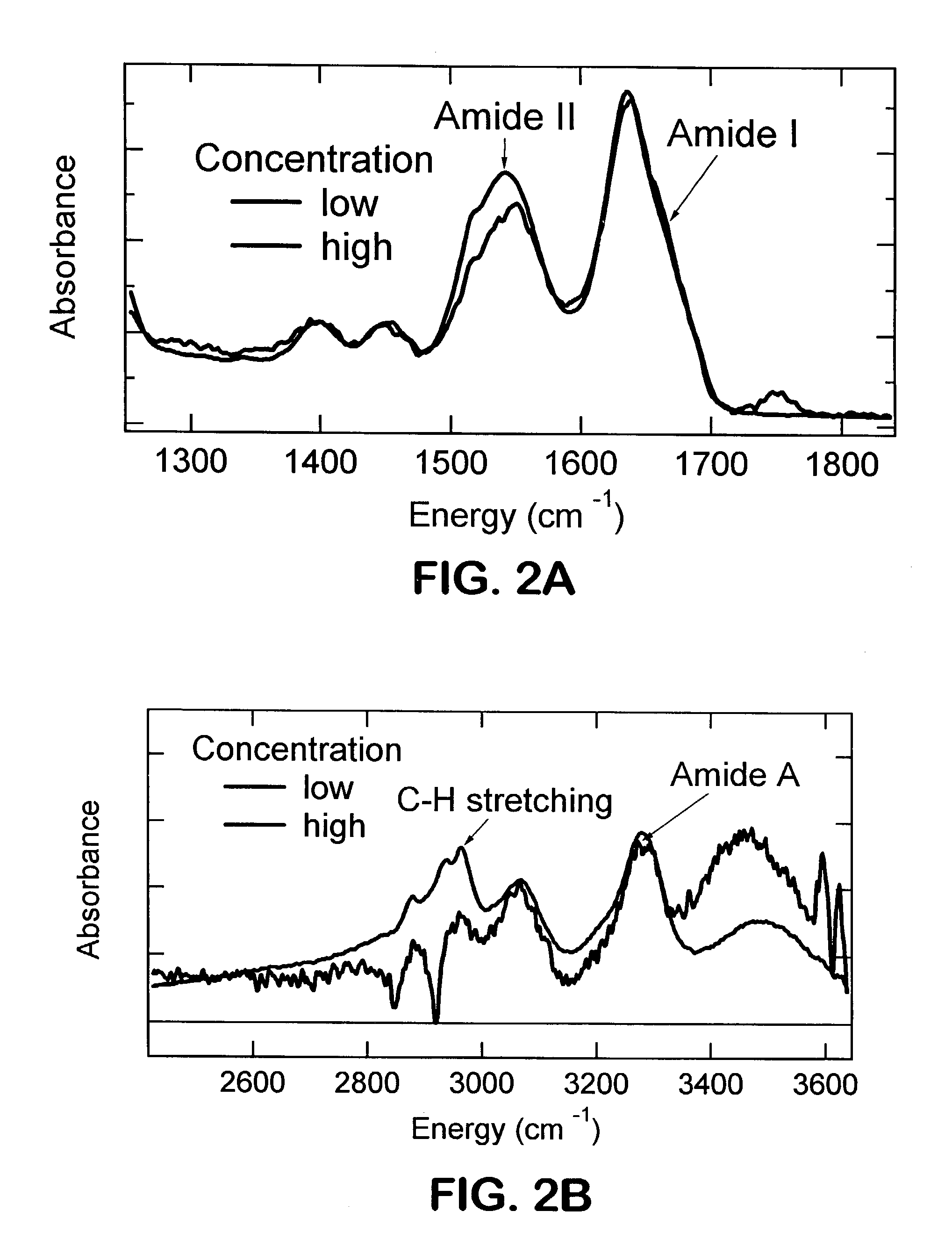
[0269]Mid-frequency and high-frequency ATR-FTIR spectra of a β-sheet forming protein were acquired as seen in FIGS. 2A and 2B. The spectral region shown includes both amide I with a peak at 1636 cm−1 and amide II with a peak at about 1550 cm−1, but also two other peaks at about 1400 and about 1450 cm−1, respectively. Discrepancies between the high and low concentration experiments are observed at about 1750 cm−1 and in the relative intensity of the amide II band. These could be attributable to the hydration state of the protein. In the high frequency region two germanium peaks appear in the low concentration spectrum (shown in blue) as negative spikes at about 2850 cm−1 and about 2910 cm−1. However, the same bands are clearly present in both low concentration and high concentration spectra. The Ge peaks can be removed by a background subtraction procedure.

example 2

Acquisition of IR Spectra from a Protein Having β-Sheet Structure

[0270]Infrared spectra were acquired for immunoglobulin G, a β-sheet forming protein. Acquired spectra are depicted in FIGS. 3A and 3B. Mid-frequency single-pass ATR-FTIR and high-frequency ATR-FTIR spectra of a β-sheet forming protein are depicted in FIGS. 3A and 3B, respectively. The spectral region shown includes both amide I with a peak at 1636 cm−1 and amide II with a peak at 1550 cm−1, but also two other peaks at 1400 and 1450 cm−1, respectively.

[0271]

TABLE 1Comparison of Multi-Gaussian Fits For Six Samples ofImmunoglobulin GStandardATR 1ATR 2ATR 3ATR 4ATR 5ATR 6Deviation1515.81515.271515.661515.041516.071517.390.780244161547.261546.871547.621546.751547.451546.970.360647571620.21620.621619.081619.711620.391618.540.781076791636.61636.781637.151637.051636.81635.910.404533831667.851669.091668.671668.11668.181665.711.09888472StandardD2O 1D2O 2D2O 3D2O 4D2O 5D2O 6Deviation1634.511602.331623.631622.481622.921609.2910.5...

example 3

Evaluation of Methods

[0272]The reproducibility of the present inventive method applied to proteins in H2O solution was compared to transmission FTIR of proteins in H2O and D2O solution. By demonstrating the simultaneous observation of amide I, II, III, A and B bands, the results indicated that protein spectra from the single-pass ATR-FTIR technique yielded more information than protein spectra acquired by other FTIR techniques.

[0273]To validate the single-pass ATR-FTIR method, the mid-infrared spectra of a representative set of proteins in D2O solution were acquired using both a conventional transmission FTIR technique and the single-pass ATR-FTIR technique. Comparisons were made for the following proteins: myoglobin, hemoglobin, cytochrome C, ribonuclease A, lysozyme, chymotrypsin, trypsin, and concanavalin. Since the protein studies were performed in D2O solution, the position of the amide I′ band was compared.

[0274]The amide I′ bands from the two different techniques were very si...


PUM


Abstract

Apparatus and method for acquiring an infrared spectrum of a sample having or suspected to have an amide I band, an amide II band, an amide III band, an amide A band, an OH stretching region or a combination thereof. A representative method includes providing a sample; providing an internal reflecting element (IRE) with a functionalized tip; contacting the sample with the IRE to form a sample-IRE interface; directing a beam of infrared (IR) radiation through the IRE under conditions such that the IR radiation interacts with the sample-IRE interface once; recording a reflectance profile over a range of preselected frequencies, whereby an infrared spectrum of the of a sample having or suspected of having an amide I band, an amide II band, an amide III band, an amide A band, an OH stretching region or a combination thereof, disposed in an aqueous solution is acquired. Representative apparatus includes an internal reflecting element (IRE) comprising a reflection face located on the IRE at a region of intended contact between the IRE and a solublized sample; an infrared radiation source for supplying an evanescent wave of infrared radiation and directing the same from the outside of the IRE to the inside thereof so as to cause the infrared radiation to be incident on the reflection face, wherein the infrared radiation is reflected from the reflection face once; a sample cell; a functionalized tip comprising a surface-immobilized probe that partially or completely fills the volume exposed to the evanescent wave; and a detector for detecting the once-reflected infrared radiation.

Description

CROSS REFERENCE TO RELATED APPLICATIONS[0001]This application is based on and claims priority to U.S. Provisional Patent Application Ser. No. 60 / 317,209, filed Sep. 4, 2001, herein incorporated by reference in its entirety.TECHNICAL FIELD[0002]The present invention relates generally to FTIR-ATR spectroscopic methods and techniques, and specifically to optimizing performance for detection of structure and binding of DNA and protein samples. The methods include automated and high throughput FTIR-ATR procedures.ABBREVIATIONS[0003]ATRattenuated total reflectanceFTIRFourier transform infrared spectroscopyFTIR-ATRattenuated total reflection Fourier transforminfraredIgGimmunoglobulin GIRinfraredIREinternal reflecting elementMCTmercury cadmium telluriumNiNTAnickel-nitrilotriacetic acidPCRprincipal component regressionPDMSpolydimethylsiloxanePLSpartial least squaresPTFEpolytetrafluoroethyleneSECstandard error of calibrationS / Nsignal-to-noiseUVultravioletBACKGROUND ART[0004]The sequencing of ...

Claims


Application Information

Patent Timeline
IPC IPC(8): G01N21/03G01N21/01G01N21/13G01N21/35G01N33/48G01N33/543G01N33/552G01N21/55
CPCG01N21/552G01N2021/3595Y10T436/145555Y10T436/144444Y10T436/143333Y10T436/147777Y10T436/148888Y10T436/142222Y10T436/14Y10T436/141111Y10T436/146666
Inventor FRANZEN, STEFANLAPPI, SIMON E.
Owner NORTH CAROLINA STATE UNIV
Why Eureka
Try Eureka