In vivo assay and molecular markers for testing the phenotypic stability of cell populations and selected cell populations for autologous transplantation

a cell population and autologous transplantation technology, applied in the field of in vivo assays and molecular markers for testing the phenotypic stability of cell populations and selected cell populations for autologous transplantation, can solve the problems of partial thickness defects degenerating into osteoarthritis of the involved joint, severe limitation of joint function, and inability to detect the phenotypic stability of the cell population

Inactive Publication Date: 2009-01-20
TIGENIX NV
View PDF8 Cites 1 Cited by
  • Summary
  • Abstract
  • Description
  • Claims
  • Application Information

AI Technical Summary

Benefits of technology

[0030]Yet another object of the present invention is to provide a cell culture exhibiting chondrocyte phenotypic stability in which the ratio of cells showing positive and negative markers is controlled.

Problems solved by technology

These lesions disrupt the congruence between the joint surfaces and therefore can lead to OA, which can be painful and severely limit the joint function.
Despite these attempts at repair, there is no appreciable increase in the bulk of cartilage matrix and the repair process is rarely effective in healing the defects.
Although initially sometimes painless, partial-thickness defects often degenerate into osteoarthritis of the involved joint.
This procedure has led to a proven at least symptomatic amelioration.
This conceptually promising approach has still wide margins for improvement, since it is known that in vitro expansion of chondrocytes results, after a limited number of cell divisions, in a loss of their phenotypic stability (as defined by the ability of chondrocytes to form hyaline cartilage in vivo) making the cell suspension to be injected unreliable.
The introduction of non-autologous materials into a patient, however, may stimulate an undesirable immune response directed against the implanted material, leading to potential rejection of the newly-formed and engrafted cartilage tissue.
In addition, heterologous implantation risks the transmission to the subject of infectious agent(s) present in the tissue or cell line.
Monitoring the formation and development of the resulting synthetic cartilage in situ is difficult to perform and usually involves an arthroscopic or open joint examination.
Furthermore, implants containing synthetic polymer components may be unsuitable for repairing large cartilage defects since polymer hydrolysis in situ inhibits the formation of cartilage and / or its integration into the defect.
This approach however results in problems similar to those associated with the first approach hereinabove.
Furthermore there is no data so far that articular cartilage contains chondrocytic progenitors available for partial thickness defect repair.
Furthermore, the resulting cartilage remains embedded within a gel matrix making it less suitable for implantation into mammals.
However the resulting particles containing synthetic cartilage-like material are usually small and of undefined shape, and do not integrate with each other and with the surrounding cartilage within the defect.
This makes them unsuitable for implantation and repair of a predetermined articular cartilage defect.
However, it is well known that during in vitro expansion as monolayers, chondrocytes dedifferentiate and lose their ability to form hyaline cartilage in vivo.
Until now it has not been possible to prepare large patches of articular cartilage from small pieces of biopsy tissue using the anchorage-dependent procedures of U.S. Pat. No. 4,356,261.
This approach does not yield an optimal integration between the implant and the surrounding cartilage.
This far there is no evidence on the phenotypic stability of cells in such preparations.
However, the same pluripotency that makes them attractive, conveys the risk of metaplastic differentiation.
In other words there is the risk that they could differentiate in an undesired direction (e.g. bone or fat within a cartilage defect).
Such cells do not show a stable phenotype.
As already outlined above, although autologous chondrocyte transplantation (“ACT”) is becoming a widely accepted technique for repair of joint surface defects (“JSD”) it still presents some drawbacks.
To date, however, it is not known how far it is possible to expand chondrocytes without hampering their phenotypic stability and therefore their capacity to form stable hyaline cartilage in vivo, resistant to vascular invasion and endochondral bone formation.
However they do not measure the capacity of chondrocytes to form cartilage in vivo.

Method used


Image

  • In vivo assay and molecular markers for testing the phenotypic stability of cell populations and selected cell populations for autologous transplantation
  • In vivo assay and molecular markers for testing the phenotypic stability of cell populations and selected cell populations for autologous transplantation
  • In vivo assay and molecular markers for testing the phenotypic stability of cell populations and selected cell populations for autologous transplantation

Examples


example 1

Cartilage Obtainment and Cell Isolation

[0086]Articular cartilage was obtained, within 24 hours after death unless otherwise indicated from human donors not having suffered from any articular disease. After macroscopic inspection to rule out gross joint pathologies, cartilage was sliced full thickness from femoral condyles and placed in Hank's Balanced Salt Solution (“HBSS”) (available from Life Technologies) supplemented with 200 units / ml penicillin, 200 μg / ml of streptomycin, and 0.5 μg / ml of amphotericin B (Life Technologies). After two washes in HBSS during 5 minutes at 37° C., cartilage was finely minced and placed in a sterile 0.2% crude collagenase (Life Technologies) solution in Dulbecco's Modified Eagle Medium (“DMEM”) with high glucose (Life Technologies) containing 10% foetal bovine serum (“FBS”) (Biowittaker), 200 units / ml penicillin, 200 μg / ml of streptomycin, and 0.5 μg / ml of amphotericin B. After overnight incubation at 37° C., cells were washed twice in culture medium...

example 2

In Vivo Assay

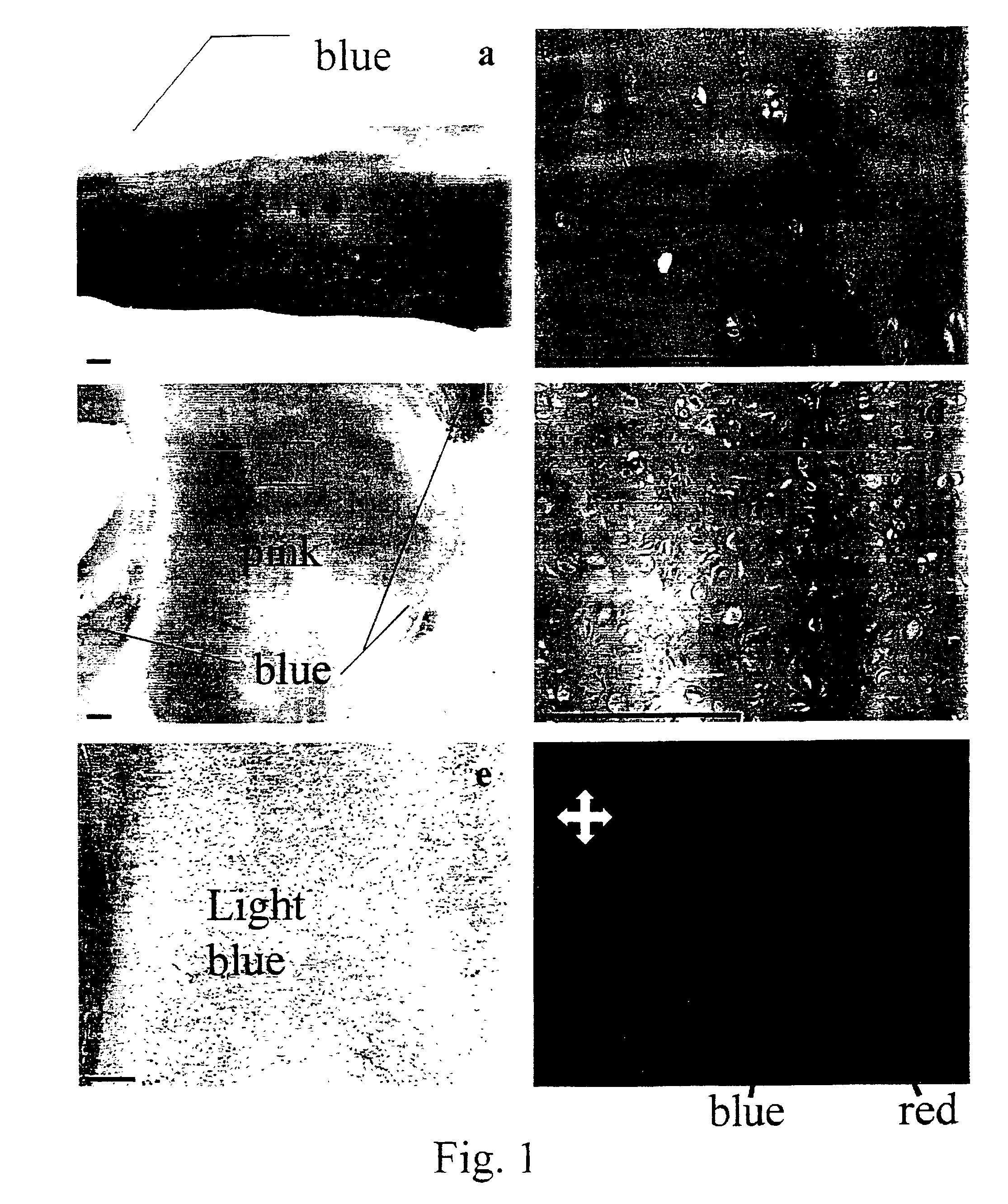
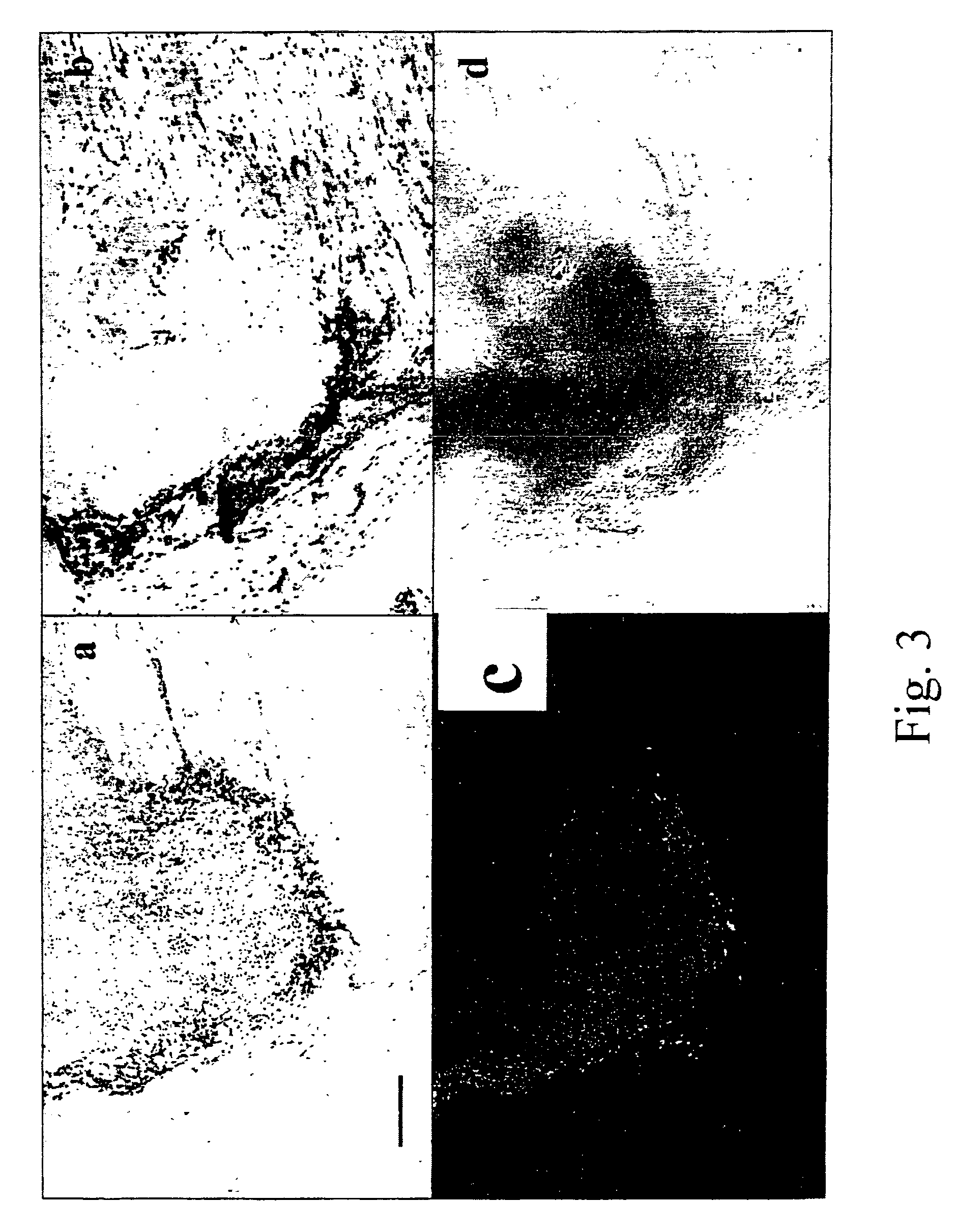
[0087]Cells isolated in example 1 were washed twice in sterile phosphate buffered saline (“PBS”), re-suspended in a volume of 100 μl of PBS and injected intramuscularly in the thigh of female, 4-5 weeks old immune-deficient nude mice. Animals were sacrificed after 3 weeks by cervical dislocation and the thigh dissected to retrieve the presence of the implant in the site of injection. Implants were weighed, and either snap-frozen and stored in liquid nitrogen for in situ hybridisation or fixed in freshly-made 4% formaldehyde for 4 hours for histology and immunohistochemistry. After fixation the samples were included in paraffin, cut in 5 μm thick sections and coloured according to standard protocols (Alcian blue pH 2.5, Toluidine blue, Masson's trichrome, Safranin O) (Manual of Histological Techniques). Different amounts of cells, from 20×106 to 5×10⁵ were used for injection in order to establish the minimum amount of cells that yielded a cartilage implant. Although the ...

example 3

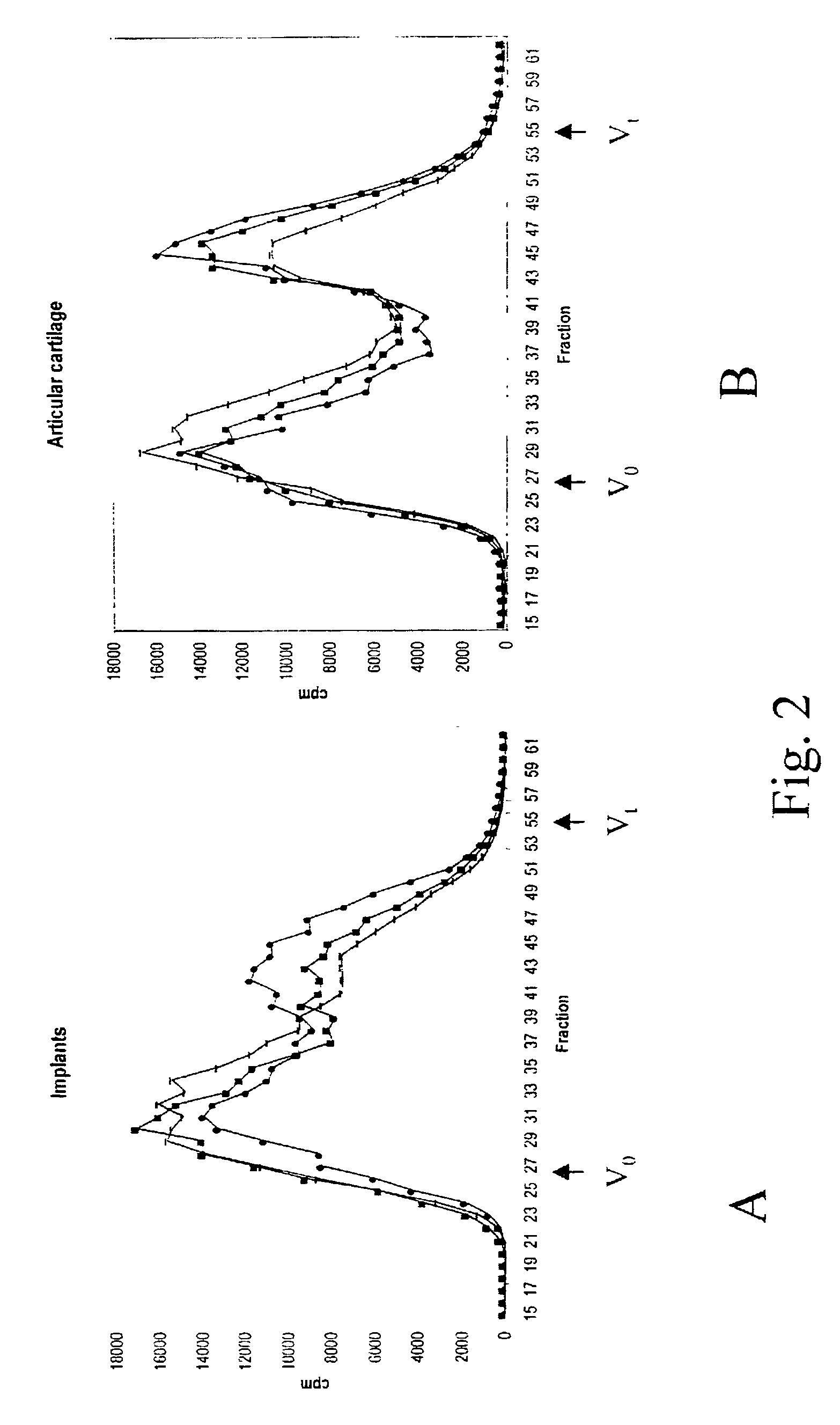
Serial Passaging Results in Impaired Chondrocyte Stability

[0093]Cartilage samples from 3 independent human donors were placed in monolayer culture. Upon passaging, an aliquot of cells was destined to duplicate injection in the in vivo assay of example 2 and to RNA isolation. Chondrocyte stability, as measured by the retrieval of a cartilage implant after 3 weeks in the site of injection, was lost between passage 1 and 3.


PUM

PropertyMeasurementUnit
volumeaaaaaaaaaa
volumeaaaaaaaaaa
pHaaaaaaaaaa

Abstract

An in vivo assay to measure anchorage-independent growth and phenotypic stability of a certain cell population comprising subcutaneous or intramuscular injection in a mammal of a cell suspension of articular chondrocytes in an iso-osmotic liquid, the same suspension comprising articular chondrocytes in an amount equivalent to at least 1×10⁶ chondrocytes as applied to immune-deficient mice. The outcome is linked to molecular markers. The present invention further relates to DNA chips and diagnostic tools comprising the latter to predict the outcome of ACT. Antibodies raised against positive and negative markers of chondrocyte stability can also be used for quality control on the chondrocytes. Therapeutical composition comprising stable chondrocytes are very useful for tissue repair.

Description

[0001]The present invention relates to the field of cartilage repair in general, and more specifically to the generation of an optimal cell population suitable for the repair of joint surface defects and the repair of the cartilage skeleton in general.BACKGROUND OF THE INVENTION[0002]Cartilage is a tissue composed by a cellular component, chondrocytes, and by an extra-cellular matrix typically rich in collagen type II and highly sulfated high molecular weight proteoglycan aggregates. The latter property confers cartilage its peculiar histochemical characteristics that are: strong staining with Alcian blue at low pH (from 0.2 to 2.5) and metachromacy with Toluidine blue and Safranin O. The abundance of type II collagen, link protein, and proteoglycan aggrecan, along with the presence of minor coliagens such as type IX and type XI collagen are hallmarks of cartilage tissue.[0003]In post-natal mammals, cartilage contributes to the structure of several organs and systems like the articu...

Claims


Application Information

Patent Timeline
Patent Type & Authority Patents(United States)
IPC IPC(8): C12Q1/00A61K39/00A61K49/00
CPCA61K49/0004A61K49/0008A61K49/0006Y10S435/975A61P19/04
Inventor LUYTEN, FRANKDE BARI, COSIMODELL'ACCIO, FRANCESCO
Owner TIGENIX NV
Why Eureka
Try Eureka