
Compositions and methods for protecting cells during cancer chemotherapy and radiotherapy

a technology of chemotherapy and radiotherapy, applied in the field of cancer therapy, can solve the problems of cancerous cell proliferation, inability to fully exploit the potency of chemotherapeutic drugs in the treatment of cancer, and toxic to mammalian cells of non-alkylating cancer chemotherapy drugs, so as to protect cells from damage

Inactive Publication Date: 2009-05-12
WISCONSIN ALUMNI RES FOUND
View PDF4 Cites 11 Cited by
  • Summary
  • Abstract
  • Description
  • Claims
  • Application Information

AI Technical Summary

Benefits of technology

The present invention provides a novel and effective strategy for protecting non-neoplastic cells from damage during cancer therapy or chemotherapy. This is achieved by stimulating the natural detoxification systems present in the cells with chemoprotective inducing agents, which activate these systems when the cells are exposed to radiation or chemotherapy. The agents can be administered to the cells in various ways, such as through a delivery vehicle or as part of a regular dietary regime. The invention is particularly useful for preventing damage to non-neoplastic cells in the skin, hair follicles, oral mucosal tissue, and gastrointestinal lining. The chemoprotective inducing agents can include a variety of compounds, such as coumarins, lactones, diterpenes, and other similar molecules. Overall, the invention provides a way to protect healthy cells while still effectively treating cancer.

Problems solved by technology

However, the potential utility of chemotherapeutic drugs in the treatment of cancer has not been fully exploited due to adverse effects associated with the nonspecific cytotoxicity of these agents.
Alkylating agents interfere with the proliferation of cancerous cells by inhibiting DNA replication.
Non-alkylating cancer chemotherapy drugs are also toxic to mammalian cells; they can inhibit multiple sites within a replicating cell, such as (1) synthesis of nucleotides required for DNA replication and (2) microtubule function required for mitosis, to name just two.
Because the toxic effects of these three commonly used agents are generally not specific to cancer cells, they also affect the growth of normal cells, particularly mitotically active normal cells.
As a result, persons being treated with one or more of these cancer therapies commonly develop numerous clinical complications.
The toxicity of cancer therapy for epithelial cells accounts for many of the side effects commonly suffered by persons undergoing a regimen of chemotherapy or radiotherapy.
These complications can be so difficult to endure that it is not uncommon for people to forego or discontinue recommended cancer therapy treatments in order to avoid the problems.
The gastrointestinal disturbances may compromise a patient's chances of recovery, because they make it difficult for him to obtain the nourishment necessary to optimize his ability to fight disease.
It appears that chemo- and radiotherapy-associated death and sloughing of GI lumenal cells results in a release of GI damage-associated molecules into the vasculature.
However, the primary destruction of the GI lining still limits the most effective use of chemotherapy.
However, in spite of these improvements, serious side effects still persist and more effective therapies are needed.
Although this condition is generally reversible and regeneration of hair growth occurs within 1-2 months after discontinuation of treatment, hair loss represents a psychologically distressing effect that can cause negative changes in body image, decreased social activity and altered interpersonal relationships and may lead to refusal of further chemotherapy.
These have included physical modules that temporarily decrease scalp blood flow and drug contact time with the hair follicle, but the patient tolerance was very poor.
These poor results led to the development of scalp cooling methods that decrease both the metabolic rate of follicular stem cells and blood flow to the follicle matrix but this strategy was found to be unsuccessful.
Minoxidil 2% solution was also found to be ineffective in treating chemotherapy induced alopecia.
This sort of therapy is limited in efficacy, inasmuch as it has been shown that intracellular GSH levels can only roughly double in a cell by adding exogenous NAC.
This therapy is limited to the particular chemotherapeutic agents that can be exported from a cell via the p-glycoprotein pump.
Thus, while treatments of the types outlined above may provide some relief from chemotherapy-induced hair loss, their utility is limited, and additional effective therapies are needed.

Method used


Image

  • Compositions and methods for protecting cells during cancer chemotherapy and radiotherapy
  • Compositions and methods for protecting cells during cancer chemotherapy and radiotherapy
  • Compositions and methods for protecting cells during cancer chemotherapy and radiotherapy

Examples


example 1

Prevention of Chemotherapy-Induced Alopecia by Topical Application of Molecules that Induce Expression of Chemoprotective Gene Products

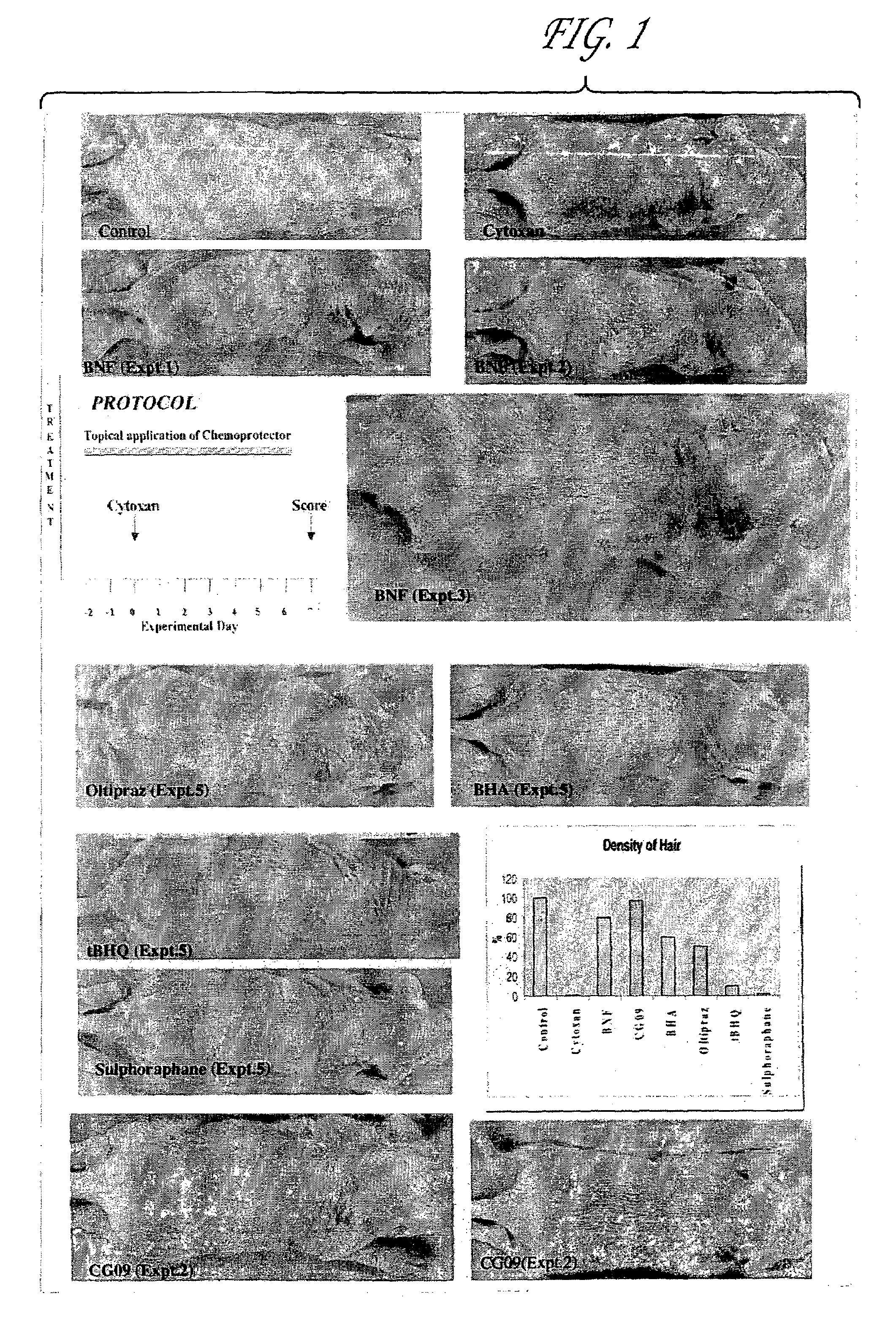
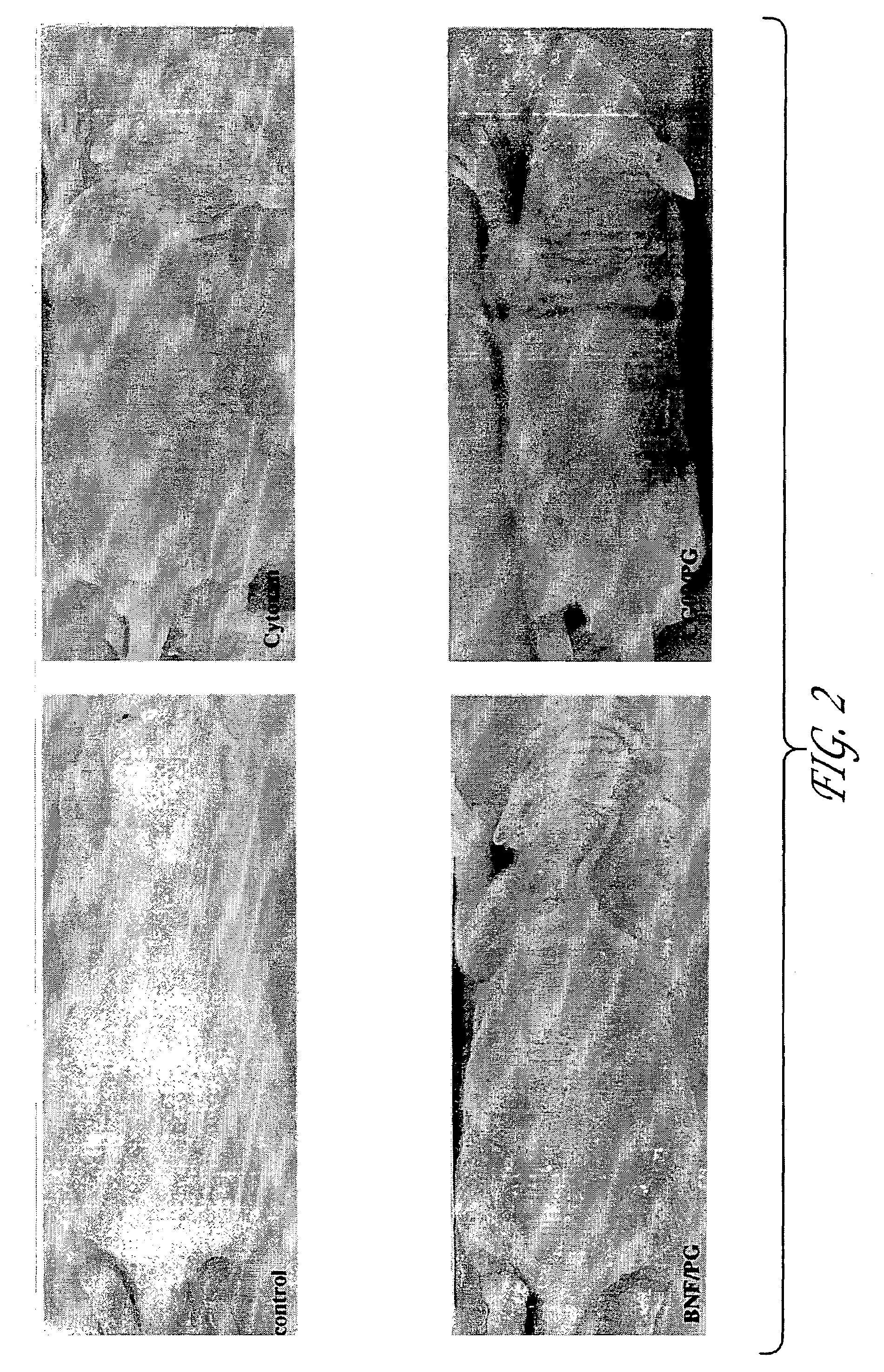
[0080]The inventors postulated that drug induced alopecia could to a large extent be successfully prevented by selective protection of hair follicles by expressing chemoprotective gene products such as GSTs, MRP, MDR, ALDH3 and the like, that are known to protect cells against cytotoxicity. Combined, increased expression of MRP and GST P1 has been found to confer high level resistance to the cytotoxicities of anticancer drugs (Morrow et al, 1998); however, expression of either MRP or GST alone was found to be less successful as these gene products work in synergy to protect cancerous cells against cytotoxicity by their coordinated action in the removal of the anticancer drugs from cells.

[0081]The experiments set forth in this example were designed with the idea of overexpressing these protective genes in hair follicle cells by delivering molecules th...

example 2

Effect of Small Molecule Inducers of Chemoprotective Genes on Chemoprotective Gene Expression in Skin

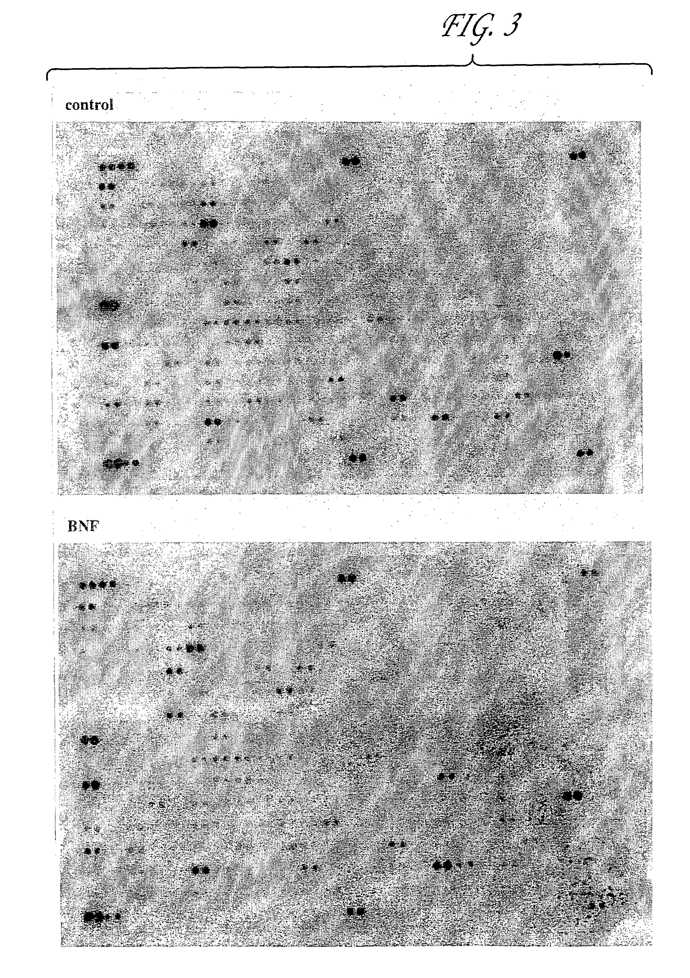
[0101]To elucidate the basic mechanism underlying this protective effect of B-NF and CG09, we studied the expression levels of chemoprotective genes such as GSTs, MDR, MRP and ALDH3, under the same topical treatment conditions as described in the previous example.

[0102]In brief, rat pups were treated with either CTX or B-NF and at different time intervals skin samples were collected and total RNA was extracted from them. The RNA was reverse transcribed to form the respective cDNAs in the presence of 32P dATP using primers specific for stress-related genes. The radiolabeled cDNA synthesized was used to probe a rat stress gene cDNA expression array consisting of 207 stress-related genes (Clontech) according to the manufacturer's protocol. The hybridized membrane was exposed to a phosphorimager screen and the image obtained was aligned with an orientation grid to identify the genes that...

example 3

Identification of Carriers for Delivery of Chemoprotective Gene Inducers to Epithelial Cells

[0105]In order to achieve efficient delivery of a chemoprotective molecule into the skin, studies were conducted using various formulations of liposomes (phospholipid-based vesicles, cationic liposomes, nonionic liposomes, non ionic / cationic liposomes, pegylated liposomes, PINC polymer, and propylene glycol and ethanol mixture (commonly used vehicle for administering minoxidil). First, these formulations were used to entrap reporter genes such as those encoding the marker proteins Luciferase or β-galactosidase, or a fluorescent probe (Nile Red), and their delivery was tracked in the target tissue by the functional assay of the marker proteins delivered or fluorescence emission in the case of Nile Red. The liposomes were prepared as set forth below.

[0106]Phospholipid based vesicles. The preparation contained the following lipid mixtures in a 1:0.5:0.1 molar ratio:[0107]1. DSPC: Cholesterol: DO...


PUM

PropertyMeasurementUnit
body weightaaaaaaaaaa
areaaaaaaaaaaa

Abstract

Compositions, pharmaceutical preparations and methods are disclosed for protecting non-neoplastic cells from damage caused by cancer chemotherapeutic agents or radiation therapy, during the course of cancer therapy or bone marrow transplant. These are based on the use of chemoprotective inducing agents that induce or increase production of cellular detoxification enzymes in target cell populations. The compositions and methods are useful to reduce or prevent hair loss, gastrointestinal distress and lesions of the skin and oral mucosa that commonly occur in patients undergoing cancer therapy. Also disclosed is a novel assay system for identifying new chemoprotective inducing agents.

Description

[0001]This is a divisional of U.S. application Ser. No. 09 / 565,714, filed May 5, 2000 now abandoned, the entire contents of which are incorporated by reference herein.[0002]Pursuant to 35 U.S.C. §202(c), it is acknowledged that the U.S. Government has certain rights in the invention described herein, which was made in part with funds from the National Institutes of Health, Grant No. CA22484.FIELD OF THE INVENTION[0003]The present invention relates to the field of cancer therapy. In particular, the invention provides novel compositions and methods for protecting non-neoplastic cells from the toxic effects of radiotherapy and cancer chemotherapeutic agents.BACKGROUND OF THE INVENTION[0004]Several publications are referenced in this application in order to more fully describe the state of the art to which this invention pertains. The disclosure of each of these publications is incorporated by reference herein.[0005]Over the past several decades, chemotherapy and radiation therapy coupl...

Claims


Application Information

Patent Timeline
Patent Type & Authority Patents(United States)
IPC IPC(8): A61K31/425G01N33/483A61K9/08A61K9/107A61K9/127A61K9/66A61K31/015A61K31/05A61K31/085A61K31/21A61K31/352A61K31/428A61K31/497A61K45/00A61K47/10A61K47/14A61K47/24A61K47/34A61P35/00A61P39/00G01N21/77G01N21/78G01N33/15G01N33/50
CPCA61K9/127A61K9/1272A61K31/497A61K35/36A61P35/00A61P39/00
Inventor FAHL, WILLIAM E.RAGHAVACHARI, NALINIZHU, MINGKINK, JOHN A.
Owner WISCONSIN ALUMNI RES FOUND
Learn More