[0003]There is broad interest in the application of
Nuclear Medicine (NM) techniques with compounds of various specificity to
functional imaging of breast lesions. The use of these compounds can be for confirmation of metastases based on the functional information, facilitating identification of cancerous lesions in women with large or especially dense breasts which particularly cause diagnostic problems in x-ray mammographic screening, and also as a way to monitor any treatment or therapy the patient receives. The
efficacy of single photon emitting tracers versus
positron emitting tracers remains debatable, yet the high sensitivities and specificities (˜90%) already achieved with
breast imaging for ≧1 cm
diameter lesions, as well as commercial availability of agents specifically targeted for
breast tumor imaging, lend credence to the
efficacy of the use of these various compounds. For example, in studies of women with suspicious mammograms, 2-dimensional
planar imaging of ≧1 cm
diameter breast tumors using single photon emitting 99mTc-labeled sestamibi or 99mTc-
methylene-diphosphonate achieved sensitivities and specificities of ˜90%. While these results are encouraging in the
specific population sample, dedicated 3-dimensional NM tomographic imaging with
single photon emission computed tomography (SPECT) or
positron emission
tomography (PET), with superior
lesion contrast and
signal-to-
noise ratio (SNR) characteristics could further improve diagnoses for this group, and potentially be applied more generally.
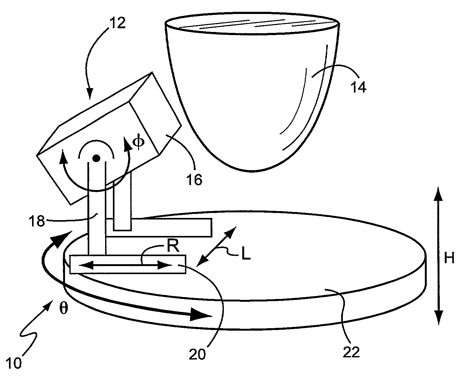
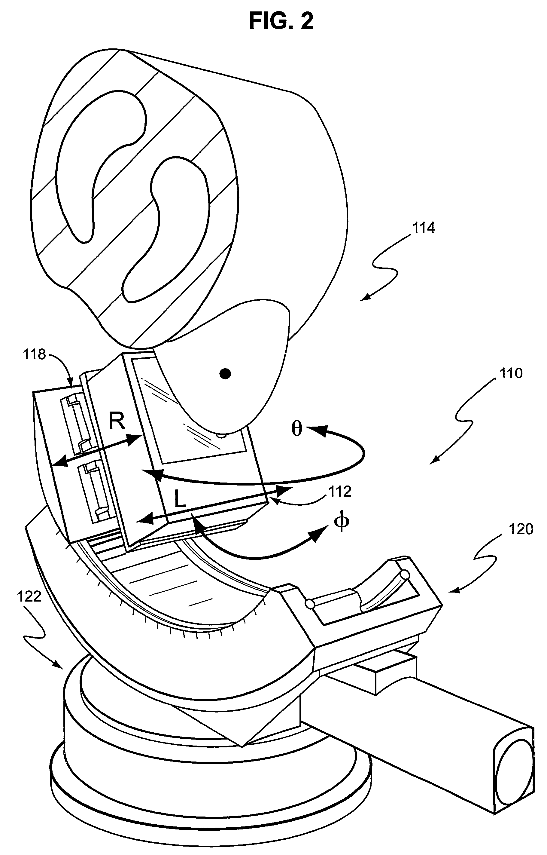
[0011]More generally, the invention is embodied in an imaging system for generating images of a body part suspended within an imaging area of the system, comprising a support having a rotation axis extending through the imaging area and at least one imaging device having an imaging device axis which passes through a first imaging device
field of view, the imaging device being mounted to the support so as to be selectively movable in three dimensions, including radial movement relative to the rotation axis, rotational movement about the rotation axis, vertical movement parallel to the rotation axis, and pivoting movement about a pivot axis perpendicular to the rotation axis, whereby the imaging device can be selectively moved along a path that defines a curved 3-dimensional surface. In an exemplary embodiment, the imaging device axis is laterally offset from the rotation axis and the support is mounted for rotational movement through at least about 180 degrees, whereby when the body part is greater than the imaging device
field of view, an entire volume of the body part can be sufficiently sampled to accurately reconstruct the emission activity distribution.
[0013]Furthermore, fully tomographic transmission data (3-dimensional) which differs from partial view planar scans (2-dimensional) can also be used in both SPECT and PET
for attenuation correction of the emission data. This highly accurate structural transmission map ultimately leads to more quantitatively accurate functional data from which parameters like metabolic rates of reaction can be determined to monitor therapeutic progress and determine
tissue necrosis versus
tumor recurrence in a patient. Simply having a
structural framework (the structural x-ray CT image) with which to identify the location of the focal radioactive uptake with NM imaging (often a diffuse or ambiguously localized region of greater
signal) may be enough to aid in
breast lesion image assessment alone.
[0015]There are various anticipated advancements gained with a high performance, dedicated tomographic system embodying the invention including improved SNR and contrast characteristics due to (1) the improved intrinsic spatial and energy resolution potentially afforded by dedicated, compact, high performance imaging systems which can therefore minimize scatter
contamination, (2) the closer achievable proximity to the object of interest with more compact imaging systems which improves
collimator-limited spatial resolution for SPECT, and (3) due to (2) the camera will preferentially view the breast and minimally view signals from other regions of the body. These advancements should result in an ability to image and 3-dimensionally localize smaller (<1 cm
diameter), non-palpable and potentially pre-metastatic tumors in a larger
population with smaller variance and bias. The use of multiple ASETT scans over time with NM techniques can guide treatments, monitor therapy, and help evaluate outcomes. The use of combined structural and
functional imaging may help even further
in patient management and care.
[0016]Both the structural and functional volumetric information could potentially be used to guide needle biopsies more accurately than with current planar approaches which have limited depth information; more accurate
needle guidance could improve the
needle localization, hence lower false positives, and overall improve diagnosis and guide decisions about treatment protocols for patients.BRIEF DESCRIPTION OF THE DRAWINGS

