Nano-fiber surface-modified xenogeneic acellular nerve graft and preparation method thereof

A surface modification, nanofiber technology, applied in fiber treatment, coating, medical science and other directions, can solve the problems of scar formation, affecting the surgical effect, unfavorable nerve connection, etc., to prevent tissue adhesion, firm bonding, and low price. Effect

Inactive Publication Date: 2017-06-30
YANTAI ZHENGHAI BIO TECH
6 Cites 8 Cited by

AI-Extracted Technical Summary

Problems solved by technology

After the above-mentioned acellular nerve graft is implanted in the body, although it can repair the nerve defect, at the same time, it will definitely promote the proliferation of fibroblasts, secrete a l...

Abstract

The invention discloses a nano-fiber surface-modified xenogeneic acellular nerve graft and a preparation method thereof. The nano-fiber surface-modified xenogeneic acellular nerve graft consists of an xenogeneic acellular nerve graft and a degradable polyester nano-fiber membrane adhered onto the surface of the xenogeneic acellular nerve graft, wherein the xenogeneic acellular nerve graft is an extracellular matrix obtained by performing acellular disposal on an isolated peripheral nerve of a healthy mammal. The nano-fiber surface-modified xenogeneic acellular nerve graft is extremely low in moisture content after freeze-drying treatment and stable in performance, and can realize no protection liquid and normal-temperature preservation after being sterilized and packaged. The nano-fiber surface-modified xenogeneic acellular nerve graft disclosed by the invention is wide in source, low in cost, free from immunogenicity, good in biocompatibility and safe to use; a microstructure of the peripheral nerve extracellular matrix is completely retained, and a natural pipeline bracket and a nutrition environment are provided for nerve axonal regeneration and nerve conduction function recovery.

Application Domain

Technology Topic

Nerve graftMicrostructure +15

Image

  • Nano-fiber surface-modified xenogeneic acellular nerve graft and preparation method thereof

Examples

  • Experimental program(5)

Example Embodiment

[0039] Example 1:
[0040] 1. Dissolve polylactic acid-caprolactone copolymer with a weight average molecular weight of 200,000 in hexafluoroisopropanol at a concentration of 0.08g/ml. After stirring at room temperature to form a uniform and stable solution, place it in In the syringe, a high voltage power supply is connected to the needle of the syringe.
[0041] 2. The roller with the xenogeneic decellularized nerve graft fixed on it is used as the collecting device, and the rotating speed is 100 rpm. Start the electrospinning process, control the solution supply flow rate at 1ml/h, apply a voltage of 15KV, and set the distance between the high voltage terminal and the ground terminal at 15cm.
[0042] 3. Washing the collected nanofiber-modified acellular nerve graft with ethanol and deionized water for 12 hours respectively, and repeating 5 times.
[0043] 4. Freeze-dry the product in a freeze dryer (at a temperature of -30°C) for 10 hours, then sterilize it with ethylene oxide and package it to obtain the final product. The schematic diagram is as follows: figure 1 shown.
[0044]In the product prepared in this embodiment, the thickness of the polylactic acid-caprolactone copolymer fiber membrane attached to the surface of the heterogeneous decellularized nerve graft is 0.05mm.

Example Embodiment

[0045] Example 2:
[0046] 1. Dissolve racemic polylactic acid with a weight average molecular weight of 300,000 in dichloromethane at a concentration of 0.075g/ml. After stirring at room temperature to form a uniform and stable solution, put it into a syringe. Connect to high voltage power supply.
[0047] 2. Take the roller fixed with the heterogeneous decellularized nerve as the collection device, and the rotation speed is 250 rpm. Start the electrospinning process, control the solution supply flow rate at 3ml/h, apply a voltage of 12.5KV, and set the distance between the high voltage end and the ground end at 10cm.
[0048] 3. The collected nanofiber-modified acellular nerve graft was washed with ethanol and deionized water for 6 hours each, and repeated 3 times.
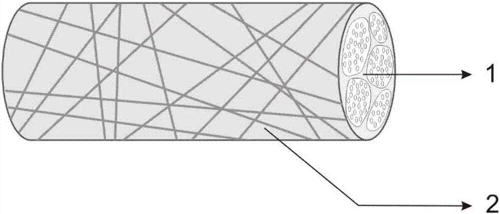
[0049] 4. Freeze-dry the product in a lyophilizer (at a temperature of -20°C) for 8 hours, and then sterilize and package it with cobalt-60 with an irradiation dose of 15KGy to obtain the final product. The schematic diagram is as follows figure 1 shown.
[0050] In the product prepared in this example, the thickness of the polylactic acid fiber membrane attached to the surface of the heterogeneous decellularized nerve graft was 0.07 mm.

Example Embodiment

[0051] Example 3:
[0052] 1. Dissolve polylactic acid-glycolic acid copolymer with a weight average molecular weight of 150,000 in acetone at a concentration of 0.15g/ml. After stirring at room temperature to form a uniform and stable solution, put it into a syringe, and the syringe needle Connect to high voltage power supply.
[0053] 2. Take the roller fixed with the xenogeneic decellularized nerve as the collection device, and the rotation speed is 300 rpm. Start the electrospinning process, control the solution supply flow rate at 2ml/h, apply a voltage of 10KV, and set the distance between the high voltage terminal and the ground terminal at 12.5cm.
[0054] 3. Washing the collected nanofiber-modified acellular nerve graft with ethanol and deionized water for 8 hours respectively, and repeating 4 times.
[0055] 4. Freeze-dry the product in a lyophilizer (at a temperature of -15°C) for 6 hours, then sterilize it with an irradiation dose of 13KGy electron beams and package it to obtain the final product. The schematic diagram is as follows: figure 1 shown.
[0056] In the product prepared in this embodiment, the thickness of the polylactic acid-caprolactone copolymer fiber membrane attached to the surface of the heterogeneous decellularized nerve graft is 0.1 mm.

PUM

PropertyMeasurementUnit
Thickness0.01 ~ 0.5mm
Mass volume concentration0.02 ~ 0.2g/ml
Thickness0.07mm
tensileMPa
Particle sizePa
strength10

Description & Claims & Application Information

We can also present the details of the Description, Claims and Application information to help users get a comprehensive understanding of the technical details of the patent, such as background art, summary of invention, brief description of drawings, description of embodiments, and other original content. On the other hand, users can also determine the specific scope of protection of the technology through the list of claims; as well as understand the changes in the life cycle of the technology with the presentation of the patent timeline. Login to view more.

Similar technology patents

Classification and recommendation of technical efficacy words

  • Strong combination
  • Evenly distributed

Method for processing high-temperature alloy furnace tube

ActiveCN101565807AStrong combinationMitigation of catalytic cokingSolid state diffusion coatingProduct gasMetallurgy
Owner:CHINA UNIV OF PETROLEUM (BEIJING)

Preparation method of macroporous aluminum oxide with diplopore distribution

InactiveCN101214454AStrong combinationOvercoming low mechanical strengthCatalyst carriersCarbon blackPetroleum
Owner:SHANGHAI SECOND POLYTECHNIC UNIVERSITY

Drug-eluting stent (DES) with multicoating

InactiveCN1465410AEvenly distributedNo crackStentsSurgeryDrugDrug-eluting stent
Owner:SHANGHAI MICROPORT MEDICAL (GROUP) CO LTD

Mould for making glass container

ActiveCN101298355AIncrease temperatureEvenly distributedVacuum and blow machinesGlass containerMaterials science
Owner:CHANGSHU WEIHENG MOLD MFG
Why Eureka
Try Eureka