Bionic tissue engineering scaffold containing micropore and nanofiber composite structure and preparation method thereof

A technology of tissue engineering scaffolds and nanofibers, applied in tissue regeneration, fiber treatment, non-woven fabrics, etc., can solve the problems of small micropores of scaffolds, difficult cells, poor mechanical properties, etc., and achieve the effect of promoting cell adhesion

Active Publication Date: 2018-02-23
TSINGHUA UNIV +1
View PDF6 Cites 12 Cited by
  • Summary
  • Abstract
  • Description
  • Claims
  • Application Information

AI Technical Summary

Problems solved by technology
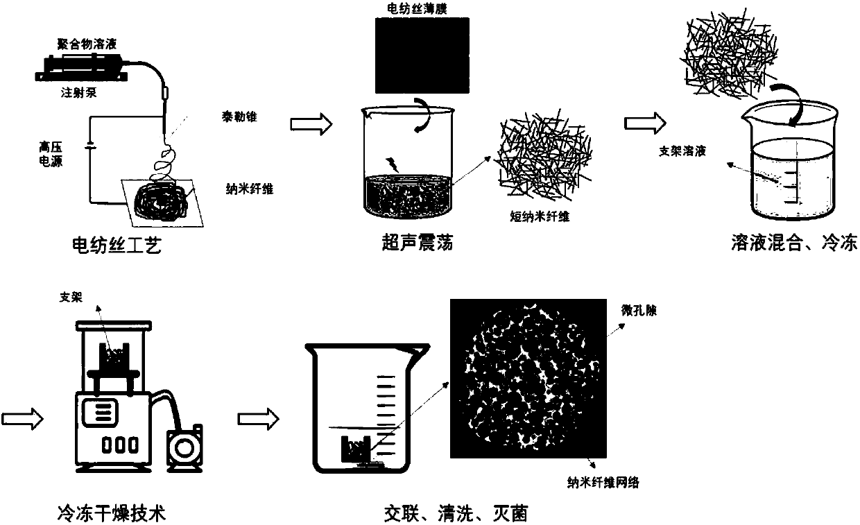
The disadvantage of the electrospinning process is that the prepared scaffold has small pores, it is difficult for cells to grow into the scaffold, and the general mechanical properties are poor.
It can be seen that none of the above existing methods can prepare biomimetic tissue engineering scaffolds with both micropores and nanofiber networks.

Method used


Image

  • Bionic tissue engineering scaffold containing micropore and nanofiber composite structure and preparation method thereof
  • Bionic tissue engineering scaffold containing micropore and nanofiber composite structure and preparation method thereof
  • Bionic tissue engineering scaffold containing micropore and nanofiber composite structure and preparation method thereof

Examples


Embodiment 1

[0049] Example 1: Preparation of a biomimetic tissue engineering scaffold with micropores and nanofiber network

[0050] Dissolve gelatin (Gelatin, 15k~25kDa) in PBS buffer solution to prepare a gelatin solution with a concentration of 0.01g / mL; dissolve sodium alginate in PBS buffer solution to prepare a concentration of 0.01g / mL alginic acid Sodium solution; the above two solutions are prepared into a uniform, bubble-free, and precipitation-free mixed solution, namely a gelatin-sodium alginate mixed solution, the mass ratio of gelatin to sodium alginate is 1:1, and the concentration of gelatin is 0.005g / mL, the concentration of sodium alginate is 0.005g / mL.

[0051] The collagen type I (Collagen, sigma) was dissolved in 1% acetic acid solution to prepare a collagen solution with a concentration of 0.08g / mL, crosslinked with 0.5wt% genipin solution, and then dissolved in hexafluoroiso In propanol (HFIP), an electrospinning equipment is used to prepare collagen fibers (diameter o...

Embodiment 2

[0054] Example 2: Preparation of a biomimetic tissue engineering scaffold with micropores and nanofiber network

[0055] Rat tail type I collagen (SIGMA company, product number C7661) was dissolved in 1.0wt% acetic acid solution to prepare a collagen solution with a mass fraction of 0.01g / mL; chitosan was dissolved in 1.0wt% acetic acid solution to prepare Prepare a chitosan solution with a mass fraction of 0.01g / mL; prepare the above two solutions into a homogeneous, bubble-free, and precipitate-free mixed solution, that is, a collagen-chitosan solution. The mass ratio of collagen to chitosan is 1 :1. The concentration of collagen is 0.005g / mL, and the concentration of chitosan is 0.005g / mL.

[0056] Dissolve polycaprolactone (PCL) in hexafluoroisopropanol (HFIP) to prepare a 0.1g / mL PCL solution, and use electrospinning equipment to prepare PCL fibers (with a diameter of 2.0±0.5μm), where the syringe is extruded The speed is 0.8ml / h, the voltage is 20kV, and the collection devic...

Embodiment 3

[0059] Example 3: Preparation of a biomimetic tissue engineering scaffold with micropores and nanofiber network

[0060] Dissolve silk fiber in 1.0wt% acetic acid solution to prepare a silk fibroin solution with a mass fraction of 0.005g / mL; dissolve chitosan in 1.0wt% acetic acid solution to prepare a mass fraction of 0.015g / mL chitosan solution; prepare the above two solutions into a uniform, bubble-free, and precipitation-free mixed solution, that is, a silk fibroin-chitosan solution. The mass ratio of silk fibroin to chitosan is 3 :1, the concentration of silk fibroin is 0.005g / mL, and the concentration of chitosan is 0.015g / mL.

[0061] Poly D, L-lactide (PDLLA) was dissolved in chloroform to prepare 0.15g / mL PDLLA solution, and PDLLA fiber (diameter 5.0±2.5μm) was prepared using electrospinning equipment, where the extrusion speed of the syringe It is 1.6ml / h, the voltage is 20kV, and the collection device is about 15cm away from the needle tip. The collected PDLLA fibers w...


PUM

PropertyMeasurementUnit
pore sizeaaaaaaaaaa
diameteraaaaaaaaaa
lengthaaaaaaaaaa

Abstract

The invention discloses a bionic tissue engineering scaffold containing a micropore and nanofiber composite structure and a preparation method thereof. The bionic tissue engineering scaffold comprisesmicropores and a nanofiber network structure. The micropores have diameters of 20-200 micrometers, the diameter of the nanofiber network structure is 10-2000nm and the length of the nanofiber networkstructure is 1-100 micrometers. The bionic tissue engineering scaffold fully simulates the structural features of a natural extracellular matrix, the micropores provide conditions for cell growth into the scaffold and high porosity is conducive to permeation and diffusion of oxygen and nutrients. The network structure of the nanometer fiber network bionic extracellular matrix can promote cell adhesion, growth, proliferation, differentiation and migration. The preparation method well solves the problems of the existing traditional scaffold preparation method and has the potential of becoming anovel tissue engineering scaffold preparation technology.

Description

Technical field [0001] The invention relates to a bionic tissue engineering scaffold containing a micropore and nanofiber composite structure and a preparation method thereof, belonging to the technical field of tissue engineering and biological manufacturing. Background technique [0002] Tissue Engineering and Regenerative Medicine (Tissue Engineering and Regenerative Medicine) is a new interdisciplinary science that repairs and replaces damaged tissues and organs by constructing artificial tissues with certain structure and function in vitro. At present, tissue engineering products have been applied in skin, bone / cartilage, bladder, blood vessels, liver, nerves and other fields, providing new possibilities for humans to treat various tissue and organ injuries. The classic strategy of tissue engineering is the three elements of cells, scaffolds and growth factors. Among them, the scaffold provides structural support for cells through the biomimetic extracellular matrix (ECM), a...

Claims


Application Information

Patent Timeline
Patent Type & Authority Applications(China)
IPC IPC(8): A61L27/26A61L27/56A61L27/50D01D5/00D04H1/728
CPCA61L27/26A61L27/50A61L27/56A61L2400/12A61L2430/40D01D5/003D01D5/0076D01D5/0092D04H1/728C08L5/04C08L5/08C08L67/04C08L89/00
Inventor 孙伟方永聪张婷莉莉安娜.利维拉尼奥尔多·博卡奇尼
Owner TSINGHUA UNIV
Why Eureka
Try Eureka