Therapeutic inhibitor of vascular smooth muscle cells

a technology of vascular smooth muscle cells and inhibitors, which is applied in the direction of nitro compound active ingredients, instruments, prostheses, etc., can solve the problems of significant morbidity and mortality, no surgical intervention or post-surgical treatment (to date) has proven effective in preventing restenosis, and the proliferation or hyperactivity of pathological cells is reduced or eliminated. , to achieve the effect of reducing or eliminating the proliferation or hyperactivity of pathological cells, reducing or eliminating the pathological

Inactive Publication Date: 2007-06-28
BOSTON SCI SCIMED INC
View PDF27 Cites 0 Cited by
  • Summary
  • Abstract
  • Description
  • Claims
  • Application Information

AI Technical Summary

Benefits of technology

[0009] In one aspect of the invention, new therapeutic methods and therapeutic conjugates are provided for inhibiting vascular smooth muscle cells in a mammalian host. The therapeutic conjugates contain a vascular smooth muscle binding protein or peptide that binds in a specific manner to the cell membranes of a vascular smooth muscle cell or an interstitial matrix binding protein / peptide that binds in a specific manner to interstitial matrix (e.g., collagen) of the artery wall, coupled to a therapeutic agent that inhibits the activity of the cell. In one embodiment, inhibition of cellular activity results in reducing, delaying, or eliminating stenosis after angioplasty or other vascular surgical procedures. The therapeutic conjugates of the invention achieve these advantageous effects by associating with vascular smooth muscle cells and pericytes, which may transform into smooth muscle cells. The therapeutic conjugate may contain: (1) therapeutic agents that alter cellular metabolism or are inhibitors of protein synthesis, cellular proliferation, or cell migration; (2) microtubule and microfilament inhibitors that affect morphology or increases in cell volume; and / or (3) inhibitors of extracellular matrix synthesis or secretion. In one representative embodiment, the conjugates include a cytotoxic therapeutic agent that is a sesquiterpenoid mycotoxin such as a verrucarin or a roridin. Other embodiments involve cytostatic therapeutic agents that inhibit DNA synthesis and proliferation at doses that have a minimal effect on protein synthesis such as protein kinase inhibitors (e.g., staurosporin), suramin, and nitric oxide releasing compounds (e.g., nitroglycerin) or analogs or functional equivalents thereof. In addition, therapeutic agents that inhibit the contraction or migration of smooth muscle cells and maintain an enlarged luminal area following, for example, angioplasty trauma (e.g., the cytochalasins, such as cytochalasin B, cytochalasin C, cytochalasin D or the like) are also contemplated for use in accordance with the present invention. Other aspects of the invention relate to vascular smooth muscle binding proteins that specifically associate with a chondroitin sulfate proteoglycan (CSPG) expressed on the membranes of a vascular smooth muscle cell, and in a preferred embodiment this CSPG has a molecular weight of about 250 kDaltons. In preferred embodiments the vascular smooth muscle binding protein binds to a CSPG target on the cell surface with an association constant of at least 10−4 M. In another preferred embodiment, the vascular smooth muscle binding protein contains a sequence of amino acids found in the Fab, Fv or CDR (complementarity determining regions) of monoclonal antibody NR-AN-01 or functional equivalents thereof.
[0010] Other aspects of the invention include methods for inhibiting stenosis, e.g., following angioplasty in a mammalian host, by administering to a human or animal subject in need of such treatment a therapeutically effective dosage of a therapeutic conjugate of the invention. In one representative embodiment, the dosage of therapeutic conjugate may be administered with an infusion catheter, to achieve a 10−3 M to 10−12 M concentration of said therapeutic conjugate at the site of administration in a blood vessel.
[0011] The present invention also contemplates therapeutic methods and therapeutic dosage forms involving sustained release of therapeutic agent to target cells. Preferably, the target cells are vascular smooth muscle cells, cancer cells, somatic cells requiring modulation to ameliorate a disease state and cells involved in immune system-mediated diseases that are accessible by local administration of the dosage form. Consequently, the methods and dosage forms of this aspect of the present invention are useful for inhibiting vascular smooth muscle cells in a mammalian host, employing a therapeutic agent that inhibits the activity of the cell (e.g., proliferation, contraction, migration or the like) but does not kill the cell and a vascular smooth muscle cell binding protein. Also, the methods and dosage forms of this aspect of the present invention are useful for inhibiting target cell proliferation or killing such target cells, employing a therapeutic agent that inhibits proliferation or is cytotoxic to the target cells and a target cell binding protein. In addition, the methods and dosage forms of this aspect of the present invention are useful for delivering cytostatic, cytocidal or metabolism modulating therapeutic agents to target cells, such as effector cells of the immune system, that are accessible by local administration of the dosage form, employing a target cell binding protein. Finally, dosage forms of the present invention are useful to reduce or eliminate pathological proliferation or hyperactivity of normal tissue (i.e., somatic cells).
[0012] For example, one specific therapeutic method and dosage form of the present method involves the placement of metallic, plastic or biodegradable intravascular stents comprising a biodegradable coating or a porous non-biodegradable coating, having dispersed therein the sustained-release dosage form. In an alternative embodiment, a biodegradable stent may also have the therapeutic agent impregnated therein, i.e., in the stent matrix. Utilization of a biodegradable stent with the therapeutic agent impregnated therein which is further coated with a biodegradable coating or with a porous non-biodegradable coating having the sustained release-dosage form dispersed therein is also contemplated. This embodiment of the invention would provide a differential release rate of the therapeutic agent, i.e., there would be a faster release of the therapeutic agent from the coating followed by delayed release, of the therapeutic agent that was impregnated in the stent matrix upon degradation of the stent matrix. Preferably, in this embodiment of the invention, the therapeutic agent is a cytochalasin, and most preferably is cytochalasin B, or a functionally equivalent analogue thereof. The intravascular stent thus provides a mechanical means of providing an increase in luminal area of a vessel, in addition to that provided via the biological stenting action of the cytochalasin B releasably embedded therein. Furthermore, this embodiment of the invention also provides an increase in the efficacy of intravascular stents by reducing or preventing intimal proliferation. This inhibition of intimal smooth muscle cells and stroma produced by the smooth muscle allows for more rapid and complete re-endothelization following the intraventional placement of the vascular stent. The increased rate of re-endothelization and stabilization of the vessel wall following stent placement would reduce the loss of luminal area and decreased blood flow which is the primary cause of vascular stent failures.
[0013] The dosage forms of the present invention are preferably either non-degradable microparticulates or nanoparticulates or biodegradable microparticulates or nanoparticulates. More preferably, the microparticles or nanoparticles are formed of a polymer containing matrix that biodegrades by random, nonenzymatic, hydrolytic scissioning. A particularly preferred structure is formed of a mixture of thermoplastic polyesters (e.g., polylactide or polyglycolide) or a copolymer of lactide and glycolide components. The lactide / glycolide structure has the added advantage that biodegradation thereof forms lactic acid and glycolic acid, both normal metabolic products of mammals.
[0014] Preferable therapeutic agents dispersed within the microparticulates or nanoparticulates are those exhibiting inhibition of a therapeutically significant target cell activity without killing the target cell, or target cell killing activity. For treatment of restenosis of vascular smooth muscle cells, useful therapeutic agents inhibit target cell activity (e.g., proliferation or migration) without killing the target cells. Preferred therapeutic moieties for this purpose are protein kinase inhibitors (e.g., staurosporin or the like), smooth muscle migration and / or contraction inhibitors (e.g., the cytochalasins, such as cytochalasin B, cytochalasin C, cytochalasin D or the like), suramin, and nitric oxide-releasing compounds, such as nitroglycerin, or analogs or functional equivalents thereof. In cancer therapy, useful therapeutic agents inhibit proliferation or are cytotoxic to the target cells. Preferred therapeutic moieties for this purpose are Roridin A and Pseudomonas exotoxin, or analogs or functional equivalents thereof. For treatment of immune system-modulated diseases, such as arthritis, useful therapeutic agents deliver cytostatic, cytocidal or metabolism-modulating therapeutic agents to target cells that are accessible by local administration of the dosage form. Preferred therapeutic moieties for this purpose are Roridin A, Pseudomonas exotoxin, suramin and protein kinase inhibitors (e.g., staurosporin), sphingosine, or analogs or functional equivalents thereof. For treatment of pathologically proliferating normal tissues (e.g., proliferative vitreoretinopathy, corneal pannus and the like), anti-proliferative agents or antimigration agents are preferred (e.g., cytochalasins, taxol, somatostatin, somatostatin analogs, N-ethylmaleimide, antisense oligonucleotides and the like).

Problems solved by technology

Stenosis following PTCA remains a significant problem, with from 25% to 35% of the patients developing restenosis within 1 to 3 months.
Restenosis results in significant morbidity and mortality and frequently necessitates further interventions such as repeat angioplasty or coronary bypass surgery.
No surgical intervention or post-surgical treatment (to date) has proven effective in preventing restenosis.
The processes responsible for stenosis after PTCA are not completely understood but may result from a complex interplay among several different biologic agents and pathways.
Compounds that reportedly suppress smooth muscle proliferation in vitro (4, 6, 7) may have undesirable pharmacological side effects when used in vivo.
Heparin is an example of one such compound, which reportedly inhibits smooth muscle cell proliferation in vitro but when used in vivo has the potential adverse side effect of inhibiting coagulation.
Heparin peptides, while having reduced anti-coagulant activity, have the undesirable pharmacological property of having a short pharmacological half-life.
First, inhibitory agents may have systemic toxicity that could create an unacceptable level of risk for patients with cardiovascular disease.
Second, inhibitory agents might interfere with vascular wound healing following surgery and that could either delay healing or weaken the structure or elasticity of the newly healed vessel wall.
Third, inhibitory agents killing smooth muscle cells could damage surrounding endothelium and / or other medial smooth muscle cells.
Fourth, delivery of therapeutically effective levels of an inhibitory agent may be problematic from several standpoints: namely, a) delivery of a large number of molecules into the intercellular spaces between smooth muscle cells may be necessary, i.e., to establish favorable conditions for allowing a therapeutically effective dose of molecules to cross the cell membrane; b) directing an inhibitory drug into the proper intracellular compartment, i.e., where its action is exerted, may be difficult to control; and, c) optimizing the association of the inhibitory drug with its intracellular target, e.g., a ribosome, while minimizing intercellular redistribution of the drug, e.g., to neighboring cells, may be difficult.
As is apparent from the foregoing, many problems remain to be solved in the use of inhibitory drugs, including cytotoxic agents, to effectively treat smooth muscle cell proliferation.

Method used


Image

  • Therapeutic inhibitor of vascular smooth muscle cells
  • Therapeutic inhibitor of vascular smooth muscle cells
  • Therapeutic inhibitor of vascular smooth muscle cells

Examples


example 1

Binding to Vascular Smooth Muscle Cells in the Blood Vessel Wall In Vivo

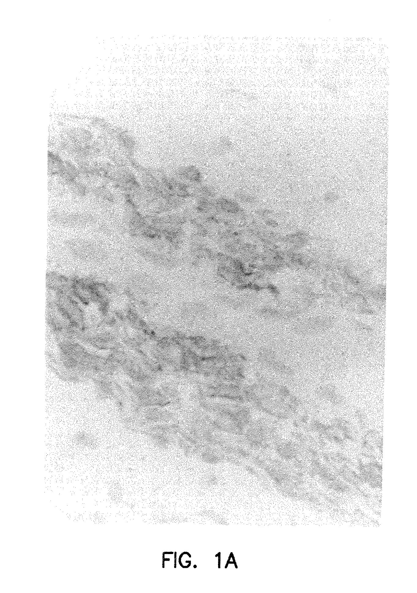
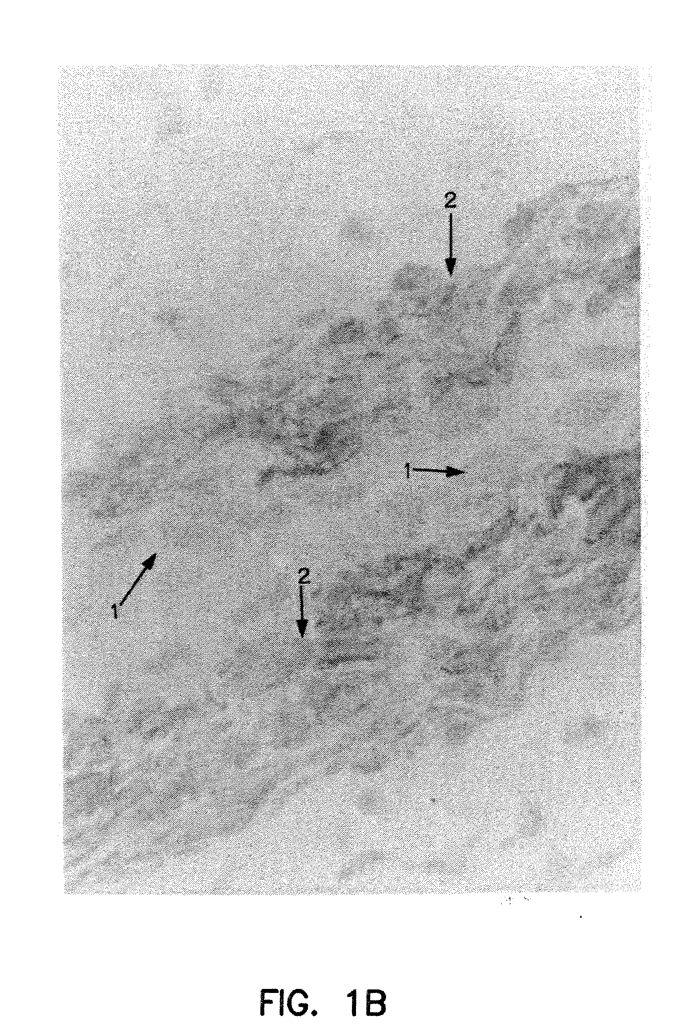
[0176]FIG. 1A illustrates the binding of NR-AN-01 (a murine IgG2b MAb) to the smooth muscle cells in the vascular wall of an artery in a 24-year old male patient, 4 days after the i.v. administration of NR-AN-01. FIG. 1A is a photomicrograph of a histological section taken through the medial region of an arterial wall of the patient after NR-AN-01 administration, where the section was reacted ex vivo with HRP-conjugated goat anti-mouse IgG. The reaction of the HRP conjugate with NR-AN-01 MAb was visualized by adding 4-chloro-1-naphthol or 3,3′-diaminobenzidine tetrahydrochloride as a peroxidase substrate (chromogen). The reaction product of the substrate forms an insoluble purple or dark brown precipitate at the reaction site (shown at #2, FIG. 1B). A counter stain was used to visualize collagenous extracellular matrix material (shown at #2, FIG. 1B) or cell nuclei (#1, FIG. 1B). Smooth muscle cells are visuali...

example 2

Therapeutic Conjugates Containing Trichothecene Therapeutic Agents

[0177] Conjugates of NR-AN-01 and Roridin A were constructed by chemically coupling a hemisuccinate derivative of the trichothecene cytotoxin (as described below) to a monoclonal antibody designated NR-AN-01. Two conjugates were prepared, one coupled at the Roridin A 2′ position and one at the 13′ position. Two schemes were used in this synthesis, as depicted in FIG. 2 and FIG. 3. The conjugate was then purified from unreacted Roridin A by PD-10 SEPHAROSE® column chromatography (Pharmacia; Piscataway, N.J.), analyzed by size exclusion high pressure liquid chromatography, and the column fractions were characterized by SDS-PAGE and isoelectric focusing (IEF), as described below.

[0178]FIG. 2 shows diagrammatically the first reaction scheme for synthesis of Roridin A hemisuccinyl succinimidate (RA-HS—NHS) through a two step process with reagents: succinic anhydride, triethylamine (NEt3) and dimethyl amino pyridine (DMAP...

example 3

Kinetics of Binding to Smooth Muscle Cells

[0200] For administration by i.v. catheter, it is desirable that the therapeutic conjugates of the invention be administered in less than 3 to 5 minutes, so that blood flow can be reestablished in the patient. Therefore, studies were conducted to determine the binding kinetics of a smooth muscle binding protein with a Ka of >109 liter / mole. Because human vascular smooth muscle cells grow slowly in culture, and baboon smooth muscle cells were found to express the human CSPG cell surface marker, BO54 baboon artery smooth muscle cells and human A375 M / M (melanoma; ATCC #CRL1619) cells bearing CSPG surface marker were used in many of the studies described in the EXAMPLES, below.

[0201] For the kinetic binding studies, A375 M / M and BO54 cells were seeded in sterile 96 well microtiter plates at 2500 cells / well. Plates were wrapped in aluminum foil, and incubated at 37° C. overnight in a humidified atmosphere of 5% CO2 / 95% air. After approximately...


PUM


Abstract

Methods are provided for inhibiting stenosis following vascular trauma or disease in a mammalian host, comprising administering to the host a therapeutically effective dosage of a therapeutic conjugate containing a vascular smooth muscle binding protein that associates in a specific manner with a cell surface of the vascular smooth muscle cell, coupled to a therapeutic agent dosage form that inhibits a cellular activity of the muscle cell. Methods are also provided for the direct and / or targeted delivery of therapeutic agents to vascular smooth muscle cells that cause a dilation and fixation of the vascular lumen by inhibiting smooth muscle cell contraction, thereby constituting a biological stent.

Description

RELATED APPLICATIONS [0001] This application is a continuation of U.S. application Ser. No. 09 / 590,002, filed Jun. 8, 2000, which is a continuation of U.S. application Ser. No. 08 / 389,712, filed Feb. 15, 1995, now U.S. Pat. No. 6,515,009, issued Feb. 4, 2003, the entire disclosure of each of which is incorporated herein by reference in its entirety.FIELD OF THE INVENTION [0002] This invention relates generally to therapeutic methods involving surgical or intravenous introduction of binding partners directed to certain target cell populations, such as smooth muscle cells, cancer cells, somatic cells requiring modulation to ameliorate a disease state and effector cells of the immune system, particularly for treating conditions such as stenosis following vascular trauma or disease, cancer, diseases resulting from hyperactivity or hyperplasia of somatic cells and diseases that are mediated by immune system effector cells. Surgical or intravenous introduction of active agents capable of ...

Claims


Application Information

Patent Timeline
Patent Type & Authority Applications(United States)
IPC IPC(8): A61K31/407A61F2/02A61F2/82A61K9/00A61K31/00A61K31/04A61K31/131A61K31/135A61K31/138A61K31/165A61K31/17A61K31/335A61K31/337A61K31/352A61K31/365A61K31/40A61K31/4025A61K31/4035A61K31/519A61K31/551A61K31/704A61K47/48A61L31/16A61P9/08
CPCA61K9/0024A61K31/00A61K31/04A61K31/131A61K31/135A61K31/138A61K31/165A61K31/17A61K31/337A61K31/352A61K31/365A61K31/40A61K31/4025A61K31/4035A61K31/407A61K31/519A61K31/551A61K31/704A61K47/48A61K47/48007A61K47/48384A61K47/48407A61K47/48438A61K47/48492A61K47/48538A61K47/48861A61K47/48876A61K47/48992A61L31/16A61L2300/416A61L2300/602A61L2300/604A61L2300/606B82Y5/00G01N2800/323A61L31/148Y10S977/906Y10S977/775Y10S977/916Y10S977/905Y10S977/808A61K47/50A61K47/51A61K47/6803A61K47/6809A61K47/6817A61K47/6831A61K47/6843A61K47/6923A61K47/6927A61K47/6957A61P35/00A61P41/00A61P43/00A61P7/00A61P9/08A61P9/10
Inventor KUNZ, LAWRENCEANDERSON, PETER
Owner BOSTON SCI SCIMED INC
Why Eureka
Try Eureka