
Use of Two-Photon Excited Fluorescence in Assays of Clinical Chemistry Analytes

a technology of clinical chemistry and fluorescence, applied in the field of clinical chemistry, can solve the problems of high cost of cuvettes for clinical chemistry assays, inconvenient use, and inconvenient use, and achieve the effects of improving the accuracy of clinical chemistry assays

Active Publication Date: 2007-12-13
ARCTIC DIAGNOSTICS
View PDF6 Cites 4 Cited by
  • Summary
  • Abstract
  • Description
  • Claims
  • Application Information

AI Technical Summary

Benefits of technology

[0102] The present invention describes the use of two-photon excited fluorescence as detection method for cli

Problems solved by technology

This means, that any variation in the optical length of the disposable cuvettes or in the optical quality of the cuvette walls causes imprecision in the final assay result.
Due to this property of photometry, and on the other hand, due to rather strict precision requirements of clinical chemistry assays, the quality requirements focused on the disposable cuvette are very demanding.
For this reason the cuvettes for clinical chemistry assays are and remain expensive.
In practice, the cuvette is the major constituent of the cost of the photometric clinical chemistry assays.
The fluorescence based assays are, however, mainly used in research laboratories, whereas their use in the clinical chemistry practice is very limited.
In research laboratories, these properties give a clear advantage, but in clinical chemistry applications, this has not been found that advantageous because the requirements for sensitivity or dynamic range of the clinical chemistry assays are most often rather modest.
However, the sensitivity is sufficient only for a limited number of analytes.
This phenomenon remained for a long time without any practical use until the intensive laser light sources became available.
The disadvantage of the two-photon excitation technology is the need of an expensive laser capable of generating intense ultra short pulses with a high repetition frequency.
This trend, however, is in contradiction to the need of intensive care, emergency care and polyclinic duty, where IVD results from single patient samples are required without delay.
However, the health care organizations usually focus their attention to the direct assay cost and are not able to count the indirect cost of IVD in the whole health care system.
In addition, variable waiting time of individual patient samples in non-controlled conditions (temperature, day light exposure) inevitably causes changes in the composition of the sample leading to inaccurate assay result and diagnosis.
Point-of-care (POC) platforms are designed for testing of single samples and are characterized by limited assay portfolio.
In larger quantities, exceeding 10 000 tests per year, POC tests become less cost effective due to high price of disposable assay component.
The technology for distributed IVD is not yet very well developed.
There is not yet any ideal and suitable methodology and instrumentation commercially available, which would allow cost effective assays for distributed IVD.
There are not many analyzers or technologies commercially available, which fulfill these requirements.
These immunoassays, however, are usually employing either turbidimetry or nephelometry detection principles (agglutination assays), which do not allow high sensitivity assays but are limited to analytes of relatively high clinical reference concentration.
Heterogeneous immunoassay principle, however, does not allow separation-free assays and is therefore not ideal for distributed IVD.
One of the most important problems with the photometric methodology is the need for expensive test cuvette.
Re-usable flow-cuvettes, however, are not commonly used today due to their sensitivity to analyte and reagent carry-over and contamination.
These quality requirements necessitate very highly skilled manufacturing technology, thus making the production of the cuvettes expensive and the corresponding assays prone to inaccuracy due to cuvette-to-cuvette variation.
For this reason the cuvettes for clinical chemistry assays are and remain expensive.
In fact, the cuvette is the major constituent of the cost of clinical chemistry assays that are based on photometry detection.
An additional problem encountered with the precision of the photometric assays is related to the sample matrix interferences.
All these substances can interfere strongly in photometric measurements unless the sample is heavily diluted before the assay.
A third problem with photometric detection relates to the assay volumes.
Such a cuvette is not optimal with immunoassays because the reagents cost of immunoassays is typically ten times higher than the cost of clinical chemistry reagents.
Consequently, clinical chemistry analyzers with photometric detection are relative large in size and require rather large reagent and diluent volumes.
This property increases the cost of the immunoassays and the advantage of low assay volumes cannot be exploited.

Method used


Image

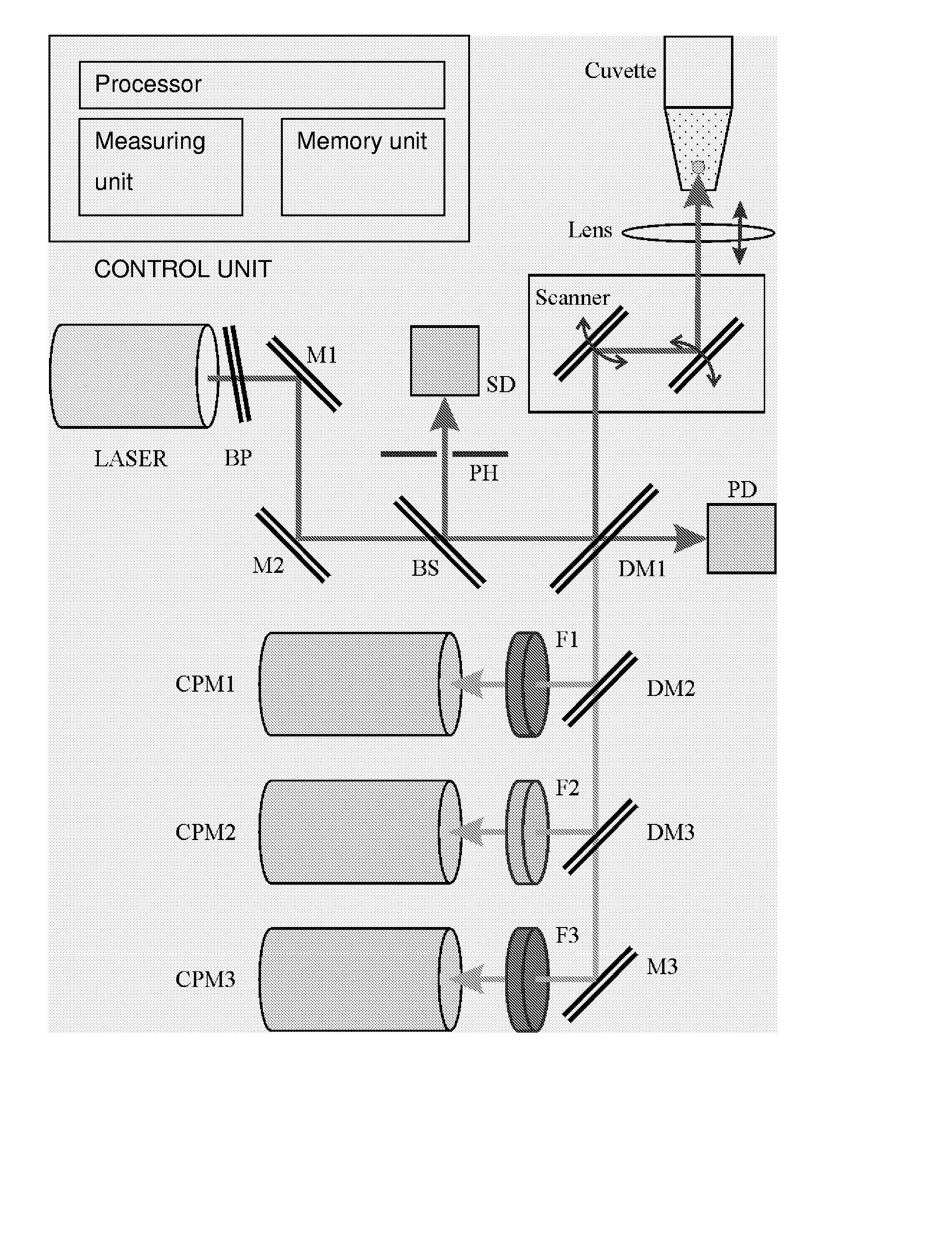
  • Use of Two-Photon Excited Fluorescence in Assays of Clinical Chemistry Analytes
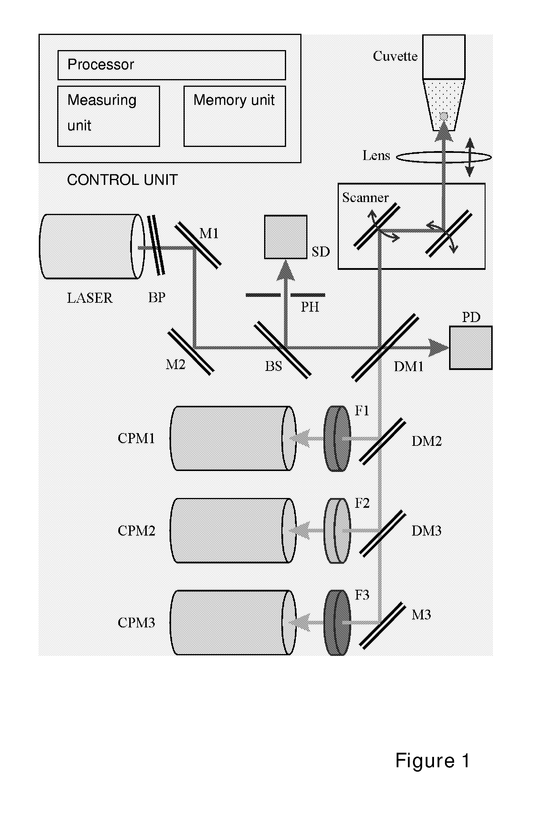
  • Use of Two-Photon Excited Fluorescence in Assays of Clinical Chemistry Analytes
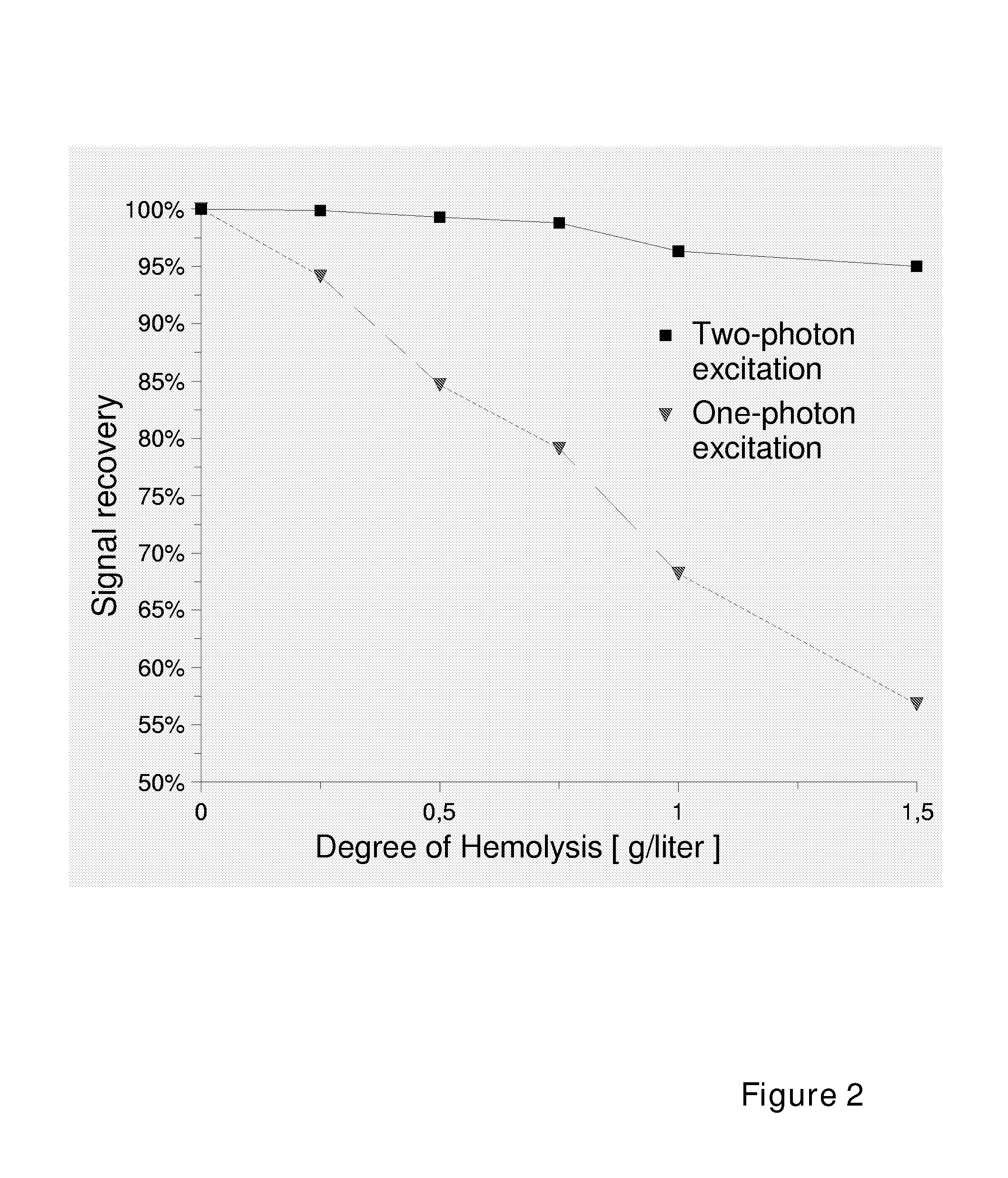
  • Use of Two-Photon Excited Fluorescence in Assays of Clinical Chemistry Analytes

Examples


example 1

[0146] Interference of Hemolysis on Signal Intensity of One-Photon Excited Fluorescence and Two-Photon Excited Fluorescence

[0147] Bovine serum albumin (BSA) was labeled with a fluorescent labeling reagent, ArcDia BF560 (Arctic Diagnostic Oy, Turku, Finland) to give label-BSA conjugate with substitution degree of 2.7. The conjugate was diluted with hemoglobin solution (variable concentration of hemoglobin, TRIS 50 mM, NaCl 150 mM, NaN3 10 mM, Tween 20 0.01%, BSA 0.5%, pH 8.0) to concentration of 100 nM. This solution was dispensed in the wells of a microtitration plate of either 384-well format (Greiner BIO-One GmbH, Code: 788096, μClear SV-plate, 2nd Gen, with black walls and plastic foil bottom) or 96-well format (Greiner BIO-One GmbH, code 655096) in aliquots of 10 and 100 μl, respectively. The 96-well plate was measured with a conventional one-photon excitation fluorometer (Ascent, Thermo Electron Oy, Vantaa, Finland) using excitation filter of 530 nm and emission filter 590 nm....

example 2

[0148] Effect of Cuvette Material on Inter-Cuvette Precision

[0149] Mouse monoclonal antibody was labeled with fluorescent label ArcDia BF 560-SE (Arctic Diagnostics Oy, Turku, Finland) according to a standard protocol [M. E. Waris et al. Anal. Biochem. 309 (2002), 67-74] to give a conjugate with substitution degree of 3.2 labels per IgG. The conjugate was diluted with buffer (TRIS 50, NaCl 150 mM, NaN3 10 mM, BSA 0.5%, Tween 20 0.01%, pH, 8.0) to concentration of 50 nM and dispensed into wells of two different microplates of 384-well format. One of the microplates was a low-cost standard microplate with plastic foil bottom window (Greiner BIO-One GmbH, Code: 788096, μClear SV-plate, 2nd Gen, with black walls and plastic foil bottom) while the other was a special plate with bottom window made of the highest quality optical glass (Greiner, SensoPlate™ 96 Well). The samples were measured with the two-photon excitation microfluorometer (ArcDia TPX Plate Reader, described in Example 1) ...

example 3

[0150] Assay of Total Bilirubin

[0151] The assay for total bilirubin was performed according to a modification of the assay protocol for total bilirubin of Sigma Diagnostics® (procedure no 605), which is based on the reaction between bilirubin and diazonium salts. The assay procedure no. 605 was followed with an exception that the addition of the alkaline tartrate reagent was omitted. Bilirubin was dissolved in a mixture containing 1 part dimethyl sulfoxide and 2 parts sodium carbonate (100 mM, aq.), and diluted further with buffer (Bovine serum albumin 40 g / l, TRIS 100 mM, pH 7.4) to give bilirubin standards of 2, 5, 10, 15 and 20 mg / dl. 2 μl of a bilirubin standard sample was dispensed in a assay cuvette (384-well plate, Greiner BIO-ONE GmbH, Code: 788096), followed by addition of caffeine solution (10 μl, caffeine 25 g / l, sodium benzoate 38 g / l, in NaAc solution, code 605-2, Sigma) and diazo reagent solution (5 μl, sulfanilic acid 1.25 mM, NaNO2 1 mM, HCl 0.05 M). The mixture was...


PUM


Abstract

The invention relates to an in vitro diagnostic method for quantification of a clinical chemistry analyte from a clinical sample wherein the clinical chemistry analyte undergoes a chemical reaction or reactions with a reagent or reagents in one or several steps, or in a reaction sequence, or catalyses a chemical reaction, or reactions, or a reaction in a reaction sequence of a reagent or reagents, in one or several steps, in a reaction system. The reaction or reactions or reaction sequence result in a change of a measurable property of a compound or compounds of said reaction or reactions or reaction sequence. Characteristic for the method is that said chemical reaction or reactions or reaction sequence results in formation of a two-photon fluorescent compound, or a change in two-photon fluorescence properties of the reaction system comprising at least one two-photon fluorescent compound, and the analyte is quantified by exciting said two-photon fluorescent compound or compounds and measuring two-photon exited fluorescence, and relating said measured fluorescence to method standardization data based on measurements obtained from reference material of said analyte. The present invention also relates to use of a fluorometric device employing two-photon fluorescence excitation for quantification of a clinical chemistry analytes. The present invention further relates to a system for quantification of clinical chemistry analytes from samples containing the analyte. Characteristic for the system is that it comprises a fluorometric device employing two-photon excited fluorescence for quantifying one or several clinical chemistry analytes, and a data processing unit with software for dedicated data reduction for quantification of the analyte or analytes using said fluorometric device. The present invention further relates to a software product for the system.

Description

FIELD OF THE INVENTION [0001] Present invention relates to clinical chemistry and to the use of two-photon excited fluorescence as detection principle for measurement of clinical chemistry analytes. BACKGROUND OF THE INVENTION [0002] The publications and other materials used herein to illustrate the background of the invention, and in particular, cases to provide additional details respecting the practice, are incorporated by reference. Clinical Chemistry Assays [0003] With clinical chemistry analytes we mean in this context metabolites or other components of serum that are commonly measured in the clinical chemistry practice. Examples of such analytes are as follows: Glucose, total cholesterol, HDL cholesterol, LDL cholesterol, triglycerides, bilirubin, creatinine, total proteins, iron, magnesium, urea, uric acid, ASAT, ALAT, amylase, LDH, GT, alkaline phosphatase, lipase and creatine kinase. In this context, clinical chemistry analytes do not include analytes, which are measured w...

Claims


Application Information

Patent Timeline
IPC IPC(8): G01N33/72G01N21/76G01N33/00G01N33/92G01N33/20G01NG01N21/64G01N33/53G01N33/58
CPCG01N21/6428G01N33/582Y10T436/19Y10T436/16Y10T436/175383Y10T436/20Y10T436/204998
Inventor SOINI, ERKKISOINI, ALEKSISOINI, JUHANIMELTOLA, NIKO
Owner ARCTIC DIAGNOSTICS
Learn More