[0083]Alternatively,
phage display libraries may be constructed from immunoglobulin variable regions that have been partially assembled
in vitro to introduce additional
antibody diversity in the
library (semi-synthetic libraries). For example,
in vitro assembled variable regions contain stretches of synthetically produced, randomized or partially randomized
DNA in those regions of the molecules that are important for
antibody specificity, e.g. CDR regions.
Phage antibodies specific for
bacteria such as staphylococci can be selected from the
library by exposing the bacteria or material thereof to a phage
library to allow binding of phages expressing
antibody fragments specific for the bacteria or material thereof. Non-bound phages are removed by washing and bound phages eluted for infection of E. coli bacteria and subsequent propagation. Multiple rounds of selection and propagation are usually required to sufficiently enrich for phages binding specifically to the bacteria or material thereof. If desired, before exposing the phage library to the bacteria or material thereof the phage library can first be subtracted by exposing the phage library to non-target material such as bacteria of a different family, species and / or strain or bacteria in a different
growth phase or material of these bacteria. These
subtractor bacteria or material thereof can be bound to a
solid phase or can be in suspension. Phages may also be selected for binding to complex antigens such as complex mixtures of bacterial proteins or (poly)peptides optionally supplemented with bacterial polysaccharides or other bacterial material. Host cells expressing one or more proteins or (poly)peptides of bacteria such as staphylococci may also be used for selection purposes. A
phage display method using these host cells can be extended and improved by subtracting non-relevant binders during screening by addition of an excess of host cells comprising no target molecules or non-target molecules that are similar, but not identical, to the target, and thereby strongly enhance the chance of finding relevant binding molecules. Of course, the subtraction may be performed before, during or after the screening with bacterial organisms or material thereof. The process is referred to as the Mabstract® process (Mabstract® is a
registered trademark of Crucell Holland B.V., see also U.S. Pat. No. 6,265,150 which is incorporated herein by reference).
[0093]Alternatively, the binding molecules, immunoconjugates,
nucleic acid molecules or compositions of the present invention can be in solution and the appropriate pharmaceutically acceptable
excipient can be added and / or mixed before or at the time of delivery to provide a unit dosage injectable form. Preferably, the pharmaceutically acceptable
excipient used in the present invention is suitable to high
drug concentration, can maintain proper fluidity and, if necessary, can
delay absorption.
[0098]In a further aspect, the binding molecules such as human
monoclonal antibodies (functional fragments and variants thereof), immunoconjugates, compositions, or pharmaceutical compositions of the invention can be used as a medicament. So, a method of treatment and / or prevention of a bacterial (
Gram-positive and / or
Gram-negative), e.g. a staphylococcal, infection using the binding molecules, immunoconjugates, compositions, or pharmaceutical compositions of the invention is another part of the present invention. The above-mentioned molecules can inter alia be used in the diagnosis, prophylaxis, treatment, or combination thereof, of a bacterial infection. They are suitable for treatment of yet untreated patients suffering from a bacterial infection and patients who have been or are treated for a bacterial infection. They may be used for patients such as hospitalized infants, premature infants, burn victims, elderly patients, immunocompromised patients, immunosuppressed patients, patient undergoing an
invasive procedure, and health
care workers. Each administration may protect against further infection by the bacterial
organism for up to three or four weeks and / or will retard the onset or progress of the symptoms associated with the infection. The binding molecules of the invention may also increase the effectiveness of existing antibiotic treatment by increasing the sensitivity of the bacterium to the antibiotic, may stimulate the
immune system to
attack the bacterium in ways other than through opsonization. This activation may result in
long lasting protection to the infection bacterium. Furthermore, the binding molecules of the invention may directly inhibit the growth of the bacterium or inhibit
virulence factors required for its survival during the infection.
[0129]The anti-staphylococcal IgGs and a control IgG (CR4374) were serially diluted in opsonophagocytosis buffer in a total volume of 20 μl to obtain dilutions having an IgG concentration of 2.50 μg / ml, 1.20 μg / ml, 0.60 μg / ml, 0.30 μg / ml, 0.15 μg / ml, 0.075 μg / ml, 0.0375 μg / ml and 0.019 μg / ml. Opsonic activity of dilutions was measured in the OPA
assay in a round bottom plate that was blocked with 1% BSA in PBS. As a control, the
assay was performed with no IgG. A 15 μl aliquot of a bacterial suspension containing 5.4×10⁶ cells was added to each well of the plate. When a bacterial suspension from S. aureus strain Cowan or S. epidermidis was used, the IgG / bacterium suspension was first incubated for 30 minutes at 37° C. while the plate was horizontally shaking (1300 rpm) in a Heidolph titramax 1000. Next, 15 μl of the differentiated HL-60 cells (total: 75×103 cells) were added to each well of the plate and the plate was incubated while shaking at 37° C. for 30-45 minutes. The final volume in the well was 50 μl. The reaction was stopped by adding 50 μl of wash buffer containing 4% v / v
formaldehyde. The content in each well was resuspended and transferred to
polystyrene disposable tubes for flow cytometric analysis. The samples were stored in the dark at 4° C. until analyzation. The tubes were vortexed for 3 seconds before sampling in the flow cytometer. To control the differentiation of the HL-60 cells the expression of the complement
receptor CD11b was measured. Fc-receptors of differentiated and non-differentiated cells were first blocked with rabbit IgG for 15 minutes on ice and the cells were subsequently labelled with CD11bAPC (BD) for 15 minutes on ice. Cells were considered properly differentiated when the mean fluorescent intensity (MFI) analyzed was at least between 10- to 100-fold higher compared to that of non-differentiated cells. Samples were assayed with a FACSCalibur immunocytometry
system (Becton Dickinson and Co., Paramus, N.J.) and were analyzed with CELLQuest
software (version 1.2 for Apple
system 7.1; Becton Dickinson). 7,000 gated HL-60 granulocytes were analyzed per tube. FAM-SE was excited at a
wavelength of 488 nm and the FAM-SE
fluorescence signal of gated viable HL-60 cells was measured for each antibody
dilution. IgGs were defined as positive in the phagocytic
assay when
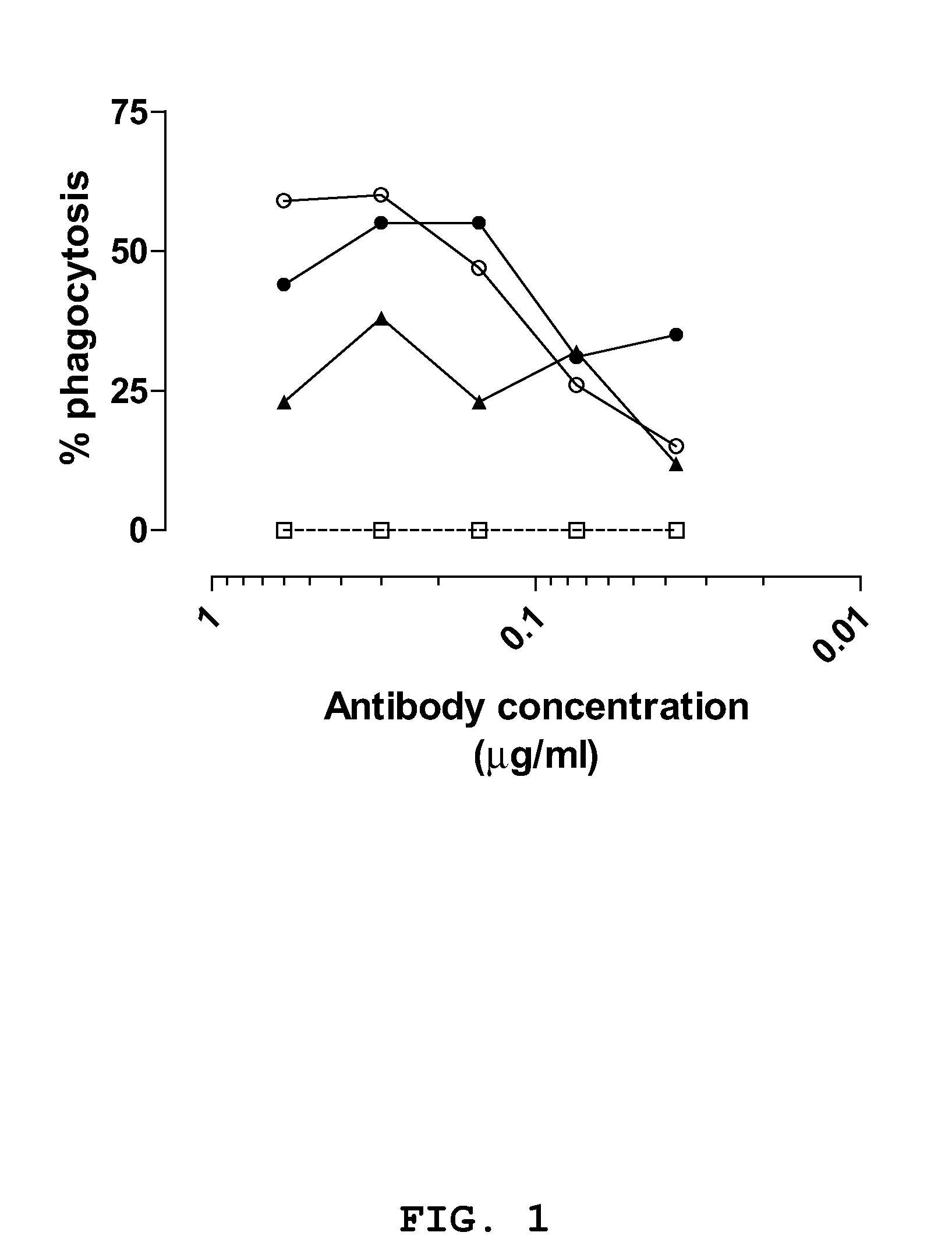
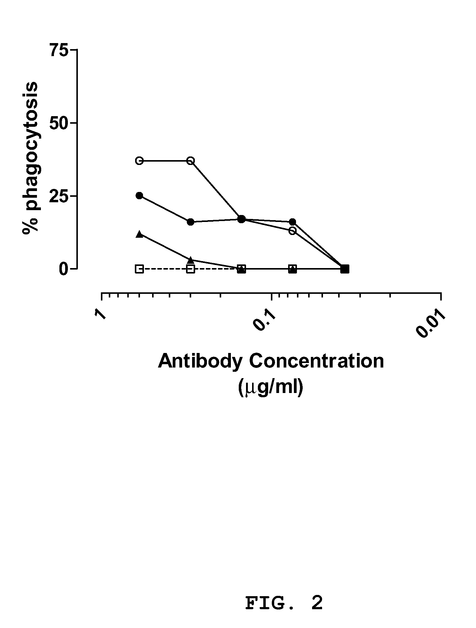
concentration dependent phagocytosis could be observed greater or equal to two times that of the control IgG. IgGs CR2430, CR5132 and CR5133 demonstrated opsonic activity against S. aureus strain Cowan in both the log (see FIG. 1) and stationary
growth phase (see FIG. 2). The three IgGs where more effective in enhancing phagocytic activity during the log phase of growth. IgGs CR5132 and CR5133 enhanced
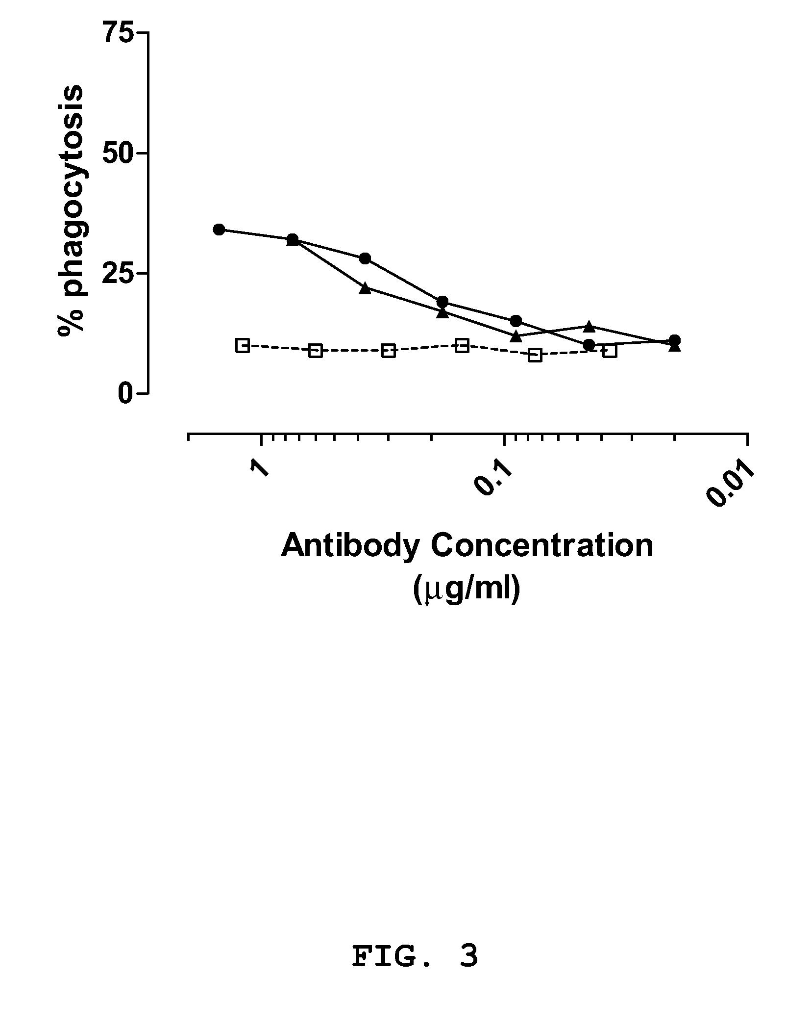
phagocytosis of S. aureus strain SA125 compared to the
negative control antibody (see FIG. 3) and antibody CR5133 significantly enhanced phagocytic activity of the differentiated HL60 cells against S. epidermidis strain SE131, when compared to the
negative control antibody (see FIG. 4).Example 8Breadth of Staphylococci
Specific IgG1 Binding Activity


