
PRO108 Antibody Compositions and Methods of Use and Use of PRO108 to Assess Cancer Risk

a technology of pro108 and composition, applied in the field of cancer risk assessment, can solve the problems of increased prostate cancer risk, etc., and achieves the effect of ovarian, colon, and reducing the risk of prostate cancer

Inactive Publication Date: 2009-09-17
DIAZYME LAB INC
View PDF1 Cites 3 Cited by
  • Summary
  • Abstract
  • Description
  • Claims
  • Application Information

AI Technical Summary

Benefits of technology

This approach provides a more sensitive and accurate method for assessing cancer risk and treating cancer, offering improved diagnostic and therapeutic options with minimal invasiveness and reasonable cost, enhancing patient outcomes.

Problems solved by technology

For example, these studies have revealed that as the number of CAG repeats decreases the transactivation ability of the gene product increases, as does the risk of prostate cancer.
Environmental / dietary risk factors which may increase the risk of prostate cancer include intake of saturated fat and calcium.
Despite the need for accurate staging of prostate cancer, current staging methodology is limited.
The wide variety of biological behavior displayed by neoplasms of the prostate has resulted in considerable difficulty in predicting and assessing the course of prostate cancer.
Techniques such as transrectal ultrasound, abdominal and pelvic computerized tomography, and MRI have not been particularly useful in predicting local tumor extension.
Unfortunately, women with ovarian cancer are typically asymptomatic until the disease has metastasized.
Reproductive factors have also been associated with an increased or reduced risk of ovarian cancer.
Current screening procedures for ovarian cancer, while of some utility, are quite limited in their diagnostic ability, a problem that is particularly acute at early stages of cancer progression when the disease is typically asymptomatic yet is most readily treated.
Pelvic examination has failed to yield adequate numbers of early diagnoses, and the other methods are not sufficiently accurate.
Moreover, the CA-125 test is prone to giving false positives in pre-menopausal women and has been reported to be of low predictive value in post-menopausal women.
Although transvaginal ultrasonography is now the preferred procedure for screening for ovarian cancer, it is unable to distinguish reliably between benign and malignant tumors, and also cannot locate primary peritoneal malignancies or ovarian cancer if the ovary size is normal.
While genetic testing for mutations of the BRCA1, BRCA2, hMSH2, and hMLH1 genes is now available, these tests may be too costly for some patients and may also yield false negative or indeterminate results.
While surgical staging is currently the benchmark for assessing the management and treatment of ovarian cancer, it suffers from considerable drawbacks, including the invasiveness of the procedure, the potential for complications, as well as the potential for inaccuracy.
Moreover, current procedures, while helpful in each of these analyses, are limited by their specificity, sensitivity, invasiveness, and / or their cost.
While effective at detecting early stage tumors, FOBT is unable to detect adenomatous polyps (premalignant lesions), and, depending on the contents of the fecal sample, is subject to rendering false positives.
Despite the advantages of these procedures, there are accompanying downsides: sigmoidoscopy, by definition, is limited to the sigmoid colon and below, colonoscopy is a relatively expensive procedure, and both share the risk of possible bowel perforation and hemorrhaging.
Double-contrast barium enema (DCBE) enables detection of lesions better than FOBT, and almost as well a colonoscopy, but it may be limited in evaluating the winding rectosigmoid region.
The CEA blood test, which involves screening the blood for carcinoembryonic antigen, shares the downside of FOBT, in that it is of limited utility in detecting colorectal cancer at an early stage.
While computerized tomography and magnetic resonance imaging are useful in staging colorectal cancer in its later stages, both have unacceptably low staging accuracy for identifying early stages of the disease, due to the difficulty that both methods have in (1) revealing the depth of bowel wall tumor infiltration and (2) diagnosing malignant adenopathy.
Rather, techniques such as transrectal ultrasound (TRUS) are preferred in this context, although this technique is inaccurate with respect to detecting small lymph nodes that may contain metastases.
Nonetheless, thirty to forty percent of patients will develop a recurrence of colon cancer following surgical resection, which in many patients is the ultimate cause of death.
All of these lead to genomic instability in colorectal cancers.
Another source of genomic instability in colorectal cancer is the defect of DNA mismatch repair (MMR) genes.
The inactivation of these proteins leads to the accumulation of mutations and causes genetic instability that represents errors in the accurate replication of the repetitive mono-, di-, tri- and tetra-nucleotide repeats, which are scattered throughout the genome (microsatellite regions).
Carcinogen metabolism enzymes such as GST, NAT, CYP and MTHFR are also associated with an increased or decreased colorectal cancer risk.
Moreover, current procedures, while helpful in each of these analyses, are limited by their specificity, sensitivity, invasiveness, and / or their cost.
However, stomach cancer persists in being responsible for considerable mortality rates in Asia, Europe and South America.
From an occupational standpoint, those who work in the metalworking, painting, fishing, ceramic, and printing industries all appear to have an elevated risk of acquiring stomach cancer.
A variation in the N-acetyltransferase 1 gene has also been linked to elevated risk of gastric cancer.
Unfortunately, the symptoms and clinical manifestations of gastric cancer typically do not appear early in the course of the disease, and the majority of patients have symptoms of the disease for six months or more prior to diagnosis.
Unfortunately, in the case of the first three tests, positive results are not necessarily obtained when gastric cancer is present, or false positives may result due to the presence of other conditions.
EUS, however, is limited for several reasons: (1) roughly 15% of patients present with non-traversable lesions, (2) there are regions of the stomach in which it is difficult to obtain high quality images, and (3) it has difficulty in discerning particular types of cancerous lesions.
CT scanning is of some utility when used in combination with other techniques, but it is too inaccurate to be used alone for several reasons: (1) it is limited in its ability to assess the tumor stage due to its inability to distinguish between the individual layers of the gastric wall, (2) it is highly inaccurate in assessing lymph node metastasis, and (3) it is generally unhelpful in assessing peritoneal or liver metastasis.
Nonetheless, other studies have indicated that MRI has little to offer to supplement a CT assessment.
The Japanese system is considerably more detailed, but in some cases may be overly complex and time consuming.
Likewise, chemotherapy has generally been of limited utility in treating gastric carcinomas, although neoadjuvant and adjuvant chemotherapy have been used with some success.
Pre- or postoperative adjuvant therapy is currently being studied due to the considerable risk for reoccurrence, as well as the fact that systemic metastasis is commonplace.
Moreover, current procedures, while helpful in each of these areas, are limited by their specificity, sensitivity, invasiveness, and / or their cost.

Method used


Image

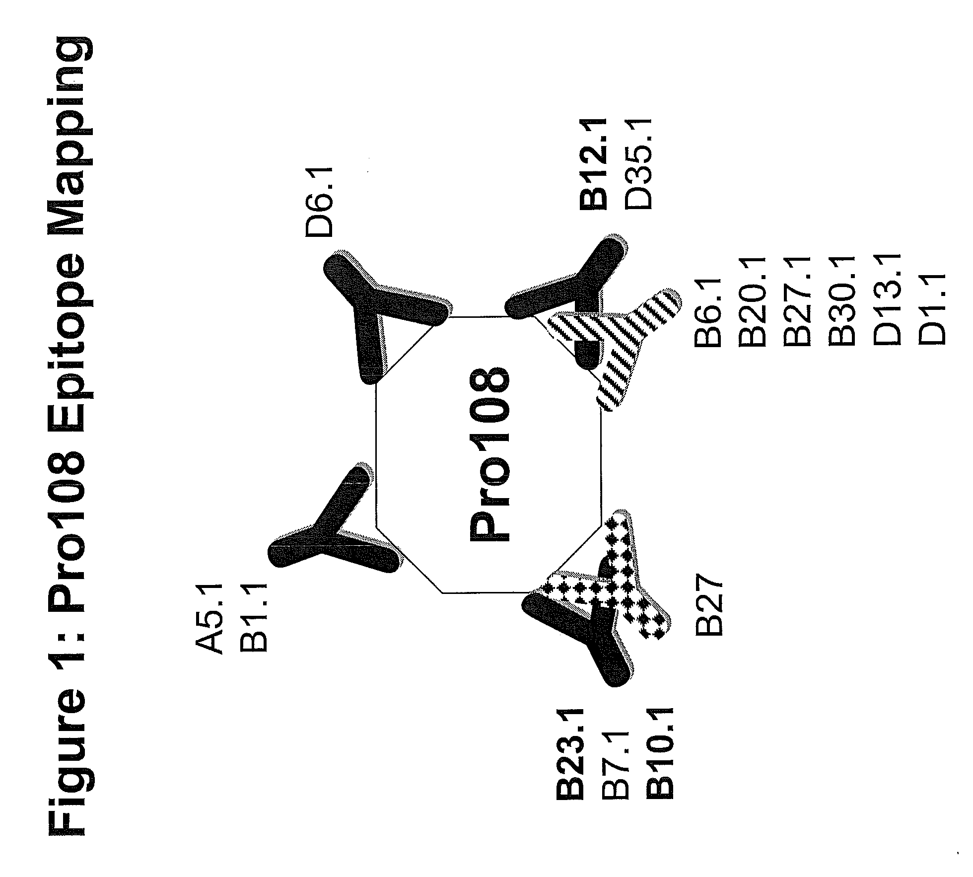
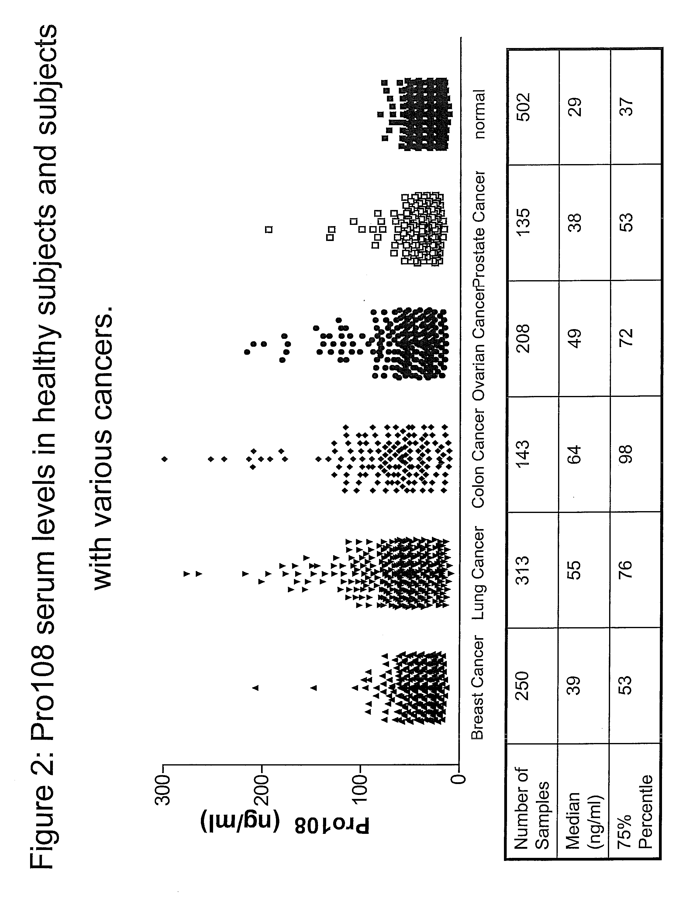
  • PRO108 Antibody Compositions and Methods of Use and Use of PRO108 to Assess Cancer Risk
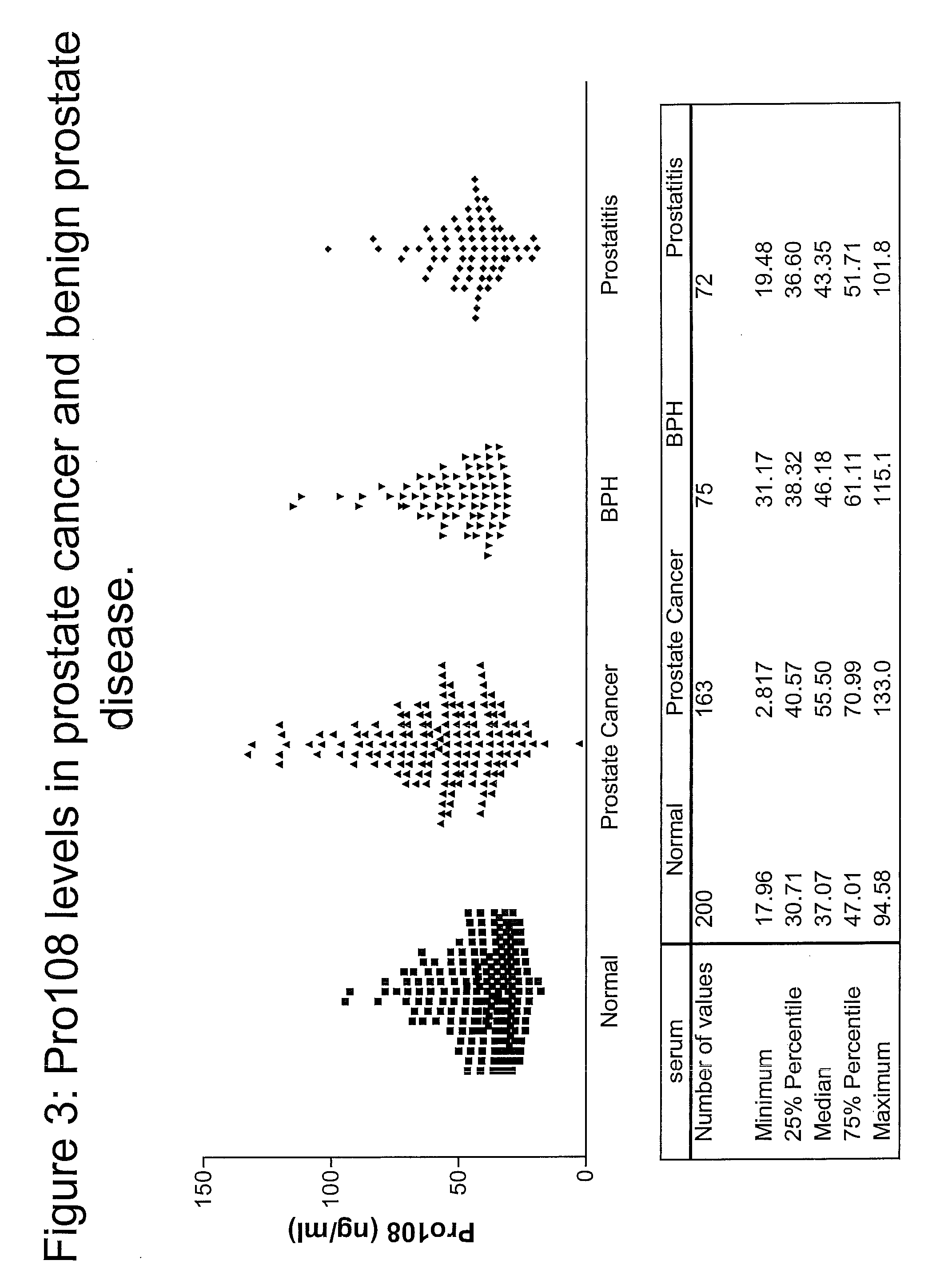
  • PRO108 Antibody Compositions and Methods of Use and Use of PRO108 to Assess Cancer Risk
  • PRO108 Antibody Compositions and Methods of Use and Use of PRO108 to Assess Cancer Risk

Examples


example 1

Production and Isolation of Monoclonal Antibody Producing Hybridomas

[0335]The following MAb / hybridomas of the present invention are described below: Pro108.A2, Pro108.A5, Pro108.B1, Pro108.B2, Pro108.B3, Pro108.B4, Pro108.B5, Pro108.B6, Pro108.B7, Pro108.B8, Pro108.B9, Pro108.B10, Pro108.B11, Pro108.B12, Pro108.B13, Pro108.B14, Pro108.B15, Pro108.B16, Pro108.B17, Pro108.B18, Pro108.B19, Pro108.B20, Pro108.B21, Pro108.B22, Pro108.B23, Pro108.B24, Pro108.B25, Pro108.B26, Pro108.B27, Pro108.B28, Pro108.B29, Pro108.B30, Pro108.B31, Pro108.B32, Pro108.B33, Pro108.B34, Pro108.B35, Pro108.B36, Pro108.B37, Pro108.B38, Pro108.B39, Pro108.B40, Pro108.B41, Pro108.B42, Pro108.B43, Pro108.B44, Pro108.B45.

[0336]If the MAb has been cloned, it will get the nomenclature “X.1,” e.g., the first clone of A7 will be referred to as A7.1, the second clone of A7 will be referred to as A7.2, etc. For the purposes of this invention, a reference to A7 will include all clones, e.g., A7.1, A7.2, etc. An alterna...

example 2

Tissue Distribution and Detection of Pro108 in Serum

Reverse Transcription-Polymerase Chain Reaction (RT-PCR)

[0359]To detect the presence and tissue distribution of Pro108 Reverse Transcription-Polymerase Chain Reaction (RT-PCR) was performed using cDNA generated from a panel of tissue RNAs. See, e.g., Sambrook et al., Molecular Cloning: A Laboratory Manual, 2d ed., Cold Spring Harbor Laboratory Press (1989) and; Kawasaki E S et al., PNAS 85(15):5698 (1988). Total RNA was extracted from a variety of tissues or cell lines and first strand cDNA is prepared with reverse transcriptase (RT). Each tissue panel includes 23 cDNAs from five cancer types (lung, ovary, breast, colon, and prostate) and normal samples of testis, placenta and fetal brain. Each cancer set is composed of three cancer cDNAs from different donors and one normal pooled sample. Using a standard enzyme kit from BD Bioscience Clontech (Mountain View, Calif.), Pro108 was detected with sequence-specific primers designed to ...

example 3

Sandwich and Checkerboard ELISA of Pro108

[0367]High binding polystyrene plates (Corning Life Sciences (MA)) were coated overnight at 4° C. with 8 ug / ml of anti-Pro108 MAb (note: later experiments used 4 ug / ml). The coating solution was aspirated off and free binding sites were blocked with 300l / well Superblock-TBS (Pierce Biotechnology, Illinois) for 1 hour at room temperature. After washing 4× with TBS+0.1% Tween20, 25 ul (note: later experiments used 20 ul) of antigen was added to each well for 90 minutes incubation. For the checkerboard experiment, each pair was tested on 50 ng / ml and 0 ng / ml of recombinant Pro108-decaHis. For each Sandwich ELISA, a standard curve of 250, 100, 50, 10, 1 and 0 ng / ml Pro108 was run in parallel with the samples. Standard Curve and samples were diluted in Assay Buffer (TBS, 1% BSA, 1% Mouse Serum, 1% Calf Serum, 0.1% Tween20) to a final volume of 100 ul. For the detection, 100 μl Biotinylated MAb (1 μg / ml) were added to each well and incubated for 1 ...


PUM


Abstract

This invention relates to a method for assessing risk of prostate cancer. Specifically, it relates to utilizing both Pro108 and Prostate Specific Antigen (PSA) in combination to determine the risk of prostate cancer. In addition, it is directed to a method for assessing risk of ovarian, colon, breast or stomach cancer utilizing Pro108 or specific antibodies to Pro108. The invention provides isolated anti-prostate, ovarian, colon, breast or stomach cancer antigen (Pro108) antibodies that bind to Pro108 on a mammalian cell in vivo. The invention also encompasses compositions comprising an anti-Pro108 antibody and a carrier. These compositions can be provided in an article of manufacture or a kit. Another aspect of the invention is an isolated nucleic acid encoding an anti-Pro108 antibody, as well as an expression vector comprising the isolated nucleic acid. Also provided are cells that produce the anti-Pro108 antibodies. The invention encompasses a method of producing the anti-Pro108 antibodies. Other aspects of the invention are a method of killing an Pro108-expressing cancer cell, comprising contacting Pro108 present in the ECM with an anti-Pro108 antibody and a method of alleviating or treating an Pro108-expressing cancer in a mammal, comprising administering a therapeutically effective amount of the anti-Pro108 antibody to the mammal.

Description

[0001]This patent application claims the benefit of priority of U.S. Provisional Patent Application Ser. No. 60 / 556,465, filed Mar. 25, 2004 and U.S. Provisional Patent Application Ser. No. 60 / 495,759, filed Aug. 15, 2004, each of which is herein incorporated by reference in its entirety.FIELD OF THE INVENTION[0002]This invention relates to a method for assessing risk for cancer. Specifically, it relates to utilizing both Pro108 (also known as Spondin 2) Prostate Specific Antigen (PSA) in combination to detect prostate cancer. In addition, it is directed to a method for assessing risk of ovarian, colon, breast or stomach cancer utilizing Pro108 or anti-Pro108 antibodies specific to Pro108. Furthermore, the present invention relates to anti-Pro108 antibody compositions and methods of inhibiting production and function or killing Pro108-expressing prostate, ovarian, colon, breast or stomach cancers cells.BACKGROUND OF THE INVENTIONProstate Cancer[0003]Prostate cancer is the most preva...

Claims


Application Information

Patent Timeline
Patent Type & Authority Applications(United States)
IPC IPC(8): A61K51/10G01N33/00C07K16/18C12N5/16C12P21/04A61K39/395G01N33/53A61P35/00C07K16/30G01N33/574
CPCC07K16/30C07K16/3069G01N33/57415G01N33/57449G01N33/57434G01N33/57446G01N33/57419A61P35/00
Inventor SIMON, IRISCORRAL, LAURALAWRENSON, CHARISKIM, NAMPILKINGTON, GLENNWOLFERT, ROBERT L.
Owner DIAZYME LAB INC
Learn More