Method for the fractionation and separation of particles by step-wise gradient density extraction

a gradient density and particle technology, applied in the field of particle fractionation, can solve the problems of high cost, complex and time-consuming, and classical fractionation methods that have not kept pace with the increased sensitivity of protein analysis, and achieve the effect of efficient process for particle separation

Inactive Publication Date: 2009-10-22
PROSPECT BIOSYST
View PDF10 Cites 6 Cited by
  • Summary
  • Abstract
  • Description
  • Claims
  • Application Information

AI Technical Summary

Benefits of technology

[0062]In accordance with the present invention, we have discovered an efficient process for the separation of particles. This process reduces the need for cumbersome steps conventionally used to produce particles of interest with a substantially defined density. Once optimized, the process is scale invariant allowing the production of analytical to preparative samples of desired particles by semi-automated or automated protocols.

Problems solved by technology

Due to the heterogeneity of organellar particles, methods for isolation and characterization of functional low-abundance proteins specific to organellar compartments currently are very expensive, complex and time-consuming.
Furthermore, these classical fractionation methods have not kept pace with the increased sensitivity in protein analysis.
Gravitational sedimentation has limited practical value for particles under a few micrometers in diameter due to the prohibitively long settling times. The sedimentation process can be accelerated by coupling driving forces including centrifugal force, magnetic force, electric force and other forces.
The longer a sample is subjected to a specific RCF, the more likely the pellet will become contaminated by smaller particles intermixed with larger particles.
The long sedimentation pathway between, and the difference in the RCF between the top and the bottom of the sample are primarily responsible for the lack of resolution and poor recoveries associated with the use of differential centrifugation.
Differential centrifugation is an effective method at separation of particles into broad size classes but not suitable for separation of particles of similar sizes.
If the centrifugation time is too short, the particles will not separate sufficiently.
Another important limitation of using this method is that the volume of the sample is typically no more than about 5% by volume of the total volume of the density gradient used in the separation.
A concentrated band of sedimentation particles can raise the density of the sample zone above that of the gradient immediately below it, leading to instability and band broadening.
Since it will take an infinite time to reach true equilibrium, the biggest limitation of isopycnic centrifugation is the damage to biological particles, particularly organelles, which is much greater than other methods.
Additionally, long gradient columns may result in hydrostatic pressures sufficient to damage cell organelles.
Hence, it is difficult to ensure that the correct shape of the gradient is obtained.
Another major disadvantage of both rate zonal and isopycnic density gradient centrifugation is defining the exact density of resulting fractions.
In addition to the disadvantage of lengthy process times, the sequential removal of substantial amounts of sample at each step and the need for the addition of solid salt significantly limits the utility of this method.
Although the applied magnetic field can possibly exceed the centrifugal force, it has not been used to accelerate particle sedimentation.
A limitation of this method is stabilizing the migrating zone of particles.
Also, uneven heating of the liquid is generated by the electric current.
However, very few of these biomarkers are ever found to be clinically relevant.
The challenges of biomarker development continue to be the processes of evaluating and validating hundreds or thousands of these biomarker candidates to find those truly indicative of diseases with utility in prognosis or diagnosis.
While the discovery process of biomarker development produces candidates on a large scale, the evaluation and validation steps of the process remain an impediment to future development.
Current bottlenecks include the lack of a system and a method which can rapidly separate candidates which are highly specific in the prognosis or diagnosis of human disease process from those with less specificity, validation steps which are slowed by the need for high sensitivity reagents, and validation processes which are expensive and time consuming.
Some of these candidates will be false positives—proteins that upon further evaluation are not truly differentially expressed within the parameters of interest.
In many cases, analysis of a single case / control sample pair can involve up to 2 weeks of instrumentation time, severely limiting the number of statistically relevant comparisons that may be run.
ELISA development, however, is costly ($100 000-$2 million per biomarker candidate), has a long development lead time (>1 year) and a high failure rate.
As such, development of ELISA assays for all potential biomarkers is highly impractical, with only a few percent of all to be used as the ultimate validation of biomarkers, there is a need for “affordable bridging technologies to facilitate testing of a large number of potential candidates” (Paulovich, A. G., et al., Proteomics Clin. Appl. 2008; 2: 1386-1402).
Currently, the majority of pathologic analyses are performed post-surgery, leaving open the possibility that elements of the metastatic disease may not be totally removed during surgery.
However, repeated surgical procedures are often required for conversion of patients to negative margin status.
Despite the high degree of accuracy of FS, there are limitations to its use.
Frozen artifact can produce inferior slides for microscopic evaluation and sampling errors can result from the heterogeneity of a tumor.
Also, accurate analysis of micrometastases at the molecular level is not possible with the use of optical microscopy.
There are no consensus guidelines for the diagnostic approach to biopsy a bone or soft tissue tumor.
Notably, there are no standard protocols for the intraoperative FS preparation of bone and soft tissue tumors.
However, this surgical method is associated with significant long-term morbidity.
While this diagnostic method is considered accurate and reliable, the use of SLN frozen sections for intraoperative diagnosis is controversial because the results are highly variable.
However, a common observation from this technique was that the false-negative rate is higher among patients with small tumors.
Therefore, the sensitivity of intraoperative examination of SLN in breast carcinoma is tumor size dependent, and false-negative results were largely due to failure to detect micrometastasis.

Method used


Image

  • Method for the fractionation and separation of particles by step-wise gradient density extraction
  • Method for the fractionation and separation of particles by step-wise gradient density extraction
  • Method for the fractionation and separation of particles by step-wise gradient density extraction

Examples


example 1

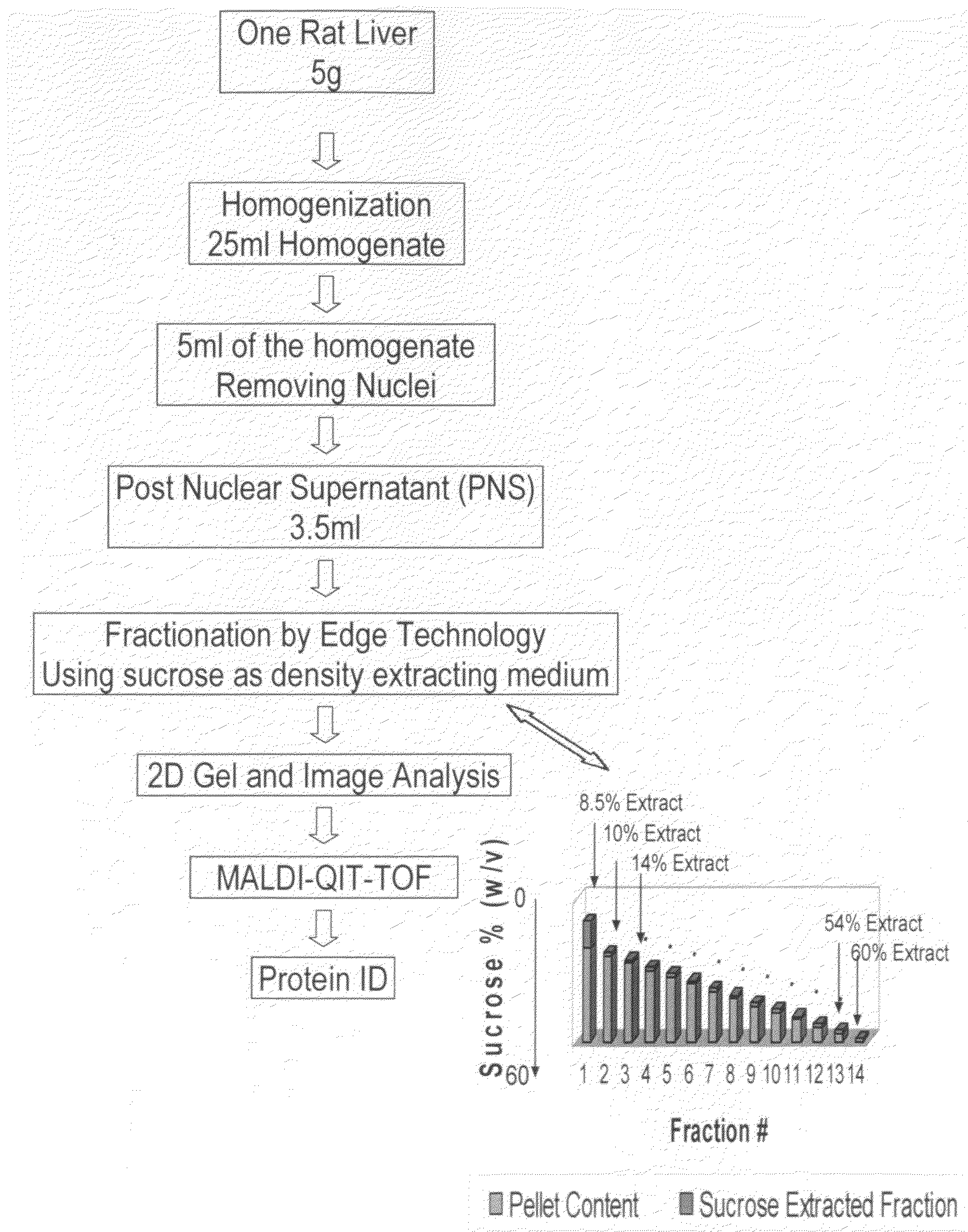
Subcellular Particle Fractionation and Proteomics Study of Rat Liver

[0098]The work flow of the rat liver subcellular fractionation and proteomics analysis is shown in FIG. 4.

[0099]I. Post Nuclear Supernatant Preparation: A rat liver post nuclear supernatant (PNS) from rat liver was prepared from a Sprague-Dawley rat (7-8 weeks of age). One frozen rat liver (about 5 g, Pel-Freez, Fayetteville, Ariz.) was thawed in 10 ml homogenization buffer (250 mM sucrose, 10 mM HEPES, 10 mM KCl, 1 mM EDTA, 10 μl protease inhibitor cocktail solution, pH 7.4) at 4° C. until the liver tissue turned soft. The thawed rat liver was diced into about 3 mm pieces with a pair of sharp scissors in the homogenization buffer. To the diced liver and homogenization buffer suspension was added an additional 10 ml of homogenization buffer. Half of the diced liver suspension was transferred to a prechilled 15 ml glass Dounce homogenizer. The rat liver was homogenized for 18-20 stokes using a loose pestle. The liver...

example 2

Virus Particle Separation from BmMLV

[0106]I: BmMLV virus preparation: Silkworm larvae (Kinshu.times.Showa strain) are injected at day 1 in the fifth instar with 150 μl of virus solution (equivalent to 1.0×10⁸ BmN cells) in phosphate-buffered saline (PBS). The virus solution is prepared as follows: 1.6×10⁸ BmN cells are homogenized in 75 ml of PBS and centrifuged at 7,000×g for 15 min at 4° C. After centrifugation, the supernatants are filtered (0.22-μm-pore-size filter) and used as the virus solution. BmN cells are harvested silkworm larvae, washed with PBS, and sonicated in 20 volumes of PBS. After low-speed centrifugation, the supernatants are filtered (0.22-μm-pore-size filter) and concentrated with an Amicon Ultra filter (Millipore).

[0107]II: BmMLV virus particle separation: The concentrated virus solution is subjected to the instant particle fractionation method using cesium chloride (CSCl) in buffer solution (10 mM Tris, 2 mM EDTA, pH 7.4) as an extracting density medium. The ...

example 3

Fractionation of Tissue Homogenates from Control and HF Diet Rats and Evaluation Potential Biomarkers: p-Synapsin I and GRP 75

[0120]I. Fractionation of Rat Brain and Heart tissue: The entire homogenization process was performed on ice. One frozen brain or heart was thawed in ice cold 1× homogenization buffer (20 mM HEPES, 1 mM Na2EDTA and 320 mM sucrose, pH 7.4 for brain; 20 mM HEPES, 10 mM KCl, 1 mM Na2EDTA and 250 mM sucrose, pH 7.4 for heart) with 0.01% v / v protease inhibitor cocktail. The tissue was dissected into 2-3 mm3 pieces and the liquid was carefully discarded. The brain tissue pieces were resuspended in 5 volumes of brain homogenization buffer and the suspension was transferred to a glass homogenizer. The tissue was homogenized up-down 10 times by a loose pestle, and then followed by another 10 up-down times with a tight pestle. The crude homogenate was transferred to several centrifuge tubes and centrifuged at 800 g force for 10 minutes to remove nuclei. The supernatant...


PUM

PropertyMeasurementUnit
timeaaaaaaaaaa
densityaaaaaaaaaa
specific densityaaaaaaaaaa

Abstract

A method for the separation of particles of different densities using a step-wise gradient density extraction method as described herein where a sample is suspended in a liquid volume of an extracting medium of specific density and the particles that have a density less than or equal to that of the extracting medium of specific density can be recovered from a horizonatally rotatable hollow disk or a removable receptacle within a horizontally rotatable hollow disk designed for such purposes while the particles that have a density greater than the extracting medium of specific density form a deposit which can be cycled through the extraction process in an iterative fashion by varying the density of the extracting medium allowing the recovery of discrete particles of differing densities from a test sample. Also disclosed herein is the use of the above method as part of a separate method or system to identify a ratio of biomarkers from different fractions of a sample homogenate or lysate which is useful in the evaluation of potential biomarkers, and for the intraoperative pathological diagnosis of positive margins of cancer and other diseases.

Description

CROSS-REFERENCE TO RELATED APPLICATION[0001]This application is a continuation-in-part of U.S. patent application Ser. No. 11 / 441,934, filed May 26, 2006, the disclosure of which is hereby incorporated by reference in its entirety.FIELD OF THE INVENTION[0002]The present invention relates to a method for the fractionation of particles, including biological particles, such as cellular compartments, cellular and subcellular particles, viruses, microorganisms, inclusion bodies, organelles from cell or tissue homogenates, organelles from cell lysates, lipoproteins and nano-particles, and non-biological particles such as micro-polymer particles (collectively “particles”), by the use of step-wise density gradient extraction, where said particles may be derived through recombinant or non-recombinant processes. Through an iterative series of substantially consecutive extractive steps, in which the density of the medium used to extract the particles is incrementally adjusted, the method descr...

Claims


Application Information

Patent Timeline
Patent Type & Authority Applications(United States)
IPC IPC(8): B03B7/00B01L11/00G06Q50/00B01L99/00G16H10/40
CPCG01N15/042G06Q50/22Y10T436/25375B01D2221/10B01D21/262G16H10/40
Inventor HORN, MARCUS JOSEPHLAN, WENKUI
Owner PROSPECT BIOSYST
Why Eureka
Try Eureka