Biological Cell Tracking With Ultrasound
- Summary
- Abstract
- Description
- Claims
- Application Information
AI Technical Summary
Benefits of technology
Problems solved by technology
Method used
Image

Examples
example i
Single Wall Polymer Contrast Microbubble Fabrication
[0266]Single wall polymer contrast microbubbles may be fabricated by double-emulsion (Water / Oil / Water) and solvent evaporation methods. Briefly, 300 mg of poly(L-lactide((PLLA) (MW 75,000-120,000) or poly(lactide-co-glycolide) (PLGA) (50:50) (IV=0.38 dl / g) are dissolved in 10-25 ml dichloromethane. 2 ml of ammonium bicarbonate (200 mg / ml) solution is emulsified with polymer solution with a sonicator. This W / O emulsion may be dropped into PVA solution (200 ml, 1% w / v) and homogenized at 10,000 rpm for 1 hour. The resulting W / O / W emulsion is continually stirred at 500-600 rpm at room temperature for another 4 hours. Finally, the resulting microbubbles are collected by centrifugation, washed five times with deionized water, and freeze-dried using a lyophilizer to sublimate the encapsulated water and get the final products. The dried microbubbles were then maintained under vacuum conditions for at least 2 hours to complete the drying p...
example ii
Polymer Contrast Microbubble Internalization
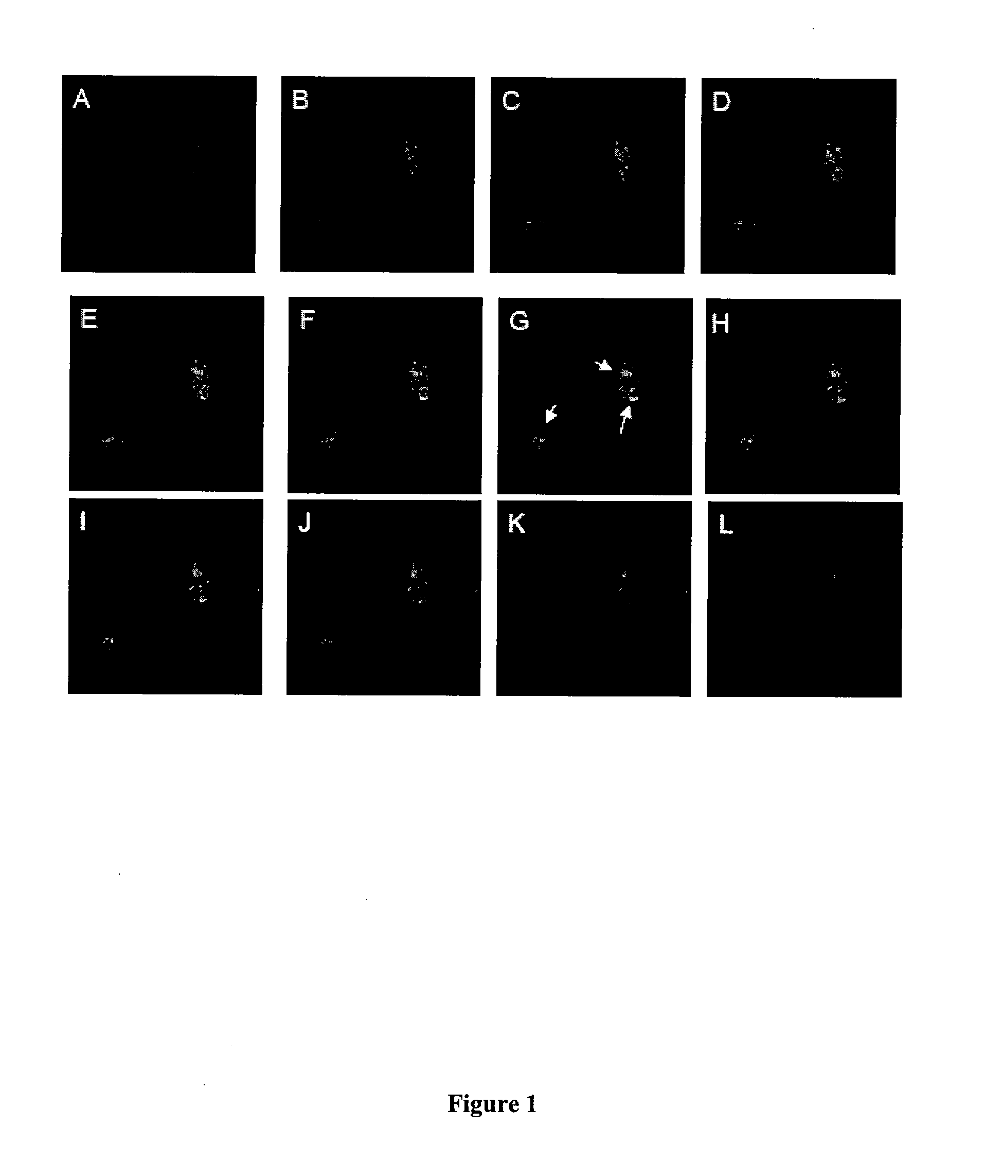
[0269]A polymer contrast microbubble suspension was transferred to a centrifuge tube and centrifuged at 1500 rpm for 10 minutes. After collecting the floating microbubbles from the supernatant and counting the number of the microbubbles with a Coulter Counter, microbubble suspension was added to a Petri dish containing media and cells which have been cultured to confluence. The Petri dish was covered with sterile paraffin film before the lid was added on top of the paraffin film. Then the whole Petri dish is turned upside down to facilitate microbubbles contact with the cells via buoyancy forces. The cells and microbubbles were kept in an incubator for 2 hours before the Petri dish is turned back to the normal position and left in the incubator overnight. After incubation, the cell-microbubble complexes were lifted using Trypsin and free microbubbles are separated by centrifugation. Several rounds of washing may be necessary to make sure t...
example iii
Preparation of a Bi-Layer Polymer Contrast Microbubble
[0271]Two solutions are prepared, the first being an aqueous solution of an outer biomaterial. The second is a solution of a polymer which is used to form the inner layer, in a relatively volatile water-immiscible liquid which is a solvent for the polymer, and a relatively non-volatile water-immiscible liquid which is a non-solvent for the polymer. The relatively volatile water-immiscible solvent is typically a C5-C7 ester, such as isopropyl acetate. The relatively non-volatile water-immiscible non-solvent is typically a C6-C20 hydrocarbon such as decane, undecane, cyclohexane, cyclooctane and the like. In the second solution containing the polymer for the inner layer, the polymer in water-immiscible solvents are combined so that the polymer fully dissolves and the two solvents are miscible with agitation. The polymer solution (organic phase) is slowly added to the biomaterial solution (aqueous phase) to form a liquid foam. Typic...
PUM
 Abstract
Description
Claims
Application Information
 - R&D
- Intellectual Property
- Life Sciences
- Materials
- Tech Scout
- Unparalleled Data Quality
- Higher Quality Content
- 60% Fewer Hallucinations
Browse by: Latest US Patents, China's latest patents, Technical Efficacy Thesaurus, Application Domain, Technology Topic, Popular Technical Reports.
© 2025 PatSnap. All rights reserved.Legal|Privacy policy|Modern Slavery Act Transparency Statement|Sitemap|About US| Contact US: help@patsnap.com