Amperometric biosensors based on redox enzymes

a biosensor and redox enzyme technology, applied in the field of amperometric biosensor electrodes, can solve the problems of affecting the sensitivity of the redox enzyme, and consuming a lot of time, and achieves low activity, low stability, and favorable kinetics

Inactive Publication Date: 2001-06-05
MONBOUQUETTE HAROLD G
View PDF1 Cites 413 Cited by
  • Summary
  • Abstract
  • Description
  • Claims
  • Application Information

AI Technical Summary

Benefits of technology

FIG. 8 illustrates a model of the sarcosine dehydrogenase immobilized in pockets of hydrophobic self-assembled monolayer on a gold electrode or glassy carbon electrode. The model reflects that the varied length of alkyl chains formed defects in the self-assembled monolayer on the gold surface to provide a very stable environment for the enzyme to interact and fixate itself upon the electrode.
As illustrated in FIG. 9, the results using this system confirmed activity of immobilized enzyme with discernible amperometric responses to varying substrate concentrations. The synthetic redox dye phenazine methosulfate was chosen for its favorable kinetics, and it is simply added to solution in this system. Since commercially prepared sarcosine dehydrogenase exhibits relatively low activity, as well as the relatively low stability, the overall content of lipids was increased in order to overcome these inherent problems. In this biosensor, a phospholipid, namely dimyristoylphosphatidylcholine, is preferred. The increased presence of phospholipids in this enzyme mimetic layer was found to improve enzyme activity and stability of the sensing electrode.
Further, it was discovered that although increasing the phospholipid content did increase the enzymatic activity, the biosensor was still unable to retain most of its activity after storage in buffer solution. As a consequence, other enzyme immobilization techniques were utilized for greater enzyme loading and higher enzyme stability/ac

Problems solved by technology

This main challenge in biosensor technology is to find an effective means to couple the biological component to the transducer.
Although an enzymatic spectrophotometric assay is available for fructose determination, the assay is time intensive, tedious and costly.
While these approaches delivered some promising results, readily oxidizable inteferents such as ascorbic acid in citrus juice were found to overwhelm the fructose signal.
When this problem is partially avoided by using lower redox potentials, relatively poor sensitivity and low current response is the result.
These assays are analytically limited in that the Jaffe reaction is not specific for creatinine.
Given this, many other substrates interfere with the assay leading to inaccurate determinations of creatine concentration in the sample.
These methods, however, are rather complex and inefficient because there is a stringent requirement for precise control of oxygen concentration in the system.
It is inconvenient, for example, to e

Method used


Image

  • Amperometric biosensors based on redox enzymes
  • Amperometric biosensors based on redox enzymes
  • Amperometric biosensors based on redox enzymes

Examples


example 2

Enzyme Immobilization

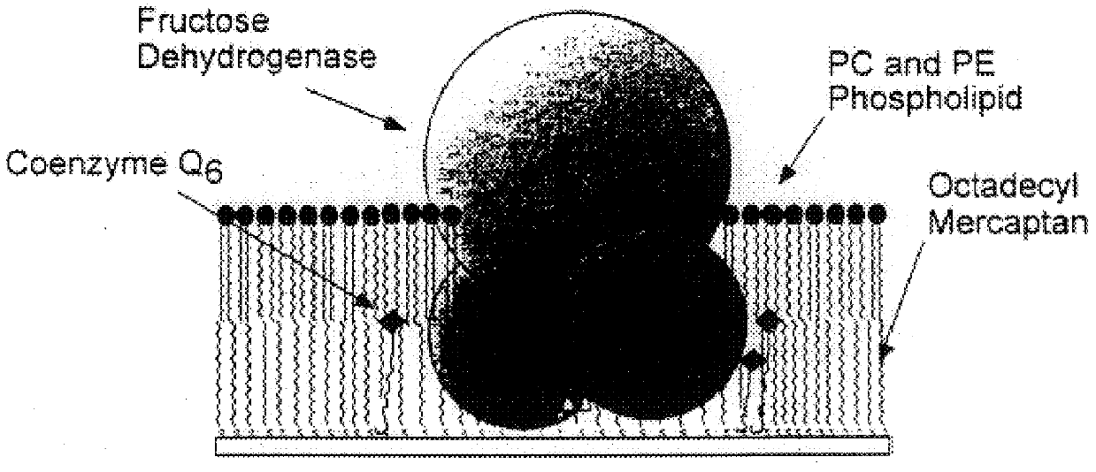
2a: Immobilization of fructose dehydrogenase and coenzyme in mixed monolayer

The following describes the method of preparing a biosensing electrode in accordance with the present invention.

First dialysis bags are prepared by cutting approximately 2.5 inch lengths of Spectra / Por CE (Cellulose Ester) Membrane 10,000 MWCO tubing (flat width 12 mm) and placing these in deionized water. The dialysis bags are soaked for at least 30 minutes changing the water at least three times. A final soak is does in cold phosphate buffer. The dialysis bags are then stored on ice.

Next a stock solution of FDH is prepared by weighing the appropriate amount of octyl glucoside to give 35 mM or 1% in 1 ml of cold phosphate buffer (10 mM, pH 4.5). FDH is allowed to warm to room temperature before use. 2.5 mg FDH is then transfered via gentle squirting of detergent solution. The stock solution is stored at 4 C between experiments and used within two weeks.

A stock solution of decylubiquinon...

example 2d

ltilayers of Immobilized Sarcosine Dehydrogenase Using DIDS

The preparation of multilayer enzyme electrodes using 4,4'-diisothiocyanato-trans-stilbene-2,2'-disulfonic acid disodium salt (DIDS) was performed using the technique described by Riklin and Willner. First, the clean gold electrode surface was immersed in a 10 mM cystamine dihydrochloride aqueous solution for 2.0 hours. The monolayer-modified electrodes were then rinsed twice with water and introduced into a cold (0.degree. C.) 100 mM potassium phosphate buffer solution (pH 7.5) that contained 20 mM DIDS for 10 minutes. The resulting electrodes were then rinsed twice with a cold phosphate buffer solution and then soaked in a sarcosine dehydrogenase solution (3.0 mg / ml) for 30 minutes at room temperature. The monolayer enzyme electrode was rinsed with phosphate buffer solution and the two-step procedure using the reaction with DIDS and sarcosine dehydrogenase was repeated to assemble the desired number of enzyme layers on the...

example 2e

tion of Creatinine Related Enzymes by Glutaraldehyde Cross Linking

To prepare a creatine sensor, approximately 1.0 mg creatine amidinohydrolase, 1.0 mg sarcosine denydrogenase, and 1.0 mg bovine serum albumin were dissolved in 120 .mu.l phosphate buffer solution, pH 7.5. 4.0 .mu.l 1.0% glutaraldehyde was added to the enzymes solution and stirred. Approximately 10 .mu.l of the resulting mixture was quickly added to each of the freshly polished and electrochemically cleaned electrodes, using a microsyringe. The enzyme layer obtained was allowed to cross-link in air, at room temperature, for 1.0 hour. The electrodes were then immersed and stored in 100 mM phosphate buffer solution, pH 7.5, at room temperature until further use. To prepare a creatinine sensor, approximately 0.5 mg creatinine amidohydrolase, 1.0 mg creatine amidinohydrolase, 1.0 mg sarcosine dehydrogenase, 1.0 mg bovine serum albumin were dissolved in 120 .mu.l phosphate buffer solution, pH 7.5. The rest of the protocol w...


PUM

PropertyMeasurementUnit
Diameteraaaaaaaaaa
Amphiphilicaaaaaaaaaa
Concentrationaaaaaaaaaa

Abstract

The invention includes various prototypical amperometric biosensors for the quantification of biological substrates such as fructose, creatinine, creatine, and sarcosine, and the methods for producing these biosensors. Also included in the invention is a minaturized version of the biosensing devices. The components of these prototypical biosensors are immobilized on a self-assembled monolayer (SAM) comprising chemisorbed alkanethiols. The deposition of an amphiphilic lipid layer to these systems increases the stability and activity of the resultant biosensor and enhances the rejection of many interferents. An additional feature of the invention is the co-deposition of the components of the sensor via a novel detergent dialysis protocol. The invention features two particular biosensor systems. One embodiment involves fructose dehydrogenase as the redox/sensor enzyme and fructose as the substrate/analyte. Another embodiment involves the measurement of the substrates/analytes, creatinine, creatine and sarcosine, using sacrosine dehydrogenase as the redox/sensor enzyme, with the involvement of creatinine amidohydrolase and/or creatine amidinohydrolase in the reaction pathway.

Description

The present relates to amperometric biosensors capable of rapidly quantifying the concentration of an analyte in a sample solution with high accuracy in a simplified manner, and to methods of producing same. In particular, the invention relates to amperometric biosensors having an immobilized, redox enzyme as a enzymatic sensing element coupled to a metal electrode.A biosensor is an analytical device incorporating biological and chemical sensing elements, either intimately connected to or integrated with a suitable transducer, which enables the conversion of concentrations of specific chemicals into electronic signals. A majority of biosensors produced thus far have incorporated an enzyme as a biological recognition component.A critical element in the design of a successful biosensor is the immobilization procedure for biological components. Generally, four main approaches to enzyme immobilization have been utilized. These include: (1) physical adsorption at a solid surface, (2) ent...

Claims


Application Information

Patent Timeline
IPC IPC(8): C12Q1/00
CPCB82Y15/00B82Y30/00C12Q1/002C12Q1/005
Inventor MONBOUQUETTE, HAROLD G.
Owner MONBOUQUETTE HAROLD G
Why Eureka
Try Eureka