System and method for radiographic imaging of tissue

a tissue radiographic imaging and tissue technology, applied in the field of medical imaging, can solve the problems of reducing the reliability of mammography as a diagnostic tool, high error rate in radiological diagnosis of cancer, and inability to visually appear tissue on the radiograph, and achieve the effect of high anatomical detail

Inactive Publication Date: 2005-08-02
VERITAS PHARM INC
View PDF43 Cites 11 Cited by
  • Summary
  • Abstract
  • Description
  • Claims
  • Application Information

AI Technical Summary

Benefits of technology

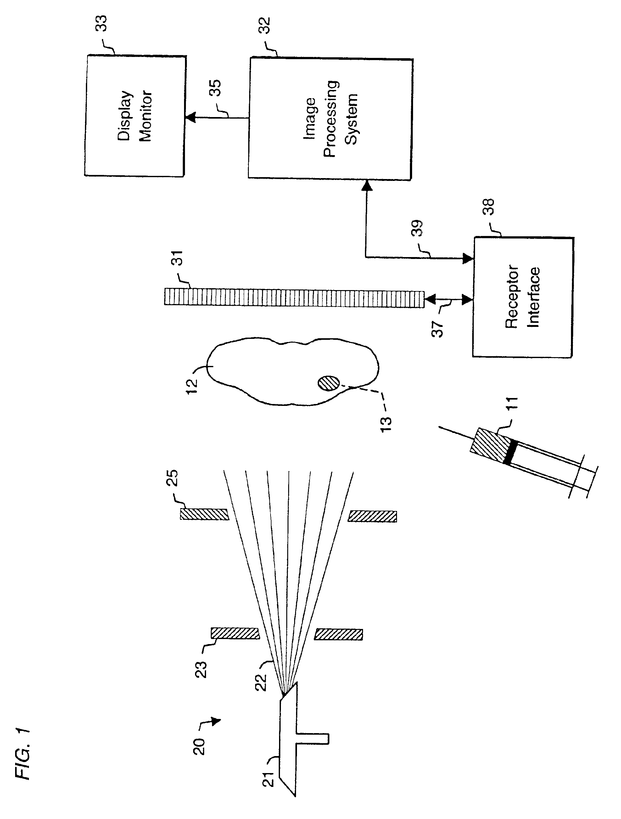
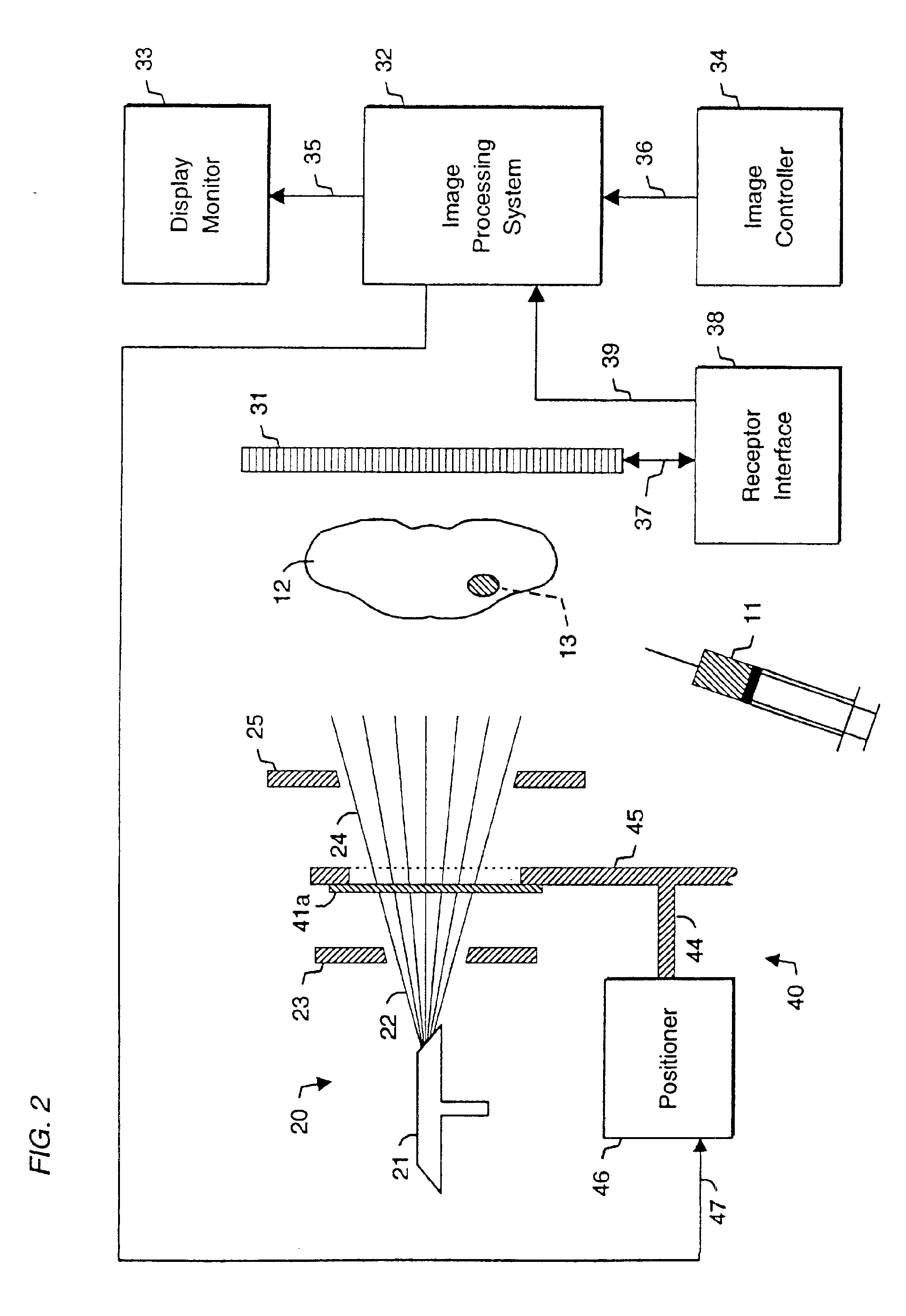
[0076]The present invention is thus capable of generating images that combine the diagnostic accuracy provided by functional imaging of tissue physiology with the high anatomical detail and spatial resolution provided by radiography.

Problems solved by technology

However, it has been repeatedly demonstrated that the visual appearance of tissue on the radiograph is not a reliable criterion for definitive diagnosis of malignancy.
Studies confirm a high error rate in the radiological diagnosis of cancer.
However, the high incidence of false positives and false negatives reduces the reliability of mammography as a diagnostic tool.
However, as previously noted, it is currently impossible to reliably differentiate malignant from benign tissue solely by visual inspection of the mammogram.
In addition to increased cost, biopsies carry the risk of infection, scanning, pain, and anxiety.
A more serious problem in diagnostic mammography is the high incidence of false negatives, in which malignant tumors that are actually present are not detected on the mammogram.
Particularly in their early stages of development, malignant tumors may not be noticed even upon careful inspection of the mammogram by an experienced radiologist.
Tumors may be too small to be detected, or their appearance may be obscured by benign tumors or cysts.
The visibility of malignant tumors may be further obscured in younger women, whose breast tissue is often dense.
Similar diagnostic problems are common in thoracic radiography, in which radiographic imaging of the lungs and heart is performed to detect abnormal tissue, particularly malignant tumors.
However, it is impossible to reliably differentiate malignant nodules from benign nodules solely by inspection of the radiograph.
As in the case of breast biopsies, these procedures are expensive and add patient morbidity risk.
As in the case of mammography, false negatives in thoracic radiography lead to less effective therapy and shorter patient survival time.
Particularly in their early stages of development, malignant lung tumors may not be detected even upon careful inspection of the radiograph.
However, this approach is fundamentally flawed because detection of malignant tissue, even in high-resolution images, still largely depends on variations in radiographic density.
But, as previously noted, radiographic density alone is not a reliable criterion for detection of malignant tissue.
The most formidable barrier to the use of PET is its cost.
Capital equipment costs of the imaging device and cyclotron are very high.
In addition, radiochemists are needed to perform complex syntheses of radiopharmaceuticals whose half-lives are typically 2 hours.
Because of the high capital equipment, materials, and labor costs, the cost of a PET scan to the patient is approximately $2500.
Because of its prohibitive cost, PET imaging is not widely available, and is not likely to be used in the foreseeable future for routine cancer diagnosis.
A second disadvantage of PET is the lower spatial resolution of PET images compared to those generated by radiography.
Relatively low spatial resolution is a limitation common to imaging modalities that depend on detector arrays for the measurement of high-energy particles emitted from radiopharmaceuticals.
Because of its low spatial resolution, PET exhibits a high rate of false negatives in the detection of small malignant tumors.
A third disadvantage of PET is the length of the imaging procedure.
Nevertheless, SPECT imaging has limitations that reduce its utility in the diagnosis of malignant tumors.
In the diagnosis of breast cancer, a major limitation of the technique is the low spatial resolution of the images, which results in a sharp decrease in sensitivity for breast tumors smaller than 1.5 cm.
A second disadvantage of SPECT in breast cancer imaging is the relatively low specificity.
In particular, Tc-99m-Sestamibi produces a significant number of false positives in patients with benign fibroadenomas.
Since fibroadenomas constitute the most frequent source of false positives, and frequently result in unnecessary biopsies, the large number of false positives produced by Tc-99m-Sestamibi reduces its value in breast cancer imaging.
It is thus inadequate for the reliable diagnosis of lung cancer.
Because these contrast agents do not enter the cytosol, they do not specifically interact with intracellular targets, and therefore cannot be used to provide images which contain information on intracellular function or physiology.
The utility of these radiographic contrast agents in the detection of functionally abnormal tissue is thereby limited.
The very slow rates of cellular entry and exit render these molecules of limited value in diagnostic imaging procedures.
This inconsistency minimizes the utility of glucose transport as a diagnostic criterion for the detection of malignant tissue.
It was not suggested that cell membrane-permeable, intracellularly localized radiographic contrast agents might be imaged using these methods, as no such imaging agents were available or known at the time.
In addition, these methods did not provide the capability of generating functional physiological images of body tissue, nor of presenting a visually aligned combination of a functional and an anatomical image of the same tissue.
This capability is not currently provided by any radiographic contrast agent.
These capabilities are not provided by any currently available single imaging modality.
These software computations are generally time-consuming and require considerable computational hardware.
It is thus evident that the present state of the diagnostic imaging art does not provide a sufficiently accurate, inexpensive, and widely available modality for detection and localization of certain types of tissue such as malignant tissue.

Method used


Image

  • System and method for radiographic imaging of tissue
  • System and method for radiographic imaging of tissue
  • System and method for radiographic imaging of tissue

Examples


example 1

2-Amino-4-(2′,4′,6′-triiodophenyl)-benzoyl-D-glucosamine (11)

[0111]

4-iodotoluene is reacted with picryl chloride in the presence of copper bronze at 215° C. to yield 2′,4′,6′-trinitro-4-methylbiphenyl (1). C7⁢H7⁢I+C6⁢H2⁢Cl⁢N3⁢ ⁢O6⁢→ΔCu⁢C13⁢H9⁢I3⁢N3⁢ ⁢O6

2′,4′,6′-trinitro-4-methylbiphenyl (1) is reacted with stannous chloride and hydrochloric acid to yield 2′,4′,6′-triamino-4-methylbiphenyl (2). C13⁢H9⁢N3⁢ ⁢O6⁢→H⁢ ⁢ClSn⁢ ⁢Cl2⁢C13⁢H15⁢N3

2′,4′,6′-triamino-4-methylbiphenyl (2) is reacted with sodium nitrite and hydrochloric acid to convert the amino groups to diazonium groups. The diazonium groups react with potassium iodide to yield 2′,4′,6′-triiodo-4-methylbiphenyl (3). C13⁢H15⁢N3⁢→H⁢ ⁢ClNa⁢ ⁢N⁢ ⁢O2⁢→K⁢ ⁢I⁢ ⁢C13⁢H9⁢I3

Aromatic nitration of 2′,4′,6′-triiodo-4-methylbiphenyl (3) is performed with nitric acid and sulfuric acid to yield a mixture of 2′,4′,6′-triiodo-2-nitro-4-methylbiphenyl (4) and 2′,4′,6′-triiodo-3-nitro-4-methylbiphenyl (5). The products are separated using column chroma...

example 2

is similar in general structure to Example 1, with the additional presence of a short alkyl chain attached to the radio-opaque tri-iodophenyl moiety at the C-3′ position to further increase lipophilicity of the molecule and enhance cell membrane permeability. The computed logP of Example 2 is 5.31.

example 3

2-Amino-4-[3′,5′-bis(N-methylcarboxamide)-2′,4′,6′-triiodophenyl]-benzoyl-D-glucosamine (24)

[0114]

Freidel-Crafts acylation is performed on 2′,4′,6′-triiodo-3′-ethyl-3-nitrobiphenyl-4-carboxylic acid (13) in the presence of AlCl3 and CH3COCl to yield 2′,4′,6′-triiodo-3′-ethyl-5′-acetyl-3-nitrobiphenyl-4-carboxylic acid (17). 2′,4′,6′-triiodo-3′-ethyl-5′-acetyl-3-nitrobiphenyl-4-carboxylic acid (17) is then reacted with a zinc mercury amalgam and hydrochloric add and heated to yield 2′,4′,6′-triiodo-3′,5′-bis(ethyl)-3-nitrobiphenyl-4-carboxylic acid (18). C15⁢ ⁢H10⁢I3⁢N⁢O4⁢→C⁢ ⁢H3⁢C⁢ ⁢O⁢ ⁢ClAl⁢ ⁢Cl3⁢→ΔZn(Hg), ⁢H⁢ ⁢Cl⁢C17⁢ ⁢H14⁢I3⁢N⁢ ⁢O4

2′,4′,6′-triiodo-3′,5′-bis(ethyl)-3-nitrobiphenyl-4-carboxylic acid (18) is reacted with thionyl chloride to yield 2′,4′,6′-triiodo-3′,5′-bis(ethyl)-3-nitrobiphenyl-4-carbonyl chloride (19). C17⁢ ⁢H14⁢I3⁢N⁢ ⁢O4⁢→S⁢ ⁢O⁢ ⁢Cl2⁢C17⁢ ⁢H13⁢Cl⁢I3⁢N⁢ ⁢O3

1,3,4,6-tetra-O-acetyl-D-glucosamine (9) is reacted with 2′,4′,6′-triiodo-3′5′-bis(ethyl)-3-nitrobiphenyl-4-c...


PUM

PropertyMeasurementUnit
timeaaaaaaaaaa
molecular weightsaaaaaaaaaa
logPaaaaaaaaaa

Abstract

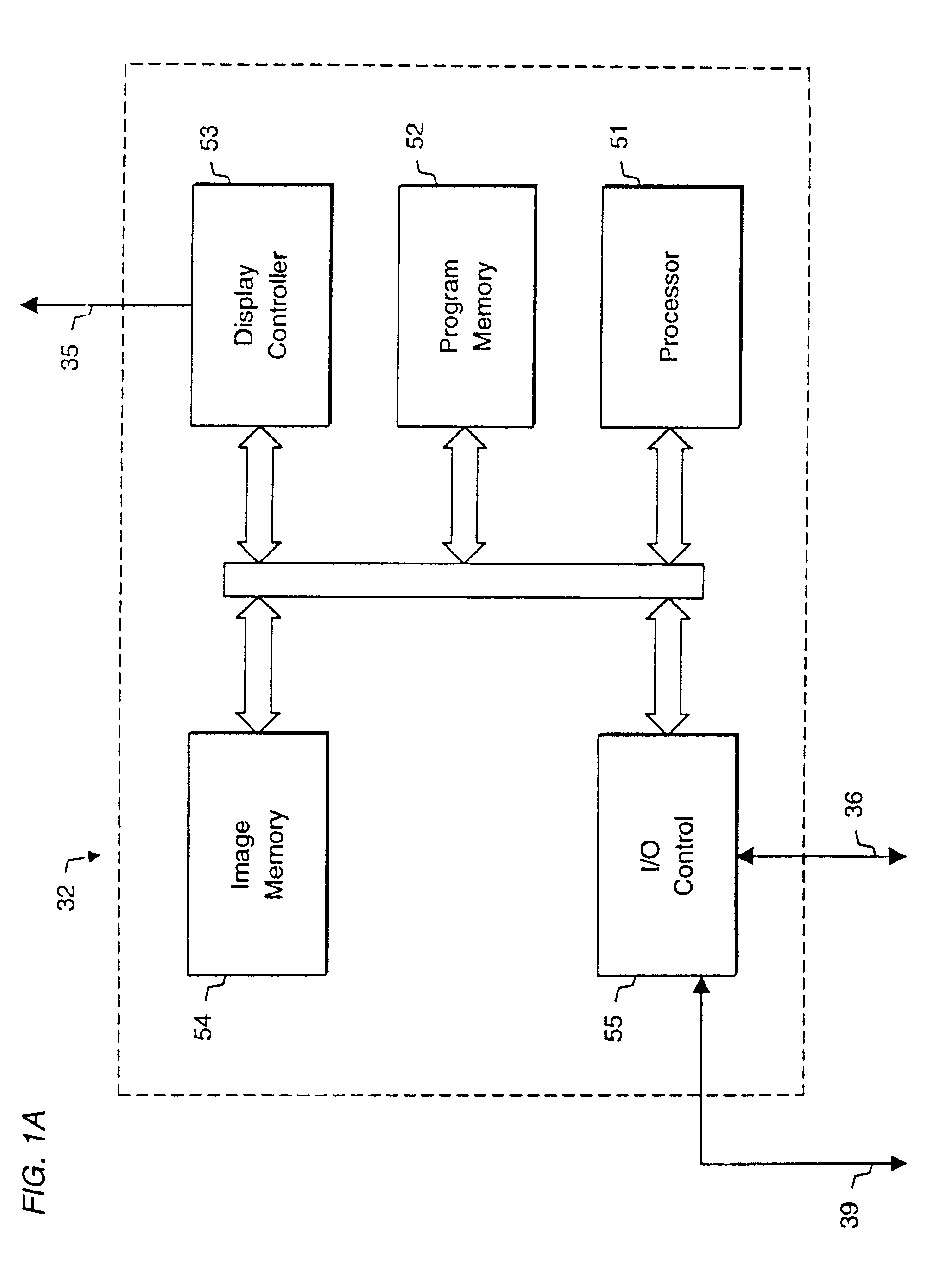
System and method for radiographic imaging of tissue using a non-radioactive, radio-opaque imaging agent that accumulates intracellularly in tissue in proportion to its functional, or physiological, activity. In one embodiment, the imaging agent is a cell-membrane permeable, radio-opaque, high affinity ligand for the intracellular enzyme hexokinase. The imaging agent is administered to a patient, and after an accumulation interval, radiographic images are acquired. The imaging agent preferentially accumulates in malignant tissue and increases its radio-opacity because of its elevated glucose metabolic rate relative to benign and normal tissue. The tissue being examined is transilluminated by X-ray beams with preselected different mean energy spectra, and a separate radiographic image is acquired during transillumination by each beam. An image processing system performs a weighted combination of the acquired images to produce a single displayed image. The image processing procedure isolates the radiographic density contributed solely by differential accumulation of the imaging agent in malignant, benign, and normal tissue. The system and method thus provides a functional image displayed with the anatomical detail and spatial resolution of a radiographic image. The viewer may interactively control the relative proportion of radiographic density contributed by imaging agent, soft tissue, and bone to the displayed image, allowing the display of functional and anatomical information in complete registration, and facilitating localization of malignant tissue in relation to nearby anatomical structures. In other embodiments, the system and method may be used to detect enzymes, nucleic acids, coenzymes, fatty acids, and other cellular targets in diagnostic imaging applications.

Description

[0001]This application is a divisional application of U.S. patent application Ser. No. 09 / 149,734, filed Sep. 8, 1998 now U.S. Pat. No. 6,226,352.BACKGROUND OF THE INVENTION[0002]A. Field of the Invention[0003]This invention relates to medical imaging and specifically to a system and method for localization of certain types of abnormal tissue, such as malignant tissue, using radiography.[0004]B. Description of the Related Art[0005]Radiography involves examination of internal organs of the body by transillumination with X-rays. The image receptor may be radiographic film, a film / intensifying screen combination, a stimulable phosphor storage plate, a fluoroscopic image intensifier, an amorphous silicon sensor array, a CCD / scintillator combination, or another type of X-ray sensitive receptor. In digital radiography, a computer digitizes the acquired image and may enhance it using image processing algorithms. The most commonly performed radiographic procedures include mammography, in wh...

Claims


Application Information

Patent Timeline
Patent Type & Authority Patents(United States)
IPC IPC(8): A61B6/03G01N33/48A61B6/00A61K49/04G01T1/00G03B42/02
CPCA61B6/4035A61B6/4042A61B6/4241A61B6/482A61B6/032A61B6/4092
Inventor SALB, JESSE
Owner VERITAS PHARM INC
Why Eureka
Try Eureka