
Endoscopic arterial pumps for treatment of cardiac insufficiency and venous pumps for right-sided cardiac support

a technology of endoscopic arterial pumps and right-sided cardiac support, which is applied in the direction of heart stimulators, prostheses, therapy, etc., can solve the problems of fewer than 3,000 donor hearts available each year, refractory to standard medical and surgical treatment, and the cost of annual treatment estimated to exceed 10 billion dollars, so as to achieve easy weaning and easy removal or replacement

Inactive Publication Date: 2006-12-05
BARBUT DENISE R +2
View PDF25 Cites 92 Cited by
  • Summary
  • Abstract
  • Description
  • Claims
  • Application Information

AI Technical Summary

Benefits of technology

[0013]In a first method for treatment of heart failure, the blood pump is mounted in an interior of the stent. The stent (or the pump, or both) may be made of a memory retaining biocompatible material, such as nitinol, which allows the stent to be compressed before deployment to facilitate insertion into a vessel, and thereafter self-expand intravascularly. Alternatively, the catheter includes an angioplasty balloon, which upon inflation expands the stent intravascularly. The distal end of the stent-deployment catheter is inserted into a peripheral artery, e.g., the subclavian artery or the femoral artery, and advanced to position the stent within the descending aorta, just beyond the takeoff of the left subclavian artery. Thus, any thromboembolic complication would be distal to the takeoff of the left subclavian artery and would not involve the brain. The stent is then expanded. The stent and the pump are released from the catheter, and the pump is activated to increase arterial blood flow downstream of the pump. Alternatively, the stent is expanded before being released. A manometer may be included in a distal end and / or a proximal end of the pump for measuring blood pressure upstream and / or downstream the pump. Radiopaque material may be mounted on the pump or the stent, thereby allowing fluoroscopic verification of the positioning.
[0022]It will be understood that there are several advantages in using the methods described herein for endoscopically deploying a blood pump / stent. For example, by using the pump (1) major surgical procedures can be avoided, (2) numerous complications associated with the existing VADs, such as cardiac arrhythmia, bleeding, thromboembolism to the brain, and infection, can be minimized or avoided, (3) the patient can be easily weaned from the pump when ventricular support is not required, (4) the pump / stent can be easily removed or replaced endoscopically like any other stent without surgical procedures, (5) patients with contraindications to the existing VADs can be treated without complication (the only contraindication to using the disclosed methods is aortic aneurysm), (6) the pump is easily placed under angiography, (7) the pump can accommodate an angioplasty, atherectomy, or other therapeutic catheter during high-risk endovascular procedures, (8) the pump can be used during stopped heart cardiac surgery, and (9) the pump can be used to treat venous insufficiency in addition to treating heart failure.

Problems solved by technology

Heart failure is responsible for at least 200,000 deaths each year, with annual costs estimated to exceed 10 billion dollars.
Although advances in medical and surgical therapy, e.g., pharmacotherapy and emergency coronary artery bypass grafting surgery, have benefited a few of these patients, the majority are refractory to standard medical and surgical treatment.
However, fewer than 3,000 donor hearts become available each year.
Even in patients accepted for transplantation, a significant waiting list mortality has been observed.
Disadvantages associated with these external VADs are that (1) multiple large surgical incisions, including median sternotomy and perforation of the ventricle, to connect the support system to the heart are required, (2) the surgical sites pose continuing risk of infection and other complications, such as hemorrhage, (3) the large console needed to house power and control systems severely limits the patient's mobility, thereby requiring hospitalization for the patients.
These devices, however, are associated with high complication rates, including (1) substantial morbidity due to the median sternotomy (Nocacor) with cardiac mobilization, often in patients who have had previous cardiac surgery, (2) morbidity due to extensive dissection of the abdominal wall to create a pocket for the pump, (3) bleeding requiring re operation (30%), (4) bleeding from anticoagulation use, (5) infection (50%) (both nosocomial and device related), (6) thromboembolism, especially to the brain (cerebral emboli were detected using transcranial doppler in two thirds of the patients with ventricular assist devices), and (7) significant bleeding associated with the extensive surgery often results in right heart failure (20%).
Unfortunately, the Acorn device also requires surgical implantation, and its efficacy has not yet been tested in humans.
However, peri-procedural morbidity associated with using this device is high, including stroke.
Systemic anticoagulation is usually necessary for its insertion and is associated with increased risk of hemorrhage.
Failure of device insertion occurs in 25% of patients, partly due to iliofemoral atheromatosis and inability to cross the aortic valve.
Apart from having high complication rates, an additional problem associated with using the existing VADs is that patients can not be weaned from the VADs without another surgical procedure to remove the implanted devices.
Existing devices are inadequate for this purpose.

Method used


Image

  • Endoscopic arterial pumps for treatment of cardiac insufficiency and venous pumps for right-sided cardiac support
  • Endoscopic arterial pumps for treatment of cardiac insufficiency and venous pumps for right-sided cardiac support
  • Endoscopic arterial pumps for treatment of cardiac insufficiency and venous pumps for right-sided cardiac support

Examples


Embodiment Construction

[0041]Although endoscopic deployment of the pump utilizing a stent is most useful in treating patients with heart failure, the methods disclosed herein can be used to treat a variety of conditions involving arterial and / or venous insufficiency. The pump can be deployed in a vessel which supplies a vital organ, e.g., in the renal artery to increase flow to the kidneys. The pump(s) can be deployed in any vessel where augmentation of blood flow is needed.

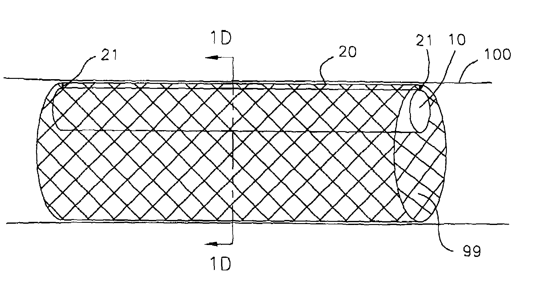
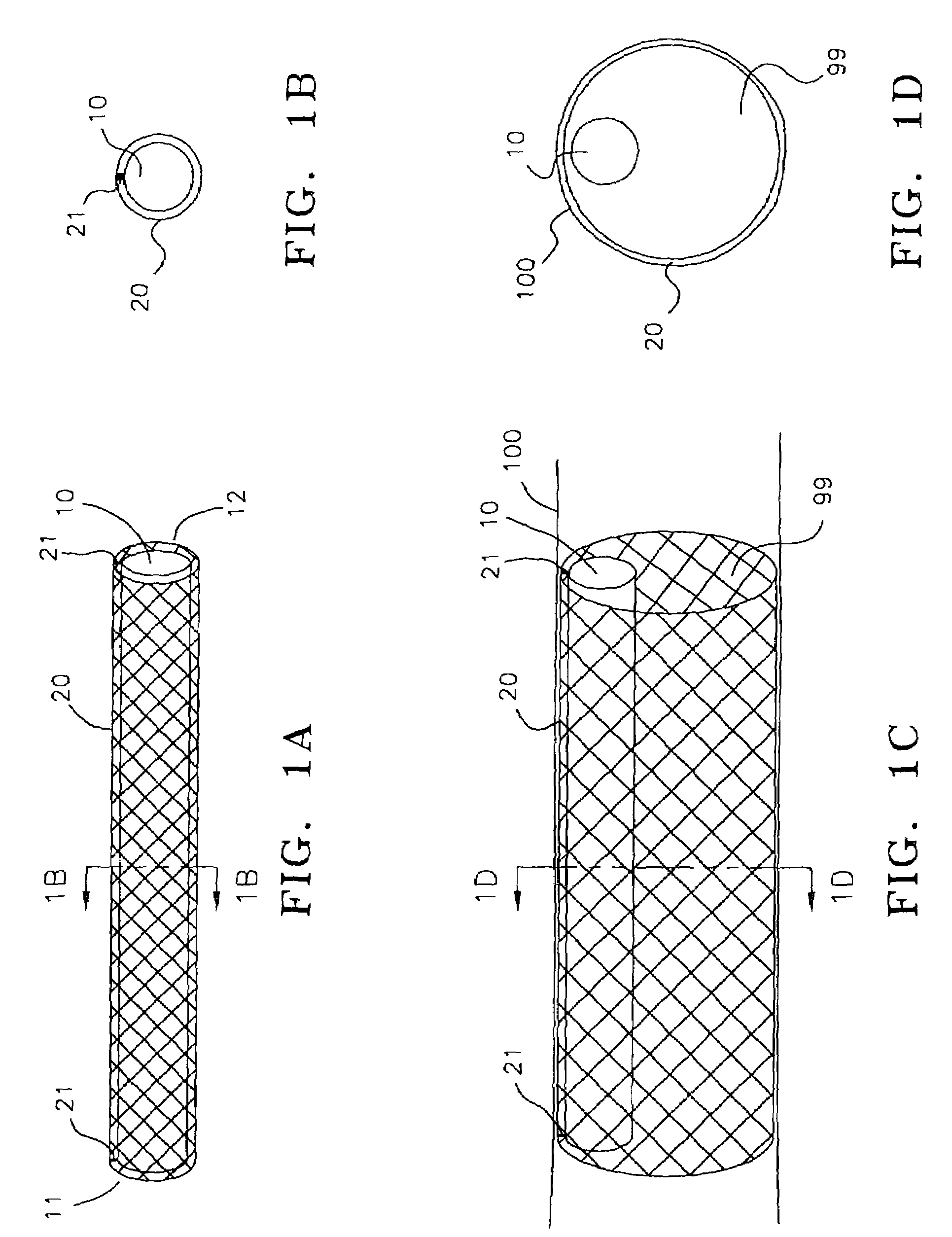
[0042]In FIG. 1A, pump 10 is disposed within stent 20. Proximal end 11 and distal end 12 of the pump are attached to stent 20 by direct bonding or through anchoring elements 21. FIG. 1B provides a cross sectional view of the pump / stent assembly. The stent is made of a memory retaining biocompatible material, e.g., nitinol, and can be placed in a collapsed condition before deployment to facilitate insertion into a vessel. The pump / stent assembly is releasably mounted on a distal end of a catheter. In treating patients with congestive he...


PUM


Abstract

Methods for using blood pumps to treat heart failure are disclosed. The pump is mounted on an interior of a stent, and the stent is releasably mounted on a distal end of a catheter. The distal end of the catheter is inserted into a peripheral artery and advanced to position at a region of interest within the descending aorta, the ascending aorta, or the left ventricle. The stent and the pump are released from the catheter, and the pump is activated to increase blood flow downstream of the pump. The pump can also be positioned in the vena cava or used to treat right-sided heart failure following the insertion of an LVAD, or to improve venous return in patients with varicose veins. Non-stent pumps are described for insertion between the pulmonary vein and aorta, and between the vena cava and pulmonary artery designed for use during cardiac surgery.

Description

[0001]This is a continuation of U.S. patent application Ser. No. 09 / 691,611, filed Oct. 17, 2000 now abandoned, which is a continuation of U.S. application Ser. No. 09 / 362,992, filed Jul. 27, 1999, now U.S. Pat. No. 6,136,025, both of which are incorporated herein by reference in their entirety.FIELD OF THE INVENTION[0002]The present invention relates generally to methods useful for treating patients with heart failure and cardiogenic shock. More specifically, the invention provides means for implanting a blood pump endoscopically in arteries, such as the descending aorta, the ascending aorta, the left ventricle, or the pulmonary vein to assist the failing left ventricle and improve perfusion to vital organs. The pump may provide continuous or intermittent flow. The device can also be implanted endoscopically in veins, such as the inferior vena cava, the right atrium or pulmonary artery for treatment of right heart failure or venous insufficiency.BACKGROUND OF THE INVENTION[0003]Hea...

Claims


Application Information

Patent Timeline
IPC IPC(8): A61M1/10A61F2/90A61M60/135A61M60/205A61M60/861A61M60/867
CPCA61F2/90A61M1/12A61M1/122A61M1/125A61M2205/32A61M60/148A61M60/861A61M60/867
Inventor BARBUT, DENISE R.PATTERSON, RUSSELL H.HEINEMANN, MURK-HEIN
Owner BARBUT DENISE R
Learn More