Multi-use multimodal imaging chelates

a multi-modal imaging and chelate technology, applied in the field of contrast agent formation, can solve the problems of limited degree to which a complete brain resection can be carried out, difficulty in visually detecting differences between normal brain tissue and malignant tissue, and inability to over-emphasize the importance of imaging and detection techniques, etc., to achieve the effect of improving brain cancer clinical outcomes, facilitating macroscopic and microscopic scale images, and easy modification of spectral properties and biospecificity

Inactive Publication Date: 2008-03-04
TEXAS TECH UNIV SYST +1
View PDF7 Cites 39 Cited by
  • Summary
  • Abstract
  • Description
  • Claims
  • Application Information

AI Technical Summary

Benefits of technology

[0023]Multiple-signature, multi-modal imaging is possible by introducing an effective amount of a cyclen-based chelate into a sample of cells. When multiple cyclen-based chelates, each capable of delivering a single unique signature, are introduced as a single contrast agent, multi-modal imagining of diseases and tissues are possible. Multi-modal imaging is possible by detecting a first signal from the cyclen-based chelate and a second signal from a second imaging agent present in a contrast agent. The preferred cyclen-based chelates are non-toxic and useful for in-vitro diagnostics and in-vivo detection. The cyclen-based chelates produce new contrast agents that are useful in bi-modal imaging and therapy tracking. Cyclen-based chelates are used as contrast agents to provide in-vitro diagnostics and in-vivo disease detection. Sub-picomolar in-vivo detection limits can be achieved for these cyclen-base chelates. Time resolved detection is possible allowing improved site discrimination. Naked eye visualization of early stage epithelia lesions can be accomplished in-vivo. Contrast agents containing cyclen-based chelates provide a new molecular imaging tool that improves cancer detection, facilitates surgical assistance and provides in-vivo and in vitro measurements previously not possible.

Problems solved by technology

Yet, it is impossible to over-emphasize the importance imaging and detection techniques play in influencing the success in treating a variety of cancers.
The degree to which a complete resection can be carried out in the brain is limited, however, by a number of factors unique to the central nervous system.
One variable that directly influences the extent of resection is the difficulty of visually detecting differences between normal brain tissue and malignant tissue.
Alternatively, patients may experience unexpected neurological morbidity if the resection is inadvertently carried into normal surrounding brain tissue.
Additionally, current tumor imaging methods are limited because they image the tumor indirectly.
Thus it is difficult to identify radiation necrosis and inflammatory changes, as well as to assess response to therapy or to provide the capability of following disease progression.
Furthermore, classical MRI contrast enhanced imaging fails to demarcate primary infiltrating glial tumors, with scans often indicating a discreet lesion border even though tumor cells typically extend several centimeters away.
Even with this dramatic improvement, MRI and CT scans cannot be used to identify the outermost border of the malignant brain tumor.
Current imaging methods do not adequately assess the extent of disease or differentiate between viable tumor infiltration, radiation necrosis, or inflammatory changes.
For these reasons, current imaging modalities are not ideal for following disease progression or assessing response to therapy.
Presently, in-vivo detection and surgical resection means are limited to either macroscopic gross visualization, white-light endoscopy or white-light microscopic visualization.
When the contrast for disease is low as it is for small foci of cancer cells, it makes surgical resection problematic.
These techniques tend to miss disease at its earliest stage because of low concentrations of the endogenous chromophores leading to contrast between normal tissue and disease.
Although MRI remains the hallmark imaging modality for examining many types of cancers, MRI does not provide a clinician with real-time intra-operative maps once surgery commences.
While there are numerous exogenous, topical or injectable agents being used to aid in the demarcating of normal tissue from disease tissue, including acetic acid, indocyanine green (ICG), methylene and toluidine blue, and 5-aminolevulinic acid (ALA), none of these agents can be used as an MRI agent.
These agents are also restricted in use due to tissue toxicity, low specificity, and / or long manifestation times, with contrast primarily based on differential permeability of the agent between normal and diseased tissue.
Generally they provide only modest diagnostic utility, primarily because of an inherent lack of specificity.
These techniques, while showing great promise, still suffer from relatively low S / N stemming from small changes in concentration of the solutes detected in early disease and the large background arising from other fluorophores, scattering, and reflected light.
While these types of markers have shown promise in a diagnostic setting, there are limitations.
Long delays for accumulation in tumors, prolonged photosensitization of skin, and phototoxicity of tissues being imaged are some examples of these limitations.
However, to date, their efficiency in intraoperative detection of tumor margins has been limited.
One drawback of this approach to cancer detection is that the chemistry required for conjugation can often be quite complicated or require great synthetic skill.
However, a major concern with using these pyclen-based lanthanide chelates is that the harvesting moiety, pyridine, requires high energy UV light (270 nm) to excite the lanthanide, which can be harmful to tissues.
Although PBRs are widely expressed throughout the body in all steroid-producing tissues, their density in the CNS is primarily limited to the ependymal cells and glia.
For various reasons, including non-ideal interstitial solute transport properties and complicated pharmokinetics, the approach of using high-affinity vector molecules as contrast agents has been limited by poor specificity when used in vivo.
However, these studies have not been fully explored nor demonstrate the potential use of PK-11195 as a vehicle for contrast enhancement in brain cancer imaging.
Furthermore, GFAP is inconsistently detectable in glioma cells.

Method used


Image

  • Multi-use multimodal imaging chelates
  • Multi-use multimodal imaging chelates
  • Multi-use multimodal imaging chelates

Examples


example 1

Method of Making Cyclen-based Lanthanide Chelates

[0072]Scheme I shows a method of preparing a first class of cyclen-based lanthanide chelates by attaching quinoline antennae to cyclen and then functionalizing the macrocycle.

[0073]

[0074]First, to a stirring solution of cyclen (3.52 g, 0.0204 mol) in chloroform (525 mL) was added 2-(Chloromethyl)-6-fluoroquinoline 19 (2 g, 0.0102 mol). The reaction was then allowed to stir until completion as determined by TLC, concentrated and purified on silica using a gradient elution system starting with 50:1 CHCl3:MeOH; 150:4:1 CHCl3:MeOH:NH4OH; 100:4:1; 50:4:1; and finally with 20:4:1 to afford 2.54 g (75%) of a pale yellow oil that solidified on standing to an off-white solid. The resulting compound is N-(6-fluoro-2-quinolylmethyl)-1,4,7,10 tetraazacyclododecane 21.

Formation of N-(6-fluoro-2-quinolylmethyl)-N′, N″, N′″-tris(methylene phosphonic acid)-1,4,7,10 tetraazacyclododecane 25, wherein Y=F

[0075]To a stirring solution of the resulting N-(...

example 2

[0085]A tri-functional cyclen-based lanthanide chelate may be used to facilitate molecular imaging and allow site-specific delivery. This tri-functional chelate is conjugated, via standard linking chemistry to a carboxylate group, is light sensitized through the antenna and is a strong ligating complex through the phosphonate pendent arms and heterocycle. This is a facile and efficient synthesis that can be used to make numerous imaging agents. In addition, the resulting 6-substituted quinaldines have light stability and are very inexpensive to prepare.

Method of Making

[0086]The general synthesis used to accomplish this task is shown in Scheme II and provides a vehicle to make numerous molecular imaging agents for targeted delivery, all of them having unique and improved absorption properties.

[0087]

[0088]As depicted in Scheme II, the synthesis of a carboxylate derivation of a cyclen-based lanthanide chelate is produced using the following method. First, 1,7-Bis(bezyloxycarbonyl)-1,4,...

example 3

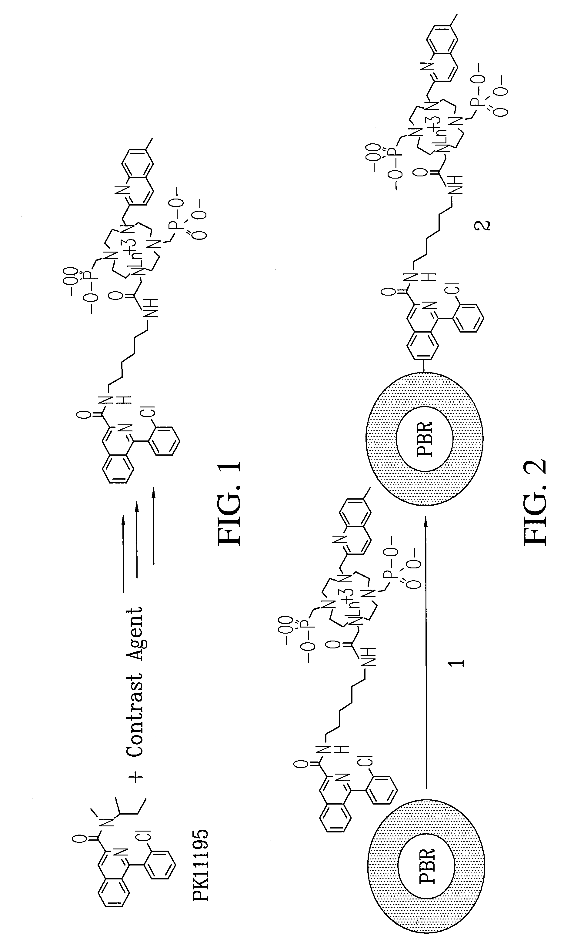
[0094]Subsequently, the cyclen-based lanthanide chelates obtained in EXAMPLE 2 can be conjugated to PK-11195 to give specificity of uptake and numerous signatures for co-registration diagnostic imaging and surgical guidance. Previous studies on PK-11195 and a closely related compound indicate that it can be conjugated, will retain it biological activity and can therefore be used as a molecular imaging agent to detect blastoglioma and study mitochondrial function.

Method of Making Conjugable PK-11195

[0095]An Ln-PK-11195 conjugated chelate (Eu-QM-CTMC-PK11195) can be prepared and will provide useful fluorescence and MR images. Scheme III illustrates a process for making a small quantity of high purity conjugable PK-11195.

[0096]

A conjugable form of PK-11195 is formed by first producing 2-Chloro-N-(2-hydroxy-1-methyl-2-phenylethyl)benzamide 75 by the following process. To an ice-cooled mixture of Norephedrine 71 HCl in CH2Cl2 (120 mL, 5.2 g, 27.8 mmol) and 40 mL of 5% NaOH was added drop...


PUM

PropertyMeasurementUnit
temperature sensitiveaaaaaaaaaa
temperatureaaaaaaaaaa
pHaaaaaaaaaa

Abstract

Cyclen-based chelates can be used as contrast agents for multi-modal imaging of tissue cells. The cyclen-based chelates are preferably polyazamacrocyclic molecules formed from 1,4,7,10 tetraazacyclododecane (“cyclen”) having varying chelating ions, phosphoester chains, and light harvesting moieties. By changing the chelating ion, phosphoester chain length and / or the light harvesting moiety different imaging techniques, such as MRI, CT, fluorescence and absorption, x-ray and NIR, may be employed to image the tissue cells. Additionally, the cyclen-based chelates may be conjugated to provide for site-specific delivery of the cyclen-based chelate to the desired tissue cells. The cyclen-based chelates may also be delivered to the tissue cells by attaching the cyclen-based chelates to a polymeric delivery vehicle. Although these cyclen-based chelates have a wide variety of application, the preferred use is for imaging of cancer cells, such as brain cancer, for improving resection of a cancerous tissue.

Description

CROSS REFERENCE TO RELATED APPLICATION[0001]This application claims the benefit, under 35 U.S.C. 119(e), of U.S. Provisional Application Nos. 60 / 316,284 and 60 / 316,303 both filed Sep. 4, 2001, the contents of which are incorporated herein by reference.BACKGROUND OF THE INVENTION[0002]1. Field of the Invention[0003]The present invention relates generally to the formation of contrast agents containing cyclen-based chelates in order to produce multimodal images of a sample of cells.[0004]2. Background Information[0005]The American Cancer Society estimates that 16,800 new intracranial tumors were diagnosed in 1999, more than double the number of diagnosed cases of Hodgkin's disease, and over half the number of cases of melanoma. Moreover, in the same year, primary cancer of the central nervous system was the cause of death in approximately 13,100 people. Despite aggressive treatment strategies including surgical resection, irradiation and chemotherapy, most patients die from the disease...

Claims


Application Information

Patent Timeline
Patent Type & Authority Patents(United States)
IPC IPC(8): A61B5/055A61K31/675A61K49/00A61K49/10
CPCA61K31/675A61K49/0002A61K49/106A61K51/0482
Inventor BORNHOP, DARRYL J.MANNING, H. CHARLESGOEBEL, TIMOTHY
Owner TEXAS TECH UNIV SYST
Why Eureka
Try Eureka