IN Vivo raman endoscopic probe

a raman endoscopic and probe technology, applied in the field of in vivo raman endoscopic probes, can solve the problems of insufficient changes, most lost years of life, and general asymptomatic lung cancer, and achieve the effect of short integration time and superior s/n ratio

Active Publication Date: 2008-06-03
BRITISH COLUMBIA CANCER AGENCY BRANCH
View PDF6 Cites 40 Cited by
  • Summary
  • Abstract
  • Description
  • Claims
  • Application Information

AI Technical Summary

Benefits of technology

[0013]The present invention comprises a novel endoscopic Raman probe. In the preferred embodiment, the probe comprises 58 collection fibers (100 μm core diameter), which will fill the CCD full vertical height and achieve spectral resolution of 8 cm−1. However, a probe comprising a smaller or larger number of collection fibers will be appropriate for use with a spectrometer having a different sized CCD. The novel probe will also preserve the round-to-parabolic linear array configuration of the Raman probe for in vivo skin measurements, as described in U.S. Pat. No. 6,486,948. The probe of the present invention therefore, will achieve similar superior S / N ratios and a short integration time of a few seconds or sub-seconds for each Raman spectral measurement.

Problems solved by technology

Lung cancer also results in the most lost years of life due to cancer death in both men and women.
However, with respect to currently available lung imaging techniques, lung cancer is generally asymptomatic until it has reached an advanced stage, when the treatment outcome is poor.
In particular, very early lung cancers are difficult to detect and localize by conventional white-light endoscopy since these cancers are only a few cell layers thick and LIP to a few millimeters in surface diameter, producing insufficient changes to make them visible under white light illumination.
However, fluorescence endoscopy technology (developed at B.C. Cancer Agency and also referred as “LIFE” technology) has less optimal specificity for lung cancer detection (66 percent for LIFE compared to 90 percent for conventional white light bronchoscopy) although it improved the sensitivity from 25 percent for white light bronchoscope to 67 percent for LIFE.
Raman spectroscopy of lung tissues, however, has only been reported on formalin-fixed parenchyma lung diseases, which provide very limited guidance to in vivo applications due to the adverse effect of formalin fixation on tissue Raman spectra.
The development of an in vivo tissue Raman probe is technically challenging due to the weak Raman signal of tissue, interference from tissue fluorescence and spectral contamination caused by the background Raman and fluorescence signals generated in the fiber itself.
Most probes published in literature and commercial products are larger than 10 mm in diameter, and therefore are not suitable for endoscopy applications.
However, the company was dissolved two years ago; therefore, the probe is no longer commercially available.
The main disadvantages of that probe are that (1) only seven collection fibers were used, which cannot fill the full vertical height of the CCD sensor in the spectrometer; therefore, it was unable to gain the maximum sensitivity; and (2) the size of the collection fibers was big (300 μm), leading to poor spectral resolution (>20 cm−1).

Method used


Image

  • IN Vivo raman endoscopic probe
  • IN Vivo raman endoscopic probe
  • IN Vivo raman endoscopic probe

Examples


Embodiment Construction

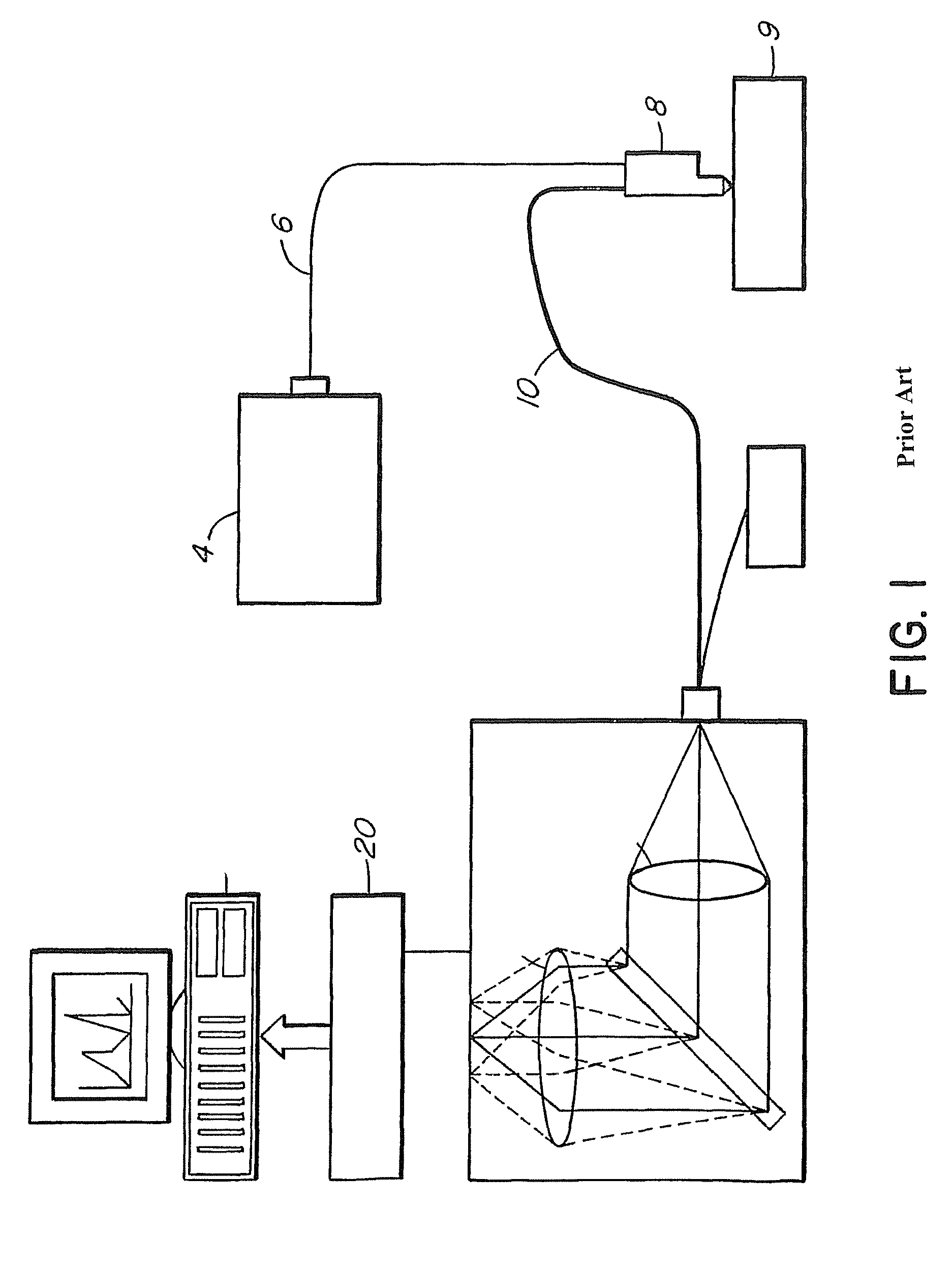
[0033]We have successfully built a rapid Raman spectroscopy system, which can obtain a Raman spectrum from in vivo skin in less than one second. This system is described and claimed in U.S. Pat. No. 6,486,948, the disclosure of which is incorporated by reference. FIG. 1 shows the block diagram of the system. It consists of an external cavity-stabilized diode laser 4 (785 nm, 300 mW; Model 8530, SDL), a transmissive imaging spectrograph 2 (HoloSpec-f / 2.2-NIR, Kaiser), a NIR-optimized, back-illuminated, deep-depletion, CCD detector 20 (LN / CCD-1024EHRB, Princeton Instruments), and a specially-designed Raman probe 8. The laser 4 is coupled to the Raman probe 8 via a 200-μm core-diameter fiber 6. The CCD 20 consists of 1024×256 pixels (27 μm×27 μm) and allowed vertical binning for improved detection sensitivity. The whole system was packed onto a movable cart for outpatient clinical data acquisition.

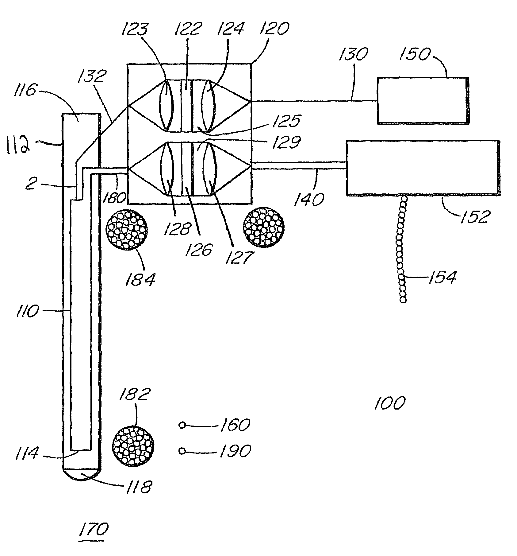
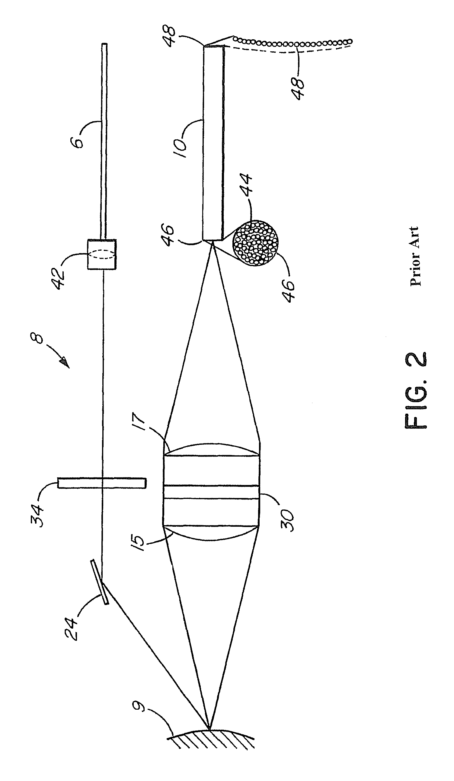
[0034]This Raman probe 8 was designed to maximize the collection of tissue Raman signals ...


PUM

PropertyMeasurementUnit
cut-off wavelengthaaaaaaaaaa
optical densityaaaaaaaaaa
diameteraaaaaaaaaa

Abstract

An apparatus for in vivo, endoscopic laser Raman spectroscopy probe and methods for its use are described. The probe comprises a fiber bundle assemble small enough to fit through an endoscope instrument channel, a novel combination of special coatings to the fiber bundle assembly, including a short-pass filter on the illumination fiber and a long-pass filter on the collection fiber bundle, a novel filter adapter comprising collimating lenses, focusing lenses, a band-pass filter, and a notch filter, and a round-to-parabolic linear array fiber bundle. The apparatus further comprises a laser to deliver illumination light and a spectrometer to analyze Raman-scattered light from a sample. The analysis of Raman spectra from in vivo measurements can discover molecular and structural changes associated with neoplastic transformations and lead to early diagnosis and treatment of cancer.

Description

CROSS-REFERENCE[0001]This application claims priority from provisional application No. 60 / 441,566, filed on Jan. 21, 2003.BACKGROUND OF THE INVENTION[0002]Lung cancer is the leading cause of death from cancer in North America, and it has the second most common cancer incidence among both men and women. One in 11 Canadian men will develop lung cancer, and 1 in 12 will die from this condition, while one in 19 Canadian women will develop lung cancer, and 1 in 22 will die from this disease. Lung cancer also results in the most lost years of life due to cancer death in both men and women. The best outcome of lung cancer treatment is achieved when the lesion is discovered in the pre-invasive stage, which is also commonly referred as carcinoma in situ (CIS).[0003]Early and accurate diagnosis of lung cancer offers a better chance of cure, results in the use of less radical treatment methods, and reduces the cost of treatment. The five-year survival for all stages of lung cancer is only 11-1...

Claims


Application Information

Patent Timeline
Patent Type & Authority Patents(United States)
IPC IPC(8): A61B5/00A61B1/267G01N21/65
CPCA61B5/0071A61B5/0075A61B5/0086G01N21/65G01N2021/656
Inventor ZENG, HAISHAN
Owner BRITISH COLUMBIA CANCER AGENCY BRANCH
Why Eureka
Try Eureka