

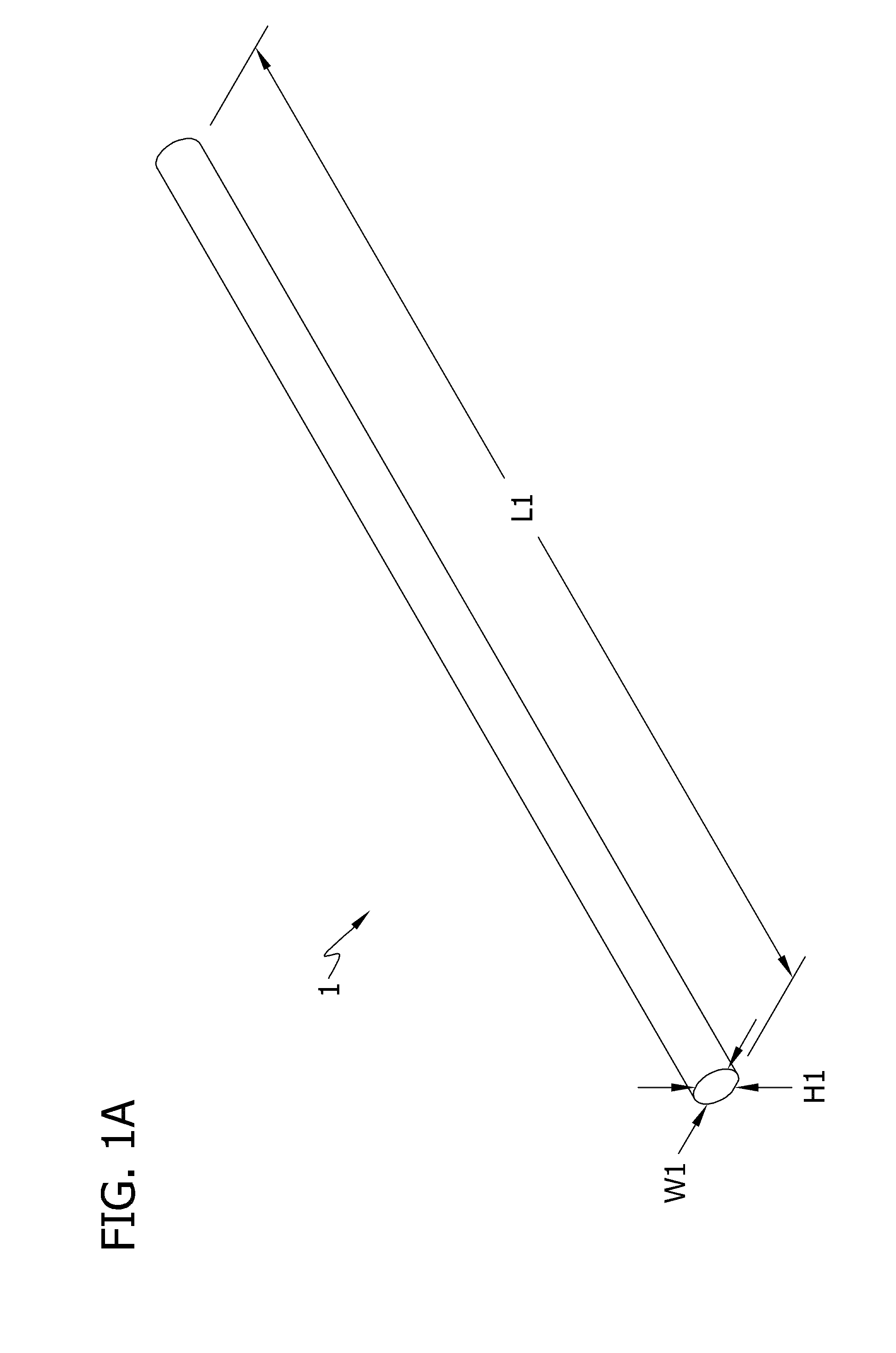
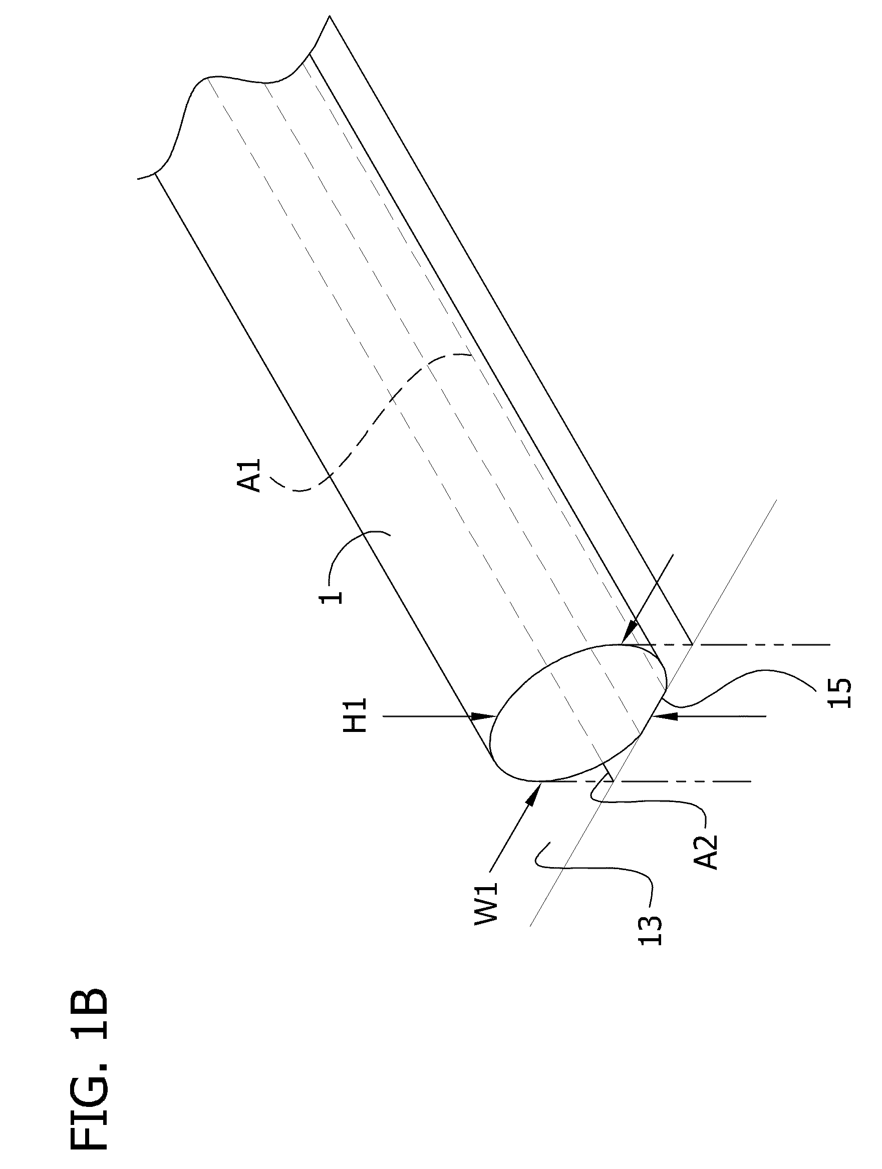
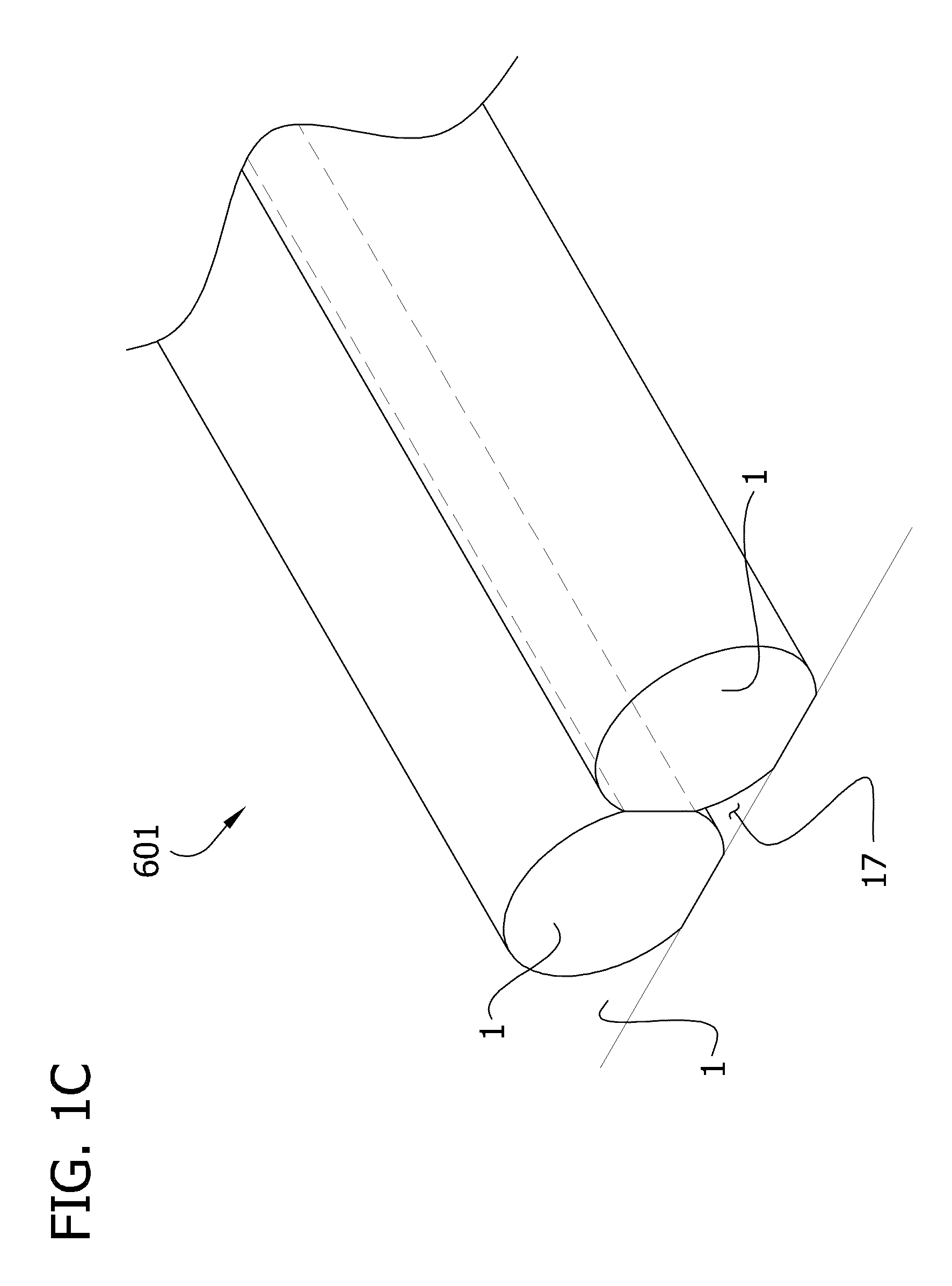
Self-assembling multicellular bodies and methods of producing a three-dimensional biological structure using the same
a multicellular body and biological structure technology, applied in the field of regenerative medicine and tissue engineering, can solve the problems of many will likely perish due to lack of replacement blood vessels, and the variety of methods to build engineered tissues has been attempted and developed with limited success, and the assembly of vascularized three-dimensional organs has not been accomplished. , to achieve the effect of sufficient integrity and convenient manipulation and handling
- Summary
- Abstract
- Description
- Claims
- Application Information
AI Technical Summary
Benefits of technology
Problems solved by technology
Method used
Image

Examples
example 1
Preparation of Multicellular Bodies and Tissue Engineering Using Pig Smooth Muscle Cells
[0132]I. Pig Smooth Muscle Cells. Pig smooth muscle cells (SMCs) were grown in the same conditions used in previous studies. The medium composition was Dulbecco's Modified Eagle Medium (DMEM) low glucose supplemented with 10% porcine serum, 10% bovine serum, 50 mg / L of proline, 20 mg / L of alanine, 50 mg / L of glycine, 50 mg / L of ascorbic acid, 12 μg / L, of Platelet Derived Growth Factor-BB (PDGF-BB), 12 μg / L of Basic Fibroblast Growth Factor (bFGF), 3.0 μg / L of CuSO4, 0.01M of HEPES buffer, and 1.0×10⁵ units / L of penicillin and streptomycin. The cells were grown on gelatin (gelatin from porcine skin) coated 10 cm Petri dishes and incubated at 37° C., 5% CO2. The SMCs were subcultured up to passage 7 before being used for multicellular body (e.g., cellular unit) preparation. Eighteen confluent Petri (i.e. cell culture) dishes were necessary to prepare 24 cellular units and 4 tubes (outside diameter ...
example 2
Alternative Procedure for Preparing Multicellular Bodies and for Tissue Engineering
I. Growth Conditions for Cells of Various Types
[0137]Chinese Hamster Ovary (CHO) cells transfected with N-cadherin were grown in Dulbecco's Modified Eagle Medium (DMEM) supplemented with 10% Fetal Bovine Serum (FBS), antibiotics (100 U / mL penicillin streptomycin and 25 μg / mL gentamicin) and 400 μg / ml geneticin. Besides gentamicin all antibiotics were purchased from Invitrogen.
[0138]Human Umbilical Vein Smooth Muscle Cells (HUSMCs) and Human Skin Fibroblasts (HSFs) were purchased from the American Type Culture Collection (CRL-2481 and CRL-2522 respectively). HUSMCs were grown in DMEM with Ham's F12 in ratio of 3:1, 10% FBS, antibiotics (100 U / mL penicillin-streptomycin and 25 μg / mL gentamicin), 20 μg / mL Endothelial Cell Growth Supplement (ECGS), and Sodium Pyruvate (NaPy) 0.1M. Human skin fibroblasts (HSFs) were grown in DMEM with Ham's F12 in ratio of 3:1, 20% FBS, antibiotics (100 U / mL penicillin / str...
example 3
Use of Gelatin and / or Fibrin in Preparing Multicellular Bodies and a Three-Dimensional Fused Tubular Structure
[0154]Human Aortic Smooth Muscle Cells (HASMCs) were purchased from Cascade Biologics (C-007-5C). HASMCs were grown in medium 231 supplemented with the Smooth Muscle Growth Supplement (SMGS). The HASMCs were grown on uncoated cell culture dishes and were maintained at 37° C. in a humidified atmosphere containing 5% CO2.
[0155]The HASMCs were trypsinized, resuspended in tissue culture media, and centrifuged as described above in Examples 1 and 2. Following centrifugation, the tissue culture medium (i.e., the supernatant) is removed, and the cells (1 confluent Petri dish) were resuspended first in 100 μl of a solution of fibrinogen (50 mg / ml in 0.9% NaCl), and then 70 μl of a solution of gelatin (20% in phosphate buffered saline (PBS)) was added. The cell suspension was centrifuged again, the supernatant was removed, and the centrifuge tube containing the cell pellet was placed...
PUM
| Property | Measurement | Unit |
|---|---|---|
| area | aaaaa | aaaaa |
| length | aaaaa | aaaaa |
| length | aaaaa | aaaaa |
Abstract
Description
Claims
Application Information