Method for preparing chitosan-silk fibroin composite nano-fiber multifunctional patch for promoting myocardial tissue regeneration and monitoring stem cells

A composite nanofiber, myocardial tissue regeneration technology, applied in the direction of bone/connective tissue cells, animal cells, vertebrate cells, etc., can solve the problem of unable to achieve sustained release of drugs or cytokines, unfavorable for artificial improvement and regulation, etc. The effect of promoting attachment and growth, inhibiting left ventricular remodeling, and increasing specific surface area

Active Publication Date: 2014-12-10
GENERAL HOSPITAL OF PLA +1
View PDF5 Cites 20 Cited by
  • Summary
  • Abstract
  • Description
  • Claims
  • Application Information

AI Technical Summary

Problems solved by technology

Due to the retention of tissue scaffolds including vasculature, decellularized biological tissue materials can simulate the microenvironment of cell growth, but it is not conducive to artificial improvement a

Method used


Image

  • Method for preparing chitosan-silk fibroin composite nano-fiber multifunctional patch for promoting myocardial tissue regeneration and monitoring stem cells
  • Method for preparing chitosan-silk fibroin composite nano-fiber multifunctional patch for promoting myocardial tissue regeneration and monitoring stem cells
  • Method for preparing chitosan-silk fibroin composite nano-fiber multifunctional patch for promoting myocardial tissue regeneration and monitoring stem cells

Examples


preparation example Construction

[0040] The preparation method of the chitosan-silk fibroin composite nanofiber multifunctional patch for promoting myocardial tissue regeneration and stem cell monitoring of the present invention comprises the following steps:

[0041] (1) Preparation of cellulose nanofiber base plate (Mat) by electrospinning technology;

[0042] (2) By layer-by-layer self-assembly (LBL), positively charged chitosan (CS) and negatively charged silk fibroin (SF) were alternately assembled layer by layer onto the surface of the cellulose nanofiber bottom plate obtained in step (1) , assemble 5.5-15.5 layers to form a chitosan-silk fibroin composite nanofibrous membrane;

[0043] (3) Plant AD-MSC or iPS-CM seed cells labeled with green fluorescent protein (GFP) and firefly luciferase (Fluc) on the surface of the composite nanofiber membrane obtained in step (2); through three-dimensional co-culture, the obtained The chitosan-silk fibroin composite nanofiber multifunctional patch for promoting my...

Embodiment 1

[0044] Example 1 Preparation of cellulose nanofiber base plate (Mat) by electrospinning technology

[0045] Example (1). The cellulose acetate (CA, Mn=3×10 4 Da, Aldrich Co., USA) is dissolved in the mixed solution of acetone and dimethylacetamide (DMAc) that the mass ratio is 2:1 with 15% mass concentration, and it is sucked into the plastic syringe with metal needle, and the inner diameter of needle is 1㎜. The distance between the needle and the roller collector covered with aluminum foil was 20 cm, and the applied DC voltage was 16 kV. The syringe is driven by a syringe pump at a speed of 0.5ml / h, the drum rotates at a linear speed of 100 m / min, the ambient temperature is 25°C, the relative humidity is 40%, and the electrospinning time is 16 hours. The prepared CA nanofiber bottom plate was dried in vacuum at room temperature for 24 hours to remove the solvent. Then, the dried CA nanofiber base plate was hydrolyzed in 0.05 mol / L sodium hydroxide solution for seven days, ...

Embodiment 2

[0048] Formation of embodiment two chitosan-silk fibroin composite nanofiber membrane

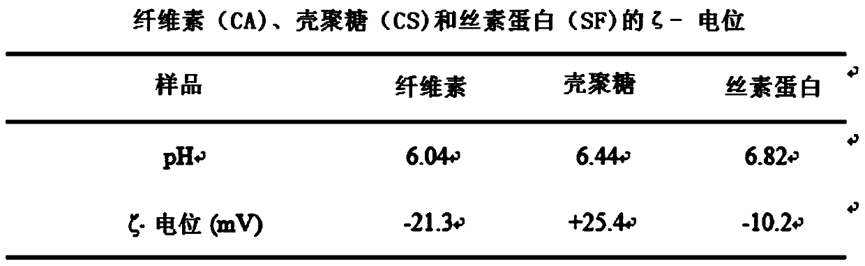
[0049] Example (1) positively charged chitosan (CS, Mw=2.0×10 5 kDa, DD=92%, Zhejiang Yuhuan Ocean Biochemical Co., China), using 2% acetic acid solution as a solvent to prepare a 1㎎ / L solution, adjusting the pH value to 5.0, negatively charged silk fibroin Protein (SF, Aladdin Chemical Reagent Co., China) was made into a 1 ㎎ / L aqueous solution, and the pH was adjusted to 5.3. The cellulose nanofiber bottom plate obtained in Example 1 was immersed in a positively charged chitosan (CS) solution for 20 minutes, and rinsed three times with 0.1 mol / L sodium chloride solution for 2 minutes each time. Then the nanofiber bottom plate was immersed in negatively charged silk fibroin (SF) solution for 20 minutes, and rinsed three times with 0.1 mol / L sodium chloride solution. 2 minutes each time. This process can make the surface of nanofibers covered with chitosan-silk fibroin (CS-SF) bimolecular...


PUM


Abstract

The invention relates to a method for preparing a chitosan-silk fibroin composite nano-fiber multifunctional patch for promoting myocardial tissue regeneration and monitoring stem cells. The method comprises the following steps: preparing a cellulose nano-fiber basal plate by an electrostatic spinning technology; alternately assembling positively charged chitosan (CS) and negatively charged silk fibroin (SF) to the surface of nano-fibers layer by layer by adopting a layer-by-layer assembly technique, and assembling 5.5-10.5 layers to form a CS-SF composite nano-fiber membrane; and planting seed cells of adipose tissue-derived stromal cells or cardiac progenitor cells labeled by green fluorescent protein and firefly luciferase on the surface of the CS-SF composite nano-fiber membrane, preparing the patch by three-dimensional co-culture. The patch has excellent biocompatibility, can be used as a cell vector, has an effect of resisting oxidative stress to improve the survival rate and treatment efficiency of stem cells, and is capable of evaluating the number, distribution and function states of transplanted stem cells, effectively preventing occurrence of post-myocardial infarction heart failure and reducing the death rate of ischemic cardiomyopathy.

Description

technical field [0001] The invention relates to a tissue engineering technology, in particular to a preparation method of a chitosan-silk fibroin composite nanofiber patch for promoting myocardial tissue regeneration and stem cell detection after acute myocardial infarction. Background technique [0002] According to the statistics of the World Health Organization in 2012, ischemic heart disease has become the number one killer of human health worldwide, accounting for 13.2% of all-cause mortality, and about half of the patients died of acute myocardial infarction. The mammalian heart is a terminally differentiated organ. Once necrotic, it is difficult to regenerate and repair itself. With the necrosis and massive loss of cardiomyocytes, it will lead to the progress of ventricular remodeling and eventually heart failure. At present, traditional treatment methods, such as coronary artery bypass surgery, drug thrombolysis, percutaneous coronary intervention, etc., can obviousl...

Claims


Application Information

Patent Timeline
IPC IPC(8): A61L27/38A61L27/20A61L27/22A61L27/50C12N5/0775C12N5/077
Inventor 曹丰邓宏斌陈江伟王亚斌
Owner GENERAL HOSPITAL OF PLA
Why Eureka
Try Eureka