Prenatal diagnostic methods

Inactive Publication Date: 2002-09-12
DUNDEE UNIV OF
View PDF0 Cites 15 Cited by
  • Summary
  • Abstract
  • Description
  • Claims
  • Application Information

AI Technical Summary

Benefits of technology

0031] A particular advantage of the present invention is that the methods identify, and can be used to isolate, embryonic and early fetal red blood cells at an early stage of gestation. Thus, it is preferred if the maternal blood sample is taken from the pregnant female at an early stage of pregnancy. In the case of a pregnant human female it is preferred if the sample is taken in the first trimester.
0032] In general the earlier in pregnancy, whether for potential fetal therapy or the option of termination, the better (ideally less than 10 weeks gestation). A further practical reason is that the means of termination of pregnancy is technically easier at earlier gestations and with less physical and psychological side-effects. There is no upper limit for intrauterine diagnosis of fetal anomalies and even late in pregnancy treatment may still be beneficial in utero or the immediate newborn period. The ontogeny of nucleated embryonic and fetal cells clearly indicates that the percentage of those cells in the embryo/fetus is higher in the first trimester than later in pregnancy. However, the total fetal blood volume increases with gestation and proportionally fetal to maternal transfusion volumes may be greater.
0033] The detailed structure of the developing human conceptus, sufficient for accurate dating in days, has only been described in detail up to 56 day post-ovulatory days and the descriptive term, embryo, will be used as the convention for this developmental staging procedure (O'Rahilly & Muller (1987) Developmental Stages in Human Embryos, Publication 637, Washington: Carnegie Institute of Washington). The descriptive term, fetus, will be used for the remainder of human intrauterine development to term (>37 completed weeks gestation), with developmental age (to the nearest week) an estimate ba

Problems solved by technology

Existing procedures such as fetal, hepatic or chorionic biopsy for diagnosis of chromosomal disorders including Down's syndrome, as well as single gene defects including cystic fibrosis are very invasive and carry a not inconsiderable risk to the foetus and a small risk to the mother.
Initial interest was directed towards trophoblastic detection systems but separation of those cells by flow cytometry has been unreliable as maternal lymphocytes appear to absorb proteins released by trophoblastic cells (Mueller et al.
(1992) J. Reprod. Med. 37,410-418 shows that the 15 transferrin receptor antigen alone is not sufficient for enrichment of fetal nucleated erythrocytes and points out that the reproducibility and reliability of the techniques are still

Method used


Image

  • Prenatal diagnostic methods

Examples


example 1

Antibodies Directed at Adult Liver Components

[0108] Antibodies directed against purified rat liver testosterone / 4-nitrophenol UDPGT are raised in Suffolk Cross Blackface sheep by a combination of intradermal and subcutaneous injection. IgG is prepared from the antiserum by a combination of ammonium sulphate precipitation and diethyl aminoethyl-cellulose chromatography (Burchell et al. (1984) Biochem. Soc. Trans. 12, 50). Typically sheep antirat liver testosterone / 4-nitrophenol UDPGT antibody preparation (RAL 1) inhibit UDPGT activity towards bilirubin, testosterone, 1-naphthol, androsterone, estrone, and morphine, and inimunoblotting confirms a broad spectrum of cross-reactivity to multiple isoforms in rat and human adult and fetal liver microsomes.

[0109] Monospecific polyclonal antisera to the catalytic subunit of the microsomal glucose-6-phosphate system, T2, and T3 are each raised in Cheviot sheep by 3 subcutaneous injections of 80 .mu.g of purified protein and Freund's complete ...

example 2

Combined Immunocytochemical and Fluorescence in Situ Hybridisation Analysis of a Material Blood Sample to Detect Trisomy 21

[0116] Blood samples and cell preparation. Peripheral venous blood samples (EDTA) are obtained from a pregnant female in the first trimester. Five ml aliquots of blood are carefully layered over 3.5 ml aliquots of Polymorphoprep (Nycomed, Norway) in 15 ml tubes which are then spun at 500 g for 30 mm at room temperature. Mononuclear cells at the plasma / Polymorphoprep interface (upper of the two bands obtained) are harvested using a Pasteur pipette and dispensed into a clean tube. The cells are washed three times using 5 ml of cold phosphate buffered saline (PBS) containing 0.5% bovine serum albumin (BSA) and 5 mM ethylenediaminetetra-acetic acid (EDTA) followed by a 10 mm spin at 400 g each time. The cell pellets are finally resuspended in PBS / BSA / EDTA at a concentration of 106 cells / ml.

[0117] Slide preparation. Aliquots (100 .mu.l) of blood mononuclear cells are...

example 3

Isolation of Fetal Cells and PCR Analysis for Sickle Cell Anaemia and Thalassaemia

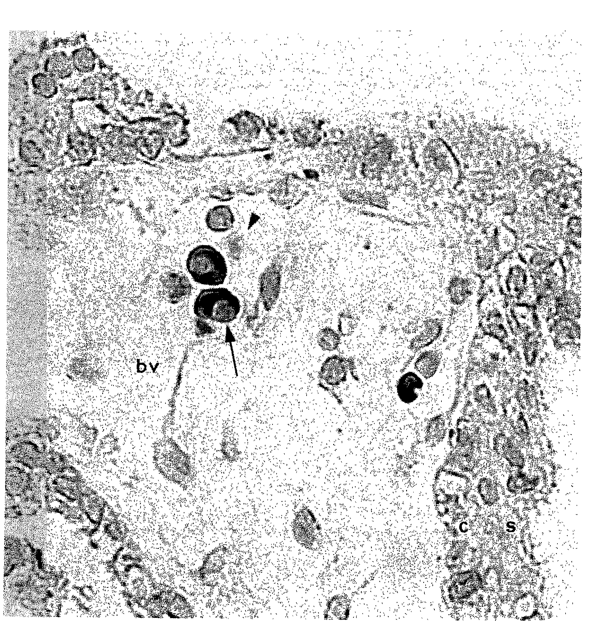
[0125] FIG. 1 (95chorion aGLUT2 1; 100X40RH) shows a human chorionic villus at 56 post-ovulatory days showing intense alpha GLUT 2 immunoreactivity in a megaloblast (arrow) and no reactivity in a normocyte (arrowhead) within a fetal chorionic blood vessel (bv). The syncytiotrophoblastic (s) and cytotrophoblastic (c) layers are minimally immunoreactive.

[0126] Fetal cells from maternal blood taken in the first trimester are isolated in the following way, and a PCR analysis for Sickle cell anaemia and thalassaemia undertaken.

[0127] Blood sample and cell separation. Peripheral blood (16-18 ml) from pregnant women in the first trimester is collected into EDTA Vacutainer tubes (Becton Dickinson, Rutherford, N.J.). The blood is then diluted 1:2 with phosphate buffered saline (PBS), with each 15 ml layered over 10 ml Ficoll-paque plus (density 1.077 g / ml, Pharmacia Biotech, Piscataway, N.J.) and centrifuged at...


PUM

PropertyMeasurementUnit
Fractionaaaaaaaaaa
Fluorescenceaaaaaaaaaa

Abstract

A method of identifying embryonic or fetal red blood cells in a sample containing maternal blood cells and embryonic or fetal red blood cells or both, the method comprising determining, which cell or cells contain or express an adult liver component. A method of isolating embryonic or fetal red blood cells from a sample containing maternal blood cells and embryonic or fetal red blood cells or both, the method comprising isolating the cells which contain or express an adult liver component. A method of determining a fetal abnormality the method comprising identifying or isolating embryonic or fetal cells according to the above methods and analysing said embryonic or early fetal cells for said abnormality. Use of a means for determining whether a cell contains or expresses an adult liver component for identifying or isolating an embryonic or fetal red blood cell.

Description

CROSS-REFERENCES[0001] This is a divisional application claiming priority from U.S. application Ser. No. 09 / 392,055, filed Sep. 8, 1999, which was in turn a continuation of International Application PCT / GB98 / 00656, with an international filing date of Mar. 3, 1998.BACKGROUND OF THE INVENTION[0002] 1. Field of the Invention[0003] The present invention relates to diagnostic methods, in particular to methods of prenatal diagnosis and to reagents for use in such methods.[0004] 2. General Background and State of the Art[0005] Prenatal diagnosis is carried out widely in hospitals throughout the world. Existing procedures such as fetal, hepatic or chorionic biopsy for diagnosis of chromosomal disorders including Down's syndrome, as well as single gene defects including cystic fibrosis are very invasive and carry a not inconsiderable risk to the foetus and a small risk to the mother.[0006] Amniocentesis, for example, involves a needle being inserted into the womb to collect cells from the e...

Claims


Application Information

Patent Timeline
IPC IPC(8): A61K35/18C07K16/40C12N5/00C12N5/06G01N33/564G01N33/573G01N33/68G01N33/80
CPCA61K35/18C12N2509/00G01N33/573G01N33/689G01N2800/36G01N2800/368
Inventor BURCHELL, ANNHUME, ROBERT
Owner DUNDEE UNIV OF
Why Eureka
Try Eureka