
Test system for determining gene toxicities

a test system and gene technology, applied in the field of test system for determining gene toxicities, can solve the problems of increasing not only the amount of labor and time invested, but also the limitations of methods, and the treatment of the compounds under investigation have not yet been clarified

Inactive Publication Date: 2004-10-07
WIESMULLER LISA
View PDF0 Cites 11 Cited by
  • Summary
  • Abstract
  • Description
  • Claims
  • Application Information

AI Technical Summary

Benefits of technology

[0114] This novel test system for the determination of genotoxicities overcomes all the disadvantages of previous detection systems: DNA damage and detection take place directly in the mammalian cells and not in the microorganisms. The detection system is transferable to living animals. The spectrum of possible DNA damage by mutagens is as wide as possible as a result of the detection of the most diverse range of recombinative repair processes and of point mutations and, in particular, also of every type of otherwise gene-inactivating events. The sensitivity of the system was maximised by the optimised use of strongly autofluorescent proteins as signals and the generation of long sequence homologies. The simple and rapid execution of the test fulfills the prerequisite for routine-type analyses on a large scale.5.

Problems solved by technology

Nevertheless, these methods have certain limitations: the detection of SCEs or of micronuclei requires the preparation, fixation, and staining of the cell material, and subsequent examination under the microscope.
Moreover, for SCE detection, treatment with colchicine or bromodeoxyuridine is additionally required before sample preparation, for the micronucleus assay, the treatment involves cytochalasin B, whose effects on the treatment with the compounds under investigation have not yet been clarified.
The greatest difficulties with the Comet assay and with the quantification of DNA repair synthesis activities seem to be the large variations of the data, so that numerous multiple determinations need to be done, as far as possible with automated data logging, to obtain statistically relevant data.
However, the need, usually connected with this method, for carrying out nucleic acid and / or protein extractions has to be mentioned as a disadvantage thereof.
It increases not only the amount of labour and time that have to be invested for the detection process, but also harbours the risk of a lack of reproducibility.
Critical disadvantages of this method are that, firstly, it was established for Saccharomyces cerevisiae rather than for mammalian cells and, secondly, fluorescence can be measured only after a series of extraction steps, i.e. not with intact cells.
The Ames assay has the critical disadvantage that Salmonella does not possess the cellular machinery of eukaryotic cells which is necessary, to convert procarcinogens into reactive metabolites.
Although attempts have been made to optimise the Ames assay by adding rat liver extracts to the assay mixture in order to simulate the human metabolism, these extracts do not necessarily contain sufficient enzymatic activities which are necessary for biotransformation (Ames, B. N. (1974) Genetics 78: 91-95).
Moreover, the methodology of the Ames assay makes automation as the basis for standardised surveillance procedures more difficult.
However, the Ames assay only measures erroneous DNA repair leading to an underestimation of DNA damage.
Moreover, larger chromosome rearrangements such as insertions, deletions or translocations are not detected by this assay.
The DEL assay, established for the HIS3 locus of the yeast Saccharomyces cerevisiae, suffers from limitations already described for other methodsm, namely that the biotransformation of procarcinogens specific for mammals is not taken into account and survival rates are measured either via cultivation under certain selection conditions or by determining the uptake of a fluorescent substance by use of cell extracts (Brennan, R. J. and Schiestl, R. H. (1999) Mutat. Res. 430: 37-45).
A major disadvantage of the so-called Big Blue mouse (Stratagene) and of transgenic mice derived from it is that the mutation indicator gene, i.e. the lacI gene which operates as a repressor of the .beta.-galactosidase expression, needs to be transduced after the genotoxic treatment of the mouse, from the genome via the .alpha.-phage into bacteria.
This method has the disadvantage that the recombinative events are detectable exclusively if they have taken place in the embryonal premelanocytes, so that statistically significant toxicity determinations would require the analysis of a large number of mice treated at the embryonal stage.

Method used


Image

  • Test system for determining gene toxicities
  • Test system for determining gene toxicities
  • Test system for determining gene toxicities

Examples


example 1

Cloning of p5-Puro-CMV-(N'-EGFP)-CMV-Red-(EGFP-EJ)

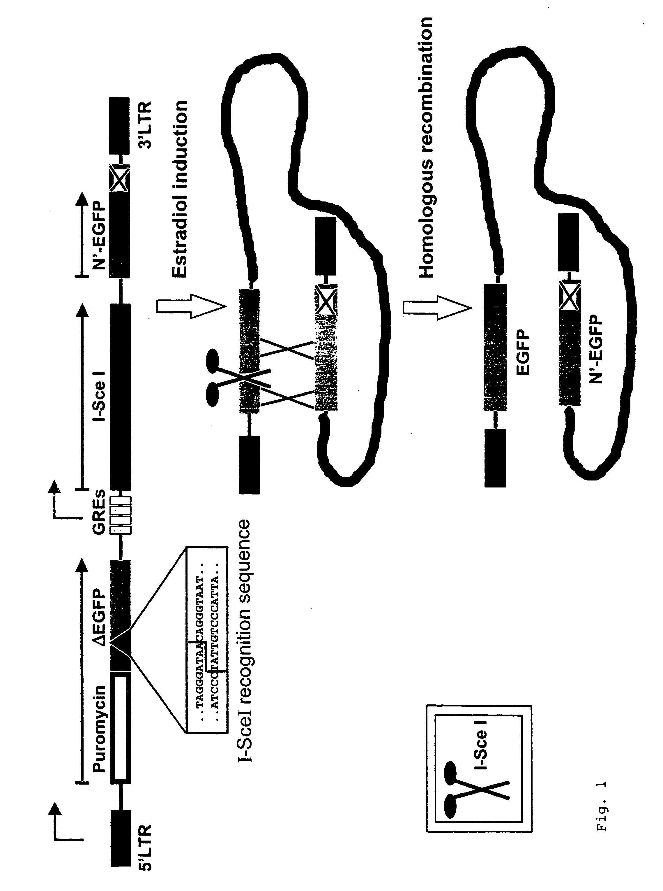
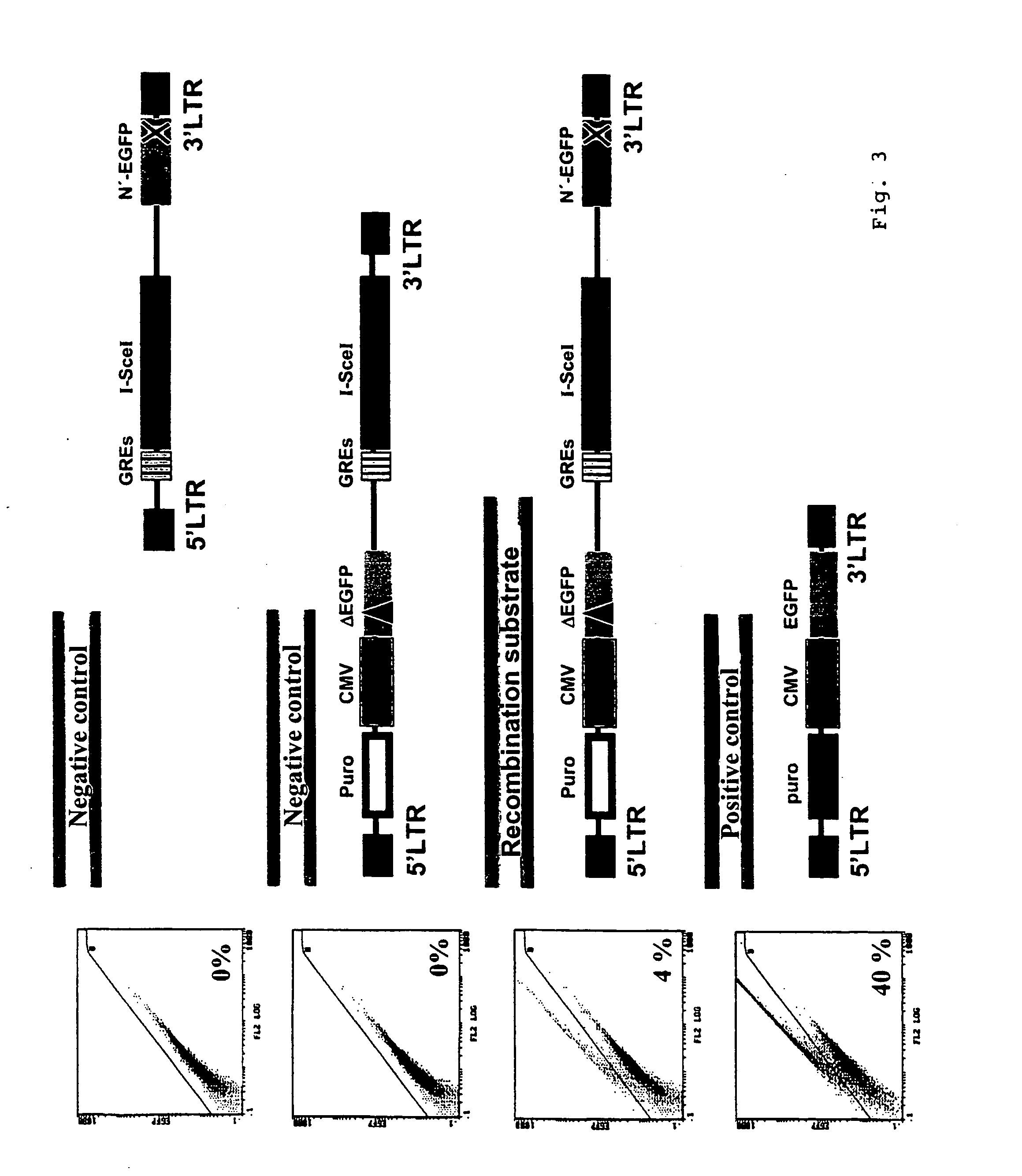
[0115] Cloning of the construct p5-Puro-CMV-(N'-EGFP)-CMV-Red-(EGFP-EJ) will be described in the following in a manner representative of the vectors in FIG. 5 (compare FIG. 5i and chapter 6 sequence protocol). The retroviral vector p5NM from Dr. Carol Stocking, Heinrich-Pette-Institut Hamburg, which belongs to the family of MPSV vectors, was used as the basis (Laker, C. et al. (1987) Proc. Natl. Acad. Sci. USA 84: 8458-8462). Restriction enzymes, Rapid DNA Ligation Kit.RTM., Klenow enzyme, T4 polymerase and shrimp alkaline phosphatase (SAP) were purchased from New England Biolabs, Schwalbach / Taunus, Roche, Mannheim, and MBI Fermentas, St. Leon-Rot. Bacteria transformations for cloning purposes were carried out using the bacteria strains XL10Gold.RTM., SURE.RTM., or SCS110 from Stratagene, Heidelberg, using standard methods (Ausubel, F. M. et al. (2000) Current Protocols in Molecular Biology, John Wiley and Sons). DNA blunt end format...

example 2

Generation of the .sub..DELTA.EGFP and EGFP-EJ Genes by PCR Mutagenesis

[0122] In a manner representative of the different mutations in the sequence coding for the chromophore amino acids within the EGFP gene, the construction of the .sub..DELTA.EGFP gene and the EGFP-EJ gene will be described here.

[0123] .sub..DELTA.EGFP was constructed via stepwise PCR amplifications. The terminal PCR primers (EGFP-13 and -15) for the complete .sub..DELTA.EGFP-PCR fragment introduced synthetic restriction enzyme recognition sites into the .DELTA.EGFP gene, namely for SexAI at the 5' end and for BamHI and EcoRI at the 3' end, so that these restriction sites were available for subsequent cloning in the reading frame of the puromycin resistance gene in pUC-Puro (compare example 1). The mutated region at the chromophore amino acids was engineered by firstly producing the sequence portion 5' and 3' to the mutation in the .DELTA.EGFP gene via 2 independent PCR reactions. The desired PCR products were pur...

example 3

Analysis of Individual KMV Cell Clones with a Genomic pGC-Sce Integrate Following I-SceI Expression After Estradiol Administration.

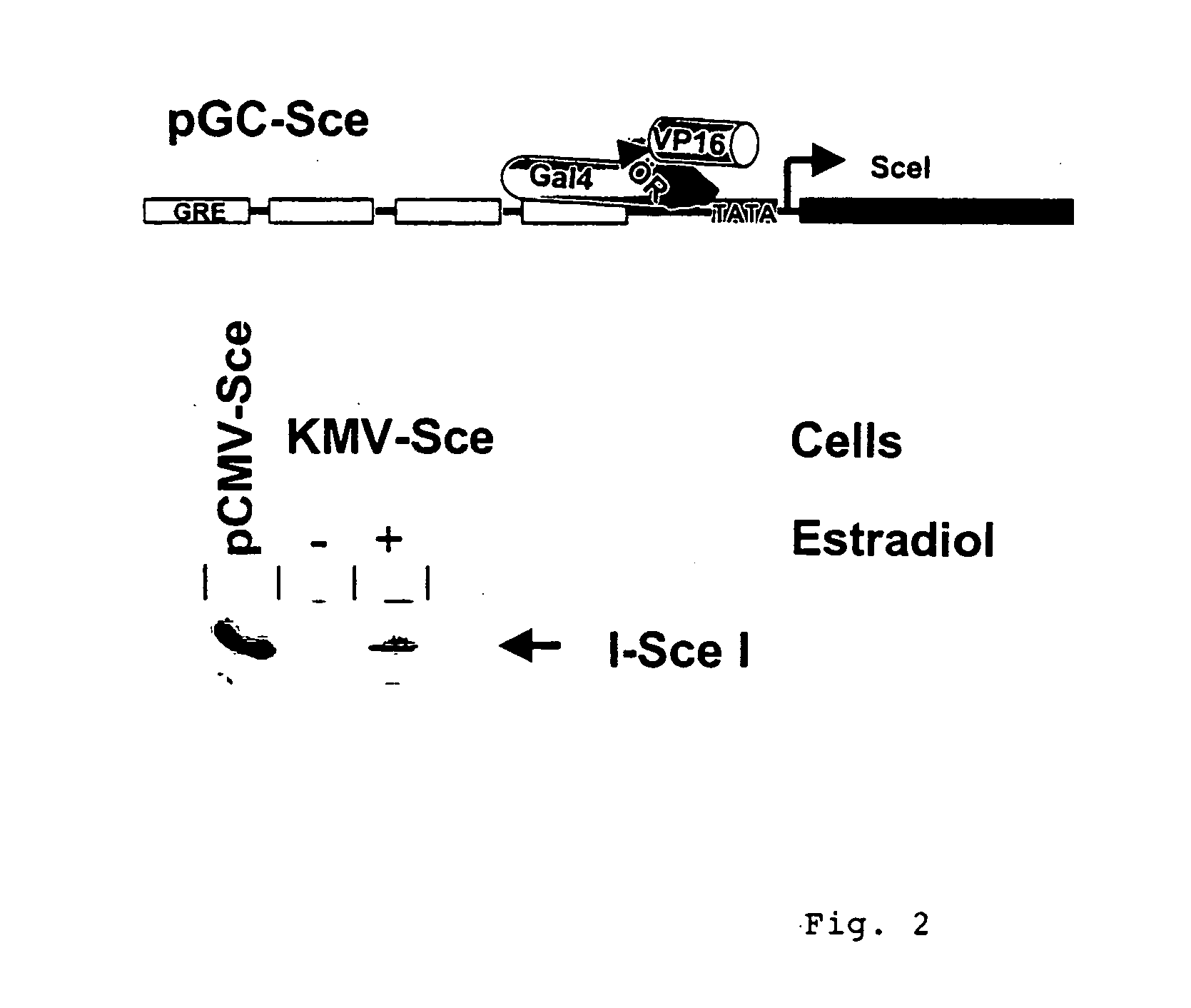
[0156] KMV cells represent K562 leukemia cells (Andersson, L. C. et al. (1979) Int. J. Cancer 23: 143-147) which express the transcription factor GalER-VP which can be activated by the administration of estradiol (Braselmann, S. et al. (1993) Proc. Natl. Acad. Sci. USA 90: 1657-1661). Following electroporation with the plasmid pGC-Sce, KMV clones were isolated with the construct integrated into genome (compare example 6). These clones were cultivated in tissue culture flasks from Nunc.TM., Wiesbaden. RPMI without phenol red (Gibco / BRL, Eggenstein) was used as the culture medium together with 10% FCS without steroid hormones (Promocell). The incubation of the cells was carried out at 37.degree. C. and in 5% CO.sub.2 in the incubator.

[0157] To activate GalER-VP and consequently indirectly the promoter in pGC-Sce via the Gal4 recognition sequences upstream ...


PUM

PropertyMeasurementUnit
wavelengthaaaaaaaaaa
excitation wavelengthaaaaaaaaaa

Abstract

The subject matter of the invention is a method which makes it possible to detect genotoxic stress in living mammalian cells by the detection of all previously documented types of DNA recombination and also of mutations in response to noxae. The detection is based on the detection of the fluorescence from intact autofluorescent proteins or of luminescence by means of intact bioluminescent enzymes or enzymes that convert chemoluminescent substrates. It is characterised by a short reaction time in the scope of hours and permits the transfer of the test system to different mammalian cells and to living experimental animals, particularly by using a retroviral vector system.

Description

1. SUBJECT MATTER OF THE INVENTION[0001] The subject matter of the invention is a method which makes the detection of genotoxic stress in living mammalian cells possible by the detection of all previously documented types of DNA recombination and also of mutations in response to noxae. The detection is based on the detection of the fluorescence from intact autofluorescent proteins or of luminescence by means of intact bioluminescent enzymes or enzymes that convert chemoluminescent substrates, it is characterised by a short reaction time in the scope of hours and permits the transfer of the test system to different mammalian cells and to living experimental animals, particularly by using a retroviral vector system in particular.2. STATE OF THE ART[0002] Excluding possible genotoxicities is one of the basic preconditions for the official approval of new food constituents, cosmetic products, and medicaments. The existing processes for identifying genotoxicities can be subdivided on the...

Claims


Application Information

Patent Timeline
Patent Type & Authority Applications(United States)
IPC IPC(8): C12N15/867C12Q1/68C12Q1/6897
CPCC12N15/86C12N2740/13043C12N2830/003C12N2840/20C12Q1/6897C12Q2563/107
Inventor WIESMULLER, LISA
Owner WIESMULLER LISA
Learn More