
Stent to be placed in vivo

a stent and in vivo technology, applied in the field of stents for in vivo placement, can solve the problems of repeated stenosis (restenosis) with high probability, difficult control of expansion size, intimal thickening, etc., and achieve low rate of restenosis, and suppression of restnosis within and around the stent.

Inactive Publication Date: 2007-02-15
KANEKA CORP
View PDF6 Cites 81 Cited by
  • Summary
  • Abstract
  • Description
  • Claims
  • Application Information

AI Technical Summary

Benefits of technology

[0038] Preferred examples of the material nondegradable in vivo used in the present invention include metal materials, such as stainless steel, a Ni—Ti alloy, a Cu—Al—Mn alloy, tantalum, a Co—Cr alloy, indium, indium oxide, and niobium. (The material nondegradable in vivo used in the present invention is not strictly required to be nondegradable in vivo, and it is sufficient that the shape can be maintained over a relatively long period of time. Hereinafter, the term “substrate” may be used as a term indicating a portion made of a material nondegradable in vivo in the present invention.) The substrate of the stent can be formed by the same method as that commonly used by a person skilled in the art in which a cylindrical metal tube is cut into a stent design by laser cutting and then electrolytically polished. However, the forming method is not limited to this, and an etching method, a method including cutting a plate metal with a laser, rounding the plate, and then welding, a method of knitting a metal wire, or the like can be also used. In the present invention, the material nondegradable in vivo is not limited to metal materials, and other usable examples include polymer materials, such as polyolefins, polyolefin elastomers, polyamides, polyamide elastomers, polyurethanes, polyurethane elastomers, polyesters, polyester elastomers, polyimides, polyamide-imides, and polyether ether ketones; and inorganic materials, such as ceramics and hydroxyapatite. A method for forming the stent substrate using such a polymer material or inorganic material does not restrict the advantage of the present invention, and any desired processing method suitable for each material can be arbitrarily selected. Since the stent of the present invention contains the nondegradable material, the strength shortage of the stent can be prevented, and variations in strength of the stent in actual use can be decreased. The nondegradable material is more preferably disposed so as to form the skeleton of the stent.
[0039] In the representative embodiment of the present invention in which only the poly (lactide-co-glycolide) is contained, the weight-average molecular weight of the poly (lactide-co-glycolide) is preferably 5,000 to 130,000. The molar ratios of lactic acid and glycolic acid which constitute the poly (lactide-co-glycolide) are preferably 50 mol % to 85 mol % and 15 mol % to 50 mol %, respectively. By controlling the weight-average molecular weight and the molar ratios of lactic acid and glycolic acid in the respective ranges described above, the biodegradation rate of the poly (lactide-co-glycolide) can be controlled, thereby realizing a low rate of restenosis.
[0040] In use of the poly (lactide-co-glycolide) having a weight-average molecular weight of 5,000 to 130,000 and the lactic acid and glycolic acid molar ratios of 50 mol % to 85 mol % and 15 mol % to 50 mol %, respectively, restenosis within and around the stent can be suppressed by a balance between tissue stimulation, degradation rate, and the like. This is remarkable in comparison to a stent not containing a poly (lactide-co-glycolide). Also, by controlling the weight-average molecular weight and the molar ratios of lactic acid and glycolic acid in the respective ranges described above, the biodegradation rate of the poly (lactide-co-glycolide) can be controlled, thereby realizing a low rate of restenosis.
[0041] The weight of the poly (lactide-co-glycolide) contained in the stent is preferably 3 μg / mm to 80 μg / mm per unit length in the axial direction of the stent, and more preferably 7 μg / mm to 65 μg / mm per unit length in the axial direction of the stent. When the weight of the poly (lactide-co-glycolide) contained in the stent is excessively small, the effect thereof is low, and the rate of restenosis is substantially the same as in a case not using the poly (lactide-co-glycolide) Conversely, when the weight is excessively large, like in a stent entirely composed of only the poly (lactide-co-glycolide), inflammatory reaction accompanying degradation of the poly (lactide-co-glycolide) becomes excessive, thereby relatively increasing the rate of restenosis. When the weight of the poly (lactide-co-glycolide) contained in the stent is 3 μg / mm to 80 μg / mm per unit length in the axial direction of the stent, as described above, the rate of restenosis is decreased, as compared with a stent not containing the poly (lactide-co-glycolide). When the weight of the poly (lactide-co-glycolide) contained in the stent is 7 μg / mm to 65 μg / mm per unit length in the axial direction of the stent, the effect becomes more significant.
[0042] In the other representative embodiment of the present invention which includes the poly (lactide-co-glycolide) and the immunosuppressive agent, the weight-average molecular weight of the poly (lactide-co-glycolide) is preferably 5,000 to 130,000. The molar ratios of lactic acid and glycolic acid which constitute the poly (lactide-co-glycolide) are preferably 50 mol % to 85 mol % and 15 mol % to 50 mol %, respectively. By controlling the weight-average molecular weight and the molar ratios of lactic acid and glycolic acid in the respective ranges described above, the biodegradation rate of the poly (lactide-co-glycolide) can be controlled, and the immunosuppressive agent contained in the stent can be efficiently transferred to a target portion to be treated. As a result, a very low rate of restenosis can be realized.
[0043] In use of the substrate including the poly (lactide-co-glycolide) having a weight-average molecular weight of 5,000 to 130,000 and the lactic acid and glycolic acid molar ratios of 50 mol % to 85 mol % and 15 mol % to 50 mol %, respectively, restenosis within and around the stent can be suppressed by a balance between tissue stimulation, degradation rate, and the like. This is remarkable in comparison to a stent not containing a poly (lactide-co-glycolide). Also, by controlling the weight-average molecular weight and the molar ratios of lactic acid and glycolic acid in the respective ranges described above, the biodegradation rate of the poly (lactide-co-glycolide) can be controlled, and the immunosuppressive agent contained in the stent can be efficiently transferred to a target portion to be treated, thereby realizing a very low rate of restenosis.

Problems solved by technology

At present, one of the serious health problems that confront us is angiostenosis due to arteriosclerosis.
However, this treatment causes repeated stenosis (restenosis) with high probability.
Next, the smooth muscle cells in intima proliferate accompanied with deposition on the substrate, thereby causing intimal thickening.
However, the polymer stent has a problem in which control of the expansion size is difficult, and the strength to hold a stenosed vessel is insufficient because the stent is entirely made of a resin, thereby causing difficulty in holding the vessel for a long time, a problem in which the stent is brittle against bending, and a problem in which the polymer used is decomposed and eluted over a long period of time.
However, the problem of insufficient stent strength and the problem of brittleness against bending remain unresolved.
Furthermore, degradation of a biodegradable polymer proceeds even in production and processing, and thus a stent entirely composed of a biodegradable polymer exhibits large variations in strength in actual use.
Although polylactic acid (PLA), polyglycolic acid (PGA), a poly (lactide-co-glycolide), and the like have excellent biocompatibility, they are known to cause inflammation in the surrounding tissues during degradation.
The above-described conventional technique has the problem of difficulty in suppressing the amount of the biodegradable polymer used, for maintaining the strength of a stent which is entirely made of the biodegradable polymer.
However, even in use of the above-described drug-coated stent, the frequency of occurrence of stenosis is still high under the present condition.

Method used


Image

  • Stent to be placed in vivo
  • Stent to be placed in vivo
  • Stent to be placed in vivo

Examples


example 1

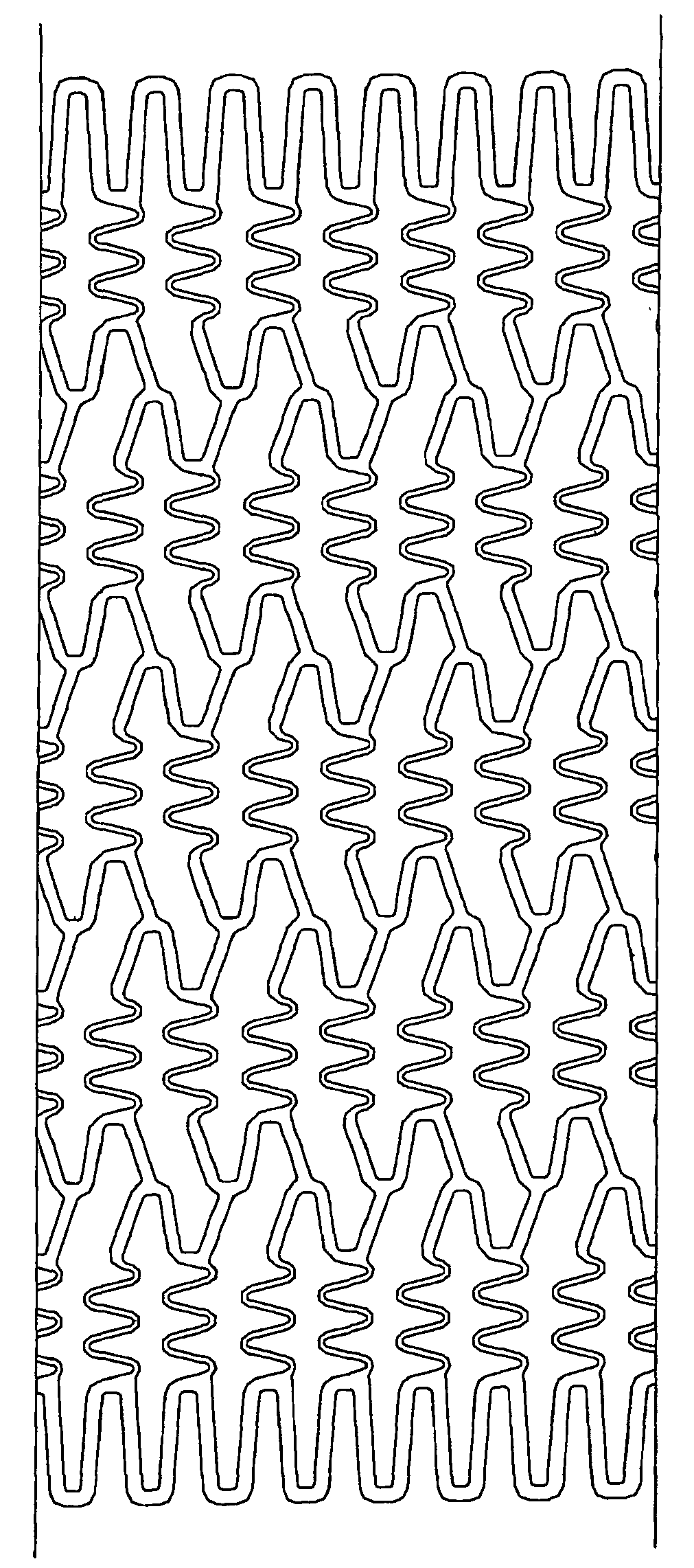
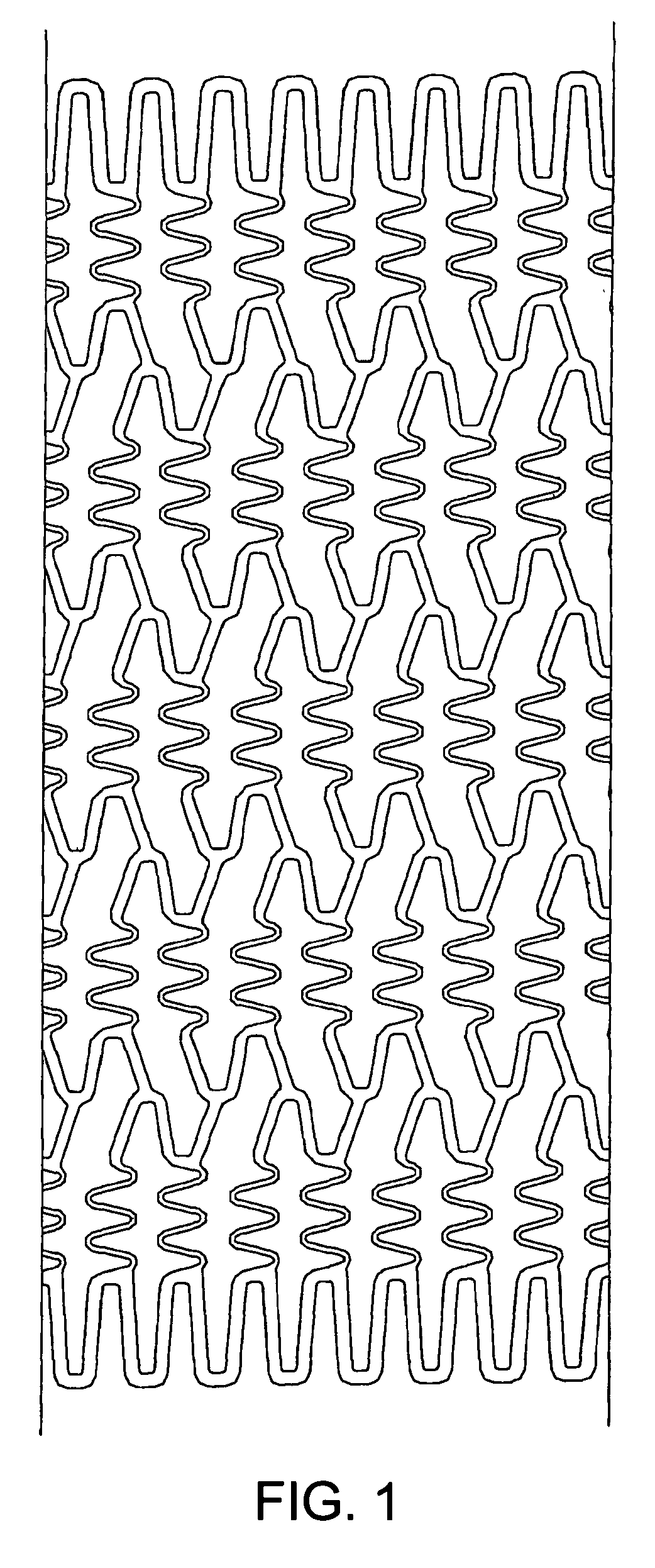
[0052] A substrate of a stent was formed by the same method as that commonly used by a person skilled in the art in which a stainless steel (SUS316L) cylindrical tube having an inner diameter of 1.50 mm and an outer diameter of 1.80 mm was cut into a stent design by laser cutting, and then electrolytically polished. FIG. 1 is a developed view of the stent used, and FIG. 2 is a schematic view. The stent had a length of 13 mm, a thickness of 120 μm, and a nominal diameter after expansion of 3.5 mm. The stent was a so-called balloon expandable type in which the stent is expanded and placed using a balloon catheter having a balloon provided near the tip thereof. The balloon expandable type stent is set in a contracted state at the balloon of the balloon catheter, delivered to a target portion, and then expanded and placed by expansion of the balloon.

[0053] A poly (lactide-co-glycolide) (SIGMA Corp., lactic acid / glycolic acid=85 / 15, weight-average molecular weight 90,000 to 126,000) was...

example 2

[0054] A stent was prepared by the same method as in Example 1 except that the spray time was controlled so that the weight of the poly (lactide-co-glycolide) per unit length in the axial direction of the substrate was 7 μg / mm (91 μg per stent).

example 3

[0055] A stent was prepared by the same method as in Example 1 except that the spray time was controlled so that the weight of the poly (lactide-co-glycolide) per unit length in the axial direction of the substrate was 65 μg / mm (845 μg per stent).


PUM


Abstract

As a treatment for angiostenosis, angioplasty (PTA or PTCA) of expanding a small-sized balloon in a vessel has been commonly conducted. However, this treatment easily causes repeated stenosis (restenosis) after the treatment. Placement of a stent in a vessel is also effective in decreasing restenosis, but this treatment may also cause restenosis. The present invention provides a stent containing a poly (lactide-co-glycolide) or both a poly (lactide-co-glycolide) and an immunosuppressive agent in at least a portion of a surface of the stent, and further containing a material nondegradable in vivo.

Description

TECHNICAL FIELD [0001] The present invention relates to a medical stent for in vivo placement for use in preventing or treating excessive vascular proliferation. BACKGROUND ART [0002] At present, one of the serious health problems that confront us is angiostenosis due to arteriosclerosis. As a treatment method for angiostenosis, angioplasty (PTA or PTCA) of expanding a small-sized balloon in a vessel has been commonly conducted as a minimally invasive treatment. However, this treatment causes repeated stenosis (restenosis) with high probability. As a method for decreasing the rate of restenosis, atherectomy, laser therapy, radiation therapy, or the like has been attempted, and another method such as a technique of placing a stent has been recently commonly employed. [0003] In order to treat various diseases caused by stenosis or occlusion of a blood vessel or another lumen in vivo, a stent is mainly used as a medical device to be placed in a stenosed or occluded site, for expanding ...

Claims


Application Information

Patent Timeline
Patent Type & Authority Applications(United States)
IPC IPC(8): A61F2/90A61F2/91A61F2/915A61M29/02
CPCA61F2/91A61F2/915A61F2002/91525A61F2230/0054A61F2002/9155A61F2002/91558A61F2002/91533A61M29/02
Inventor NISHIDE, TAKUJINAKANO, RYOJIYOSHIDA, SHINYAFUKAYA, KOHEIKAWATSU, MASAJI
Owner KANEKA CORP
Learn More