

Liposomes and liposomal compositions for vaccination and drug delivery
a technology of liposomes and compositions, applied in the direction of snake antigen ingredients, viral antigen ingredients, bacteria antigen ingredients, etc., can solve the problems of not improving the specific binding of these cells by increasing ps, pg or pa further
- Summary
- Abstract
- Description
- Claims
- Application Information
AI Technical Summary
Benefits of technology
Problems solved by technology
Method used
Image

Examples
example 1
Binding of AVE3 and AVE5 to Antigen-Presenting Cells
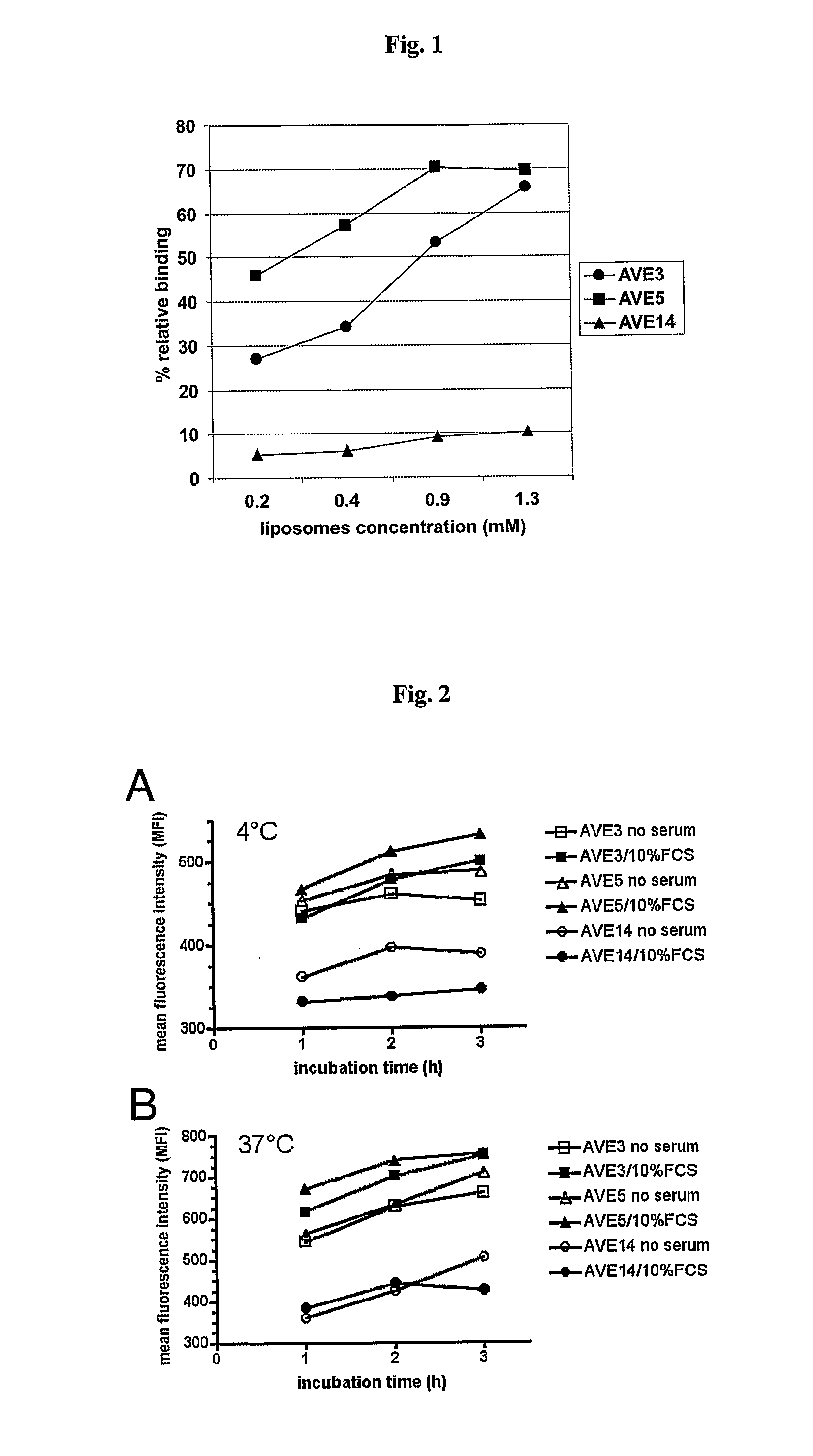
[0117] Various liposomal formulations were analyzed for binding to various types of antigen-presenting cells: AVE3 (cholesterol, DLPE, DOPS at a molar ratio of 1:1:1), AVE5 (cholesterol, DLPE, DOPG at a molar ratio of 1:1:1), or AVE14 (cholesterol, DLPE, EPC at a molar ratio of 1:1:1). All lipids were purchased from Avanti Polar Lipids (USA), Calbiochem (USA) or Lipoid GmbH (Germany) and were used without further purification. Liposomes were prepared from dried lipid films by hydration. For this purpose lipids were dissolved in chloroform or chloroform / methanol (1:1) and mixed at the indicated ratios. For binding studies 0.3 mol % rhodamine-labeled DPPE was added. Lipids were dried using a rotary evaporator and residual solvent was removed under high vacuum. Lipid films were then hydrated with 10 mmol / l Hepes pH 7.4 to a final lipid concentration of 10 μmol / ml. Liposomes were extruded 21-times through 50 nm membranes. All liposome...
example 2
Influence of Phosphatidylserine, Cholesterol, and Phosphatidylethanolamine Concentration on Binding to Antigen-Presenting Cells
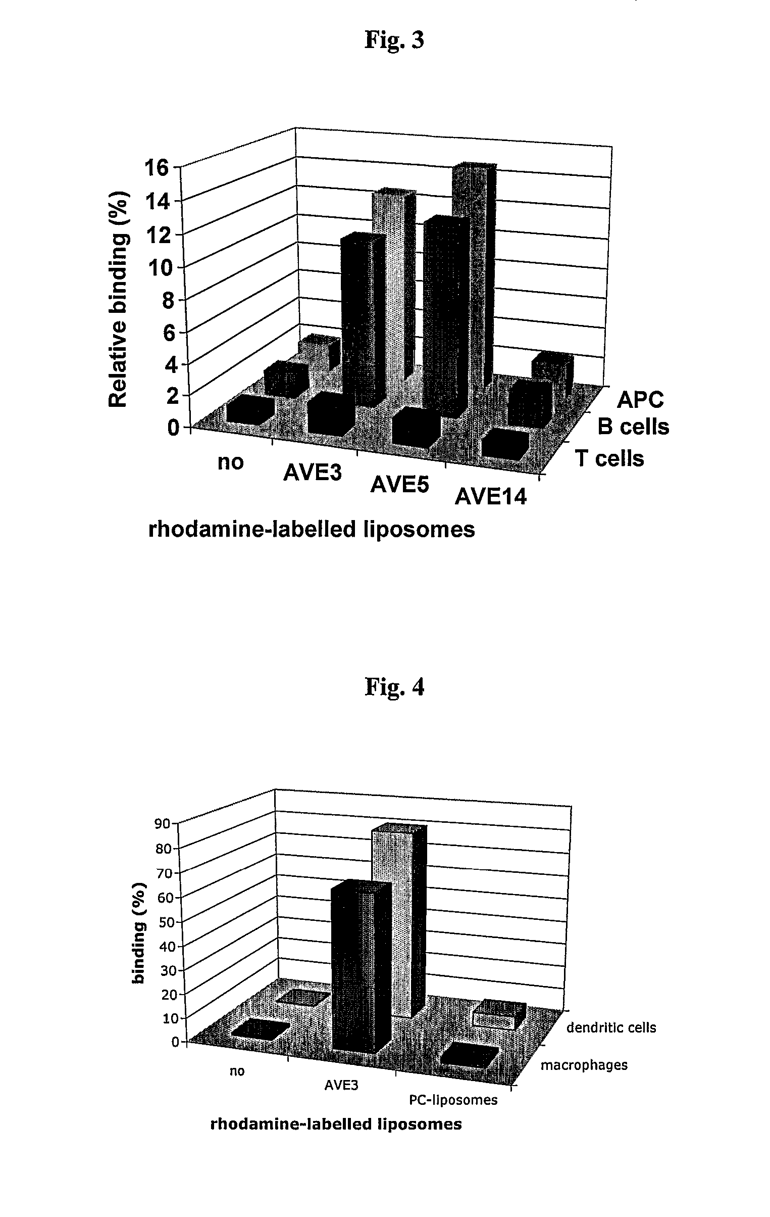
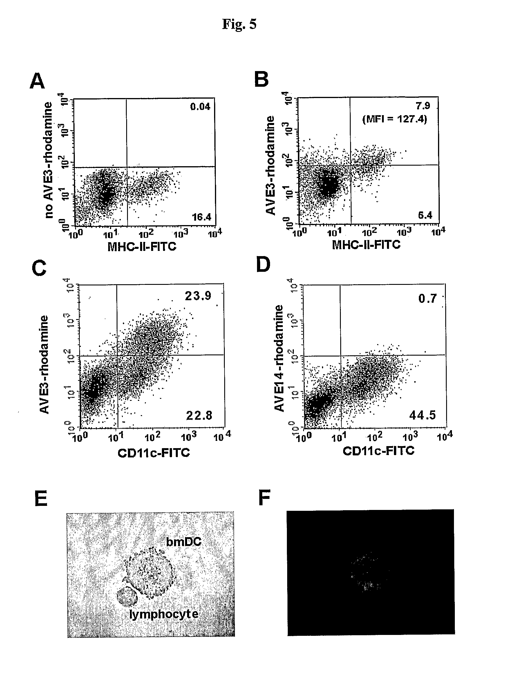
[0123] In a first experiment the influence of varying phosphatidylserine (DOPS) concentrations on binding to dendritic cells, B cells and T cells isolated from murine spleens was analyzed. In order to keep the concentrations of the other lipids constant the following liposomes were produced: AVE31 (25 mol %, cholesterol, 25 mol % DLPE, 33.3 mol % DOPS, 16.7 mol % POPC), AVE31-10PS (25 mol %, cholesterol, 25 mol % DLPE, 10 mol % DOPS, 40 mol % POPC), AVE31-50PS (25 mol %, cholesterol, 25 mol % DLPE, 50 mol % DOPS) adding 0.3 mol % rhodamine-labeled PE for detection. Murine spleen cells were incubated with liposomes at 4-8° C. DCs were identified by counter-staining with anti-CD11c antibody, T cells with anti-CD3 antibody, and B cells with anti-CD45R antibody. Strong binding was observed to DCs but also to B cells for all liposomes (5×10⁵ spleen cells incubat...
example 3
Long-Lasting Liposome Deposition at the Site of Injection and the Draining Lymph Nodes
[0127] In order to evaluate the availability and the stability of different liposomal formulations in vivo rhodamine-labeled AVE3 or AVE14 liposomes (see example 1) were injected id into the flank or into the hind footpad of C57BL / 6 mice. At different time points skin from the injection site as well as draining lymph nodes were embedded and cyrosections were prepared (FIG. 10A). A strong fluorescence signal was observed within the skin up to 7 days after AVE3 injection, whereas no significant fluorescence could be detected in the case of AVE14 liposomes (FIG. 10A). Furthermore, in the draining lymph nodes, fluorescence could be detected up to 7 days after injection only in the case of AVE3, whereas only a weak fluorescence of the neutral liposomes (AVE14) could be detected 16 hrs after injection. These findings indicate that AVE3 result in long-lasting deposition at the injection site and the drai...
PUM
| Property | Measurement | Unit |
|---|---|---|
| Diameter | aaaaa | aaaaa |
| Diameter | aaaaa | aaaaa |
| Composition | aaaaa | aaaaa |
Abstract
Description
Claims
Application Information