
Self-assembling multicellular bodies and methods of producing a three-dimensional biological structure using the same

Active Publication Date: 2010-02-18
UNIVERSITY OF MISSOURI
View PDF9 Cites 51 Cited by
  • Summary
  • Abstract
  • Description
  • Claims
  • Application Information

AI Technical Summary

Benefits of technology

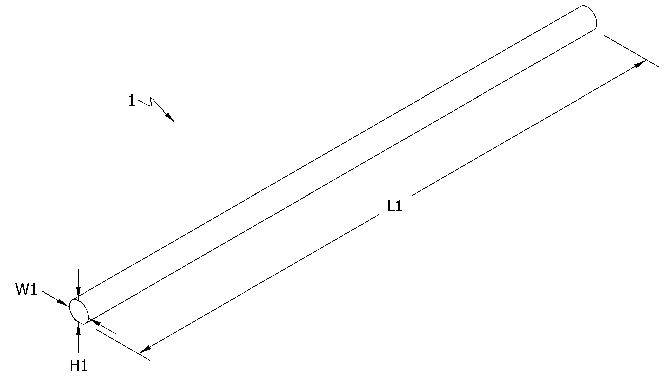
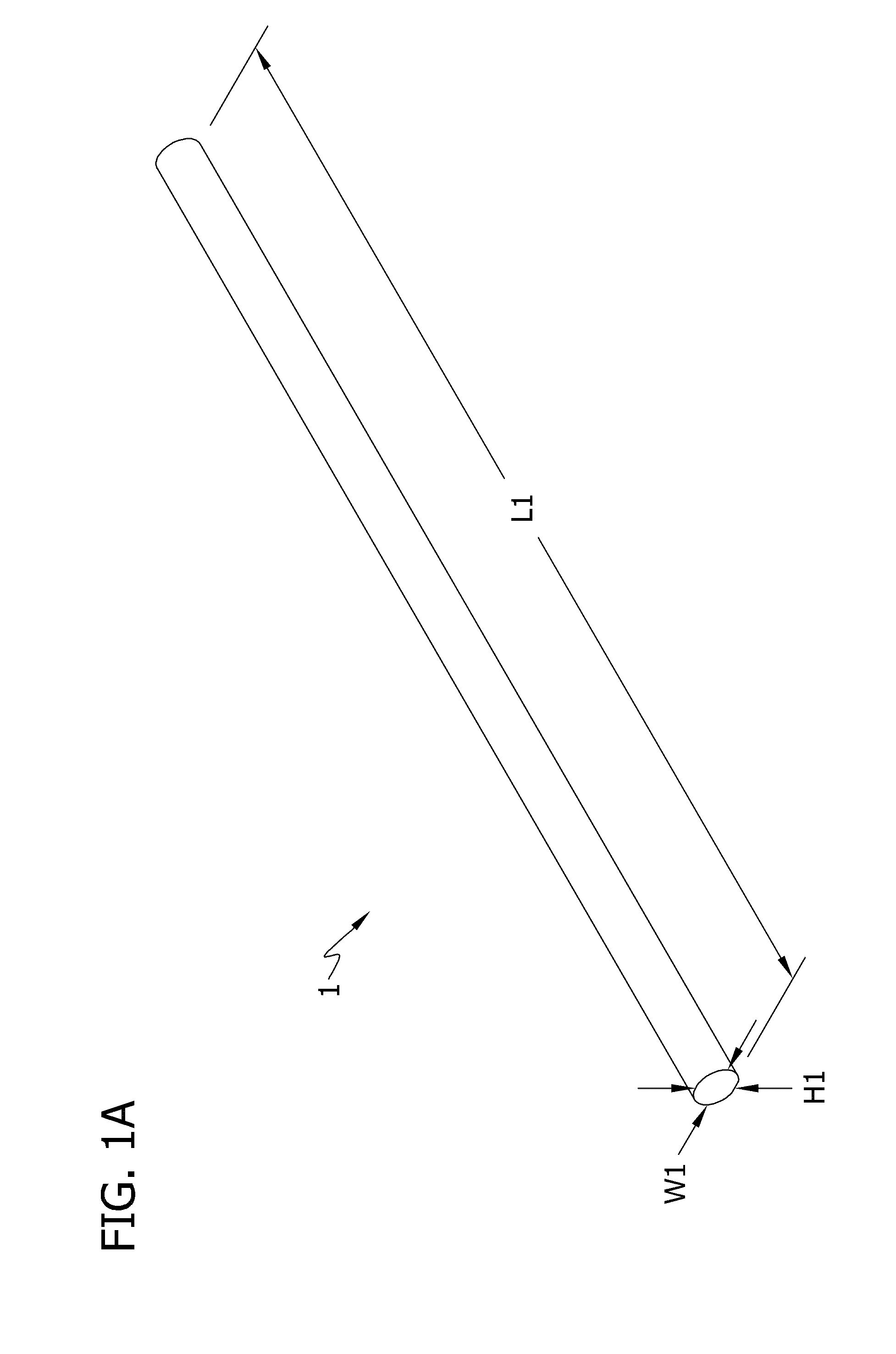
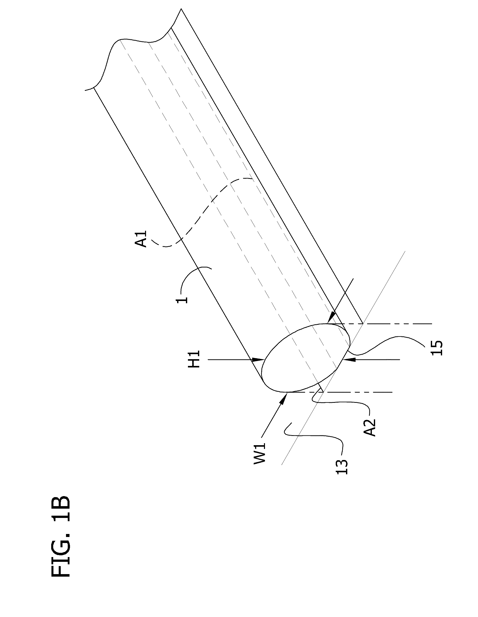
[0011]In one aspect of the invention, a multicellular body made of a plurality of cells or cell aggregates in a desired three-dimensional shape with viscoelastic consistency is described. The multicellular body comprises a plurality of cells or cell aggregates, wherein the cells or cell aggregates cohere together to form a construct in a pre-determined shape with viscoelastic consistency, desired cell density, and sufficient integrity for easy manipulation and handling.

Problems solved by technology

Many will likely perish for lack of replacement blood vessels for diseased arteries or veins or replacement abdominal organs.
A variety of methods to build engineered tissues have been attempted and developed with limited success.
However, assembly of vascularized three-dimensional organs has not been accomplished.
Prior art solutions, though promising, have presented a number of challenges.
Scaffold choice, immunogenicity, degradation rate, toxicity of degradation products, host inflammatory responses, fibrous tissue formation due to scaffold degradation, and mechanical mismatch with the surrounding tissue may affect the long term behavior of the engineered tissue construct and directly interfere with its primary biological function.
The use of scaffolds in cardiac tissue engineering has been associated with reduced cell-to-cell connection, as well as incorrect deposition and alignment of extracellular matrix (ECM; e.g., collagen and elastin), affecting scaffold biodegradation and the force-generating ability of myocardial constructs.
Besides the recurrent difficulty of producing elastic fibers in vitro, the use of scaffolds presents additional problems.
The inherent weakness of the gels may hinder the final strength of the tissue-engineered vessel.
In addition, the presence of residual polymer fragments can disrupt the normal organization of the vascular wall and even influence smooth muscle cell (SMC) phenotype.

Method used


Image

  • Self-assembling multicellular bodies and methods of producing a three-dimensional biological structure using the same
  • Self-assembling multicellular bodies and methods of producing a three-dimensional biological structure using the same
  • Self-assembling multicellular bodies and methods of producing a three-dimensional biological structure using the same

Examples


example 1

Preparation of Multicellular Bodies and Tissue Engineering Using Pig Smooth Muscle Cells

[0132]I. Pig Smooth Muscle Cells. Pig smooth muscle cells (SMCs) were grown in the same conditions used in previous studies. The medium composition was Dulbecco's Modified Eagle Medium (DMEM) low glucose supplemented with 10% porcine serum, 10% bovine serum, 50 mg / L of proline, 20 mg / L of alanine, 50 mg / L of glycine, 50 mg / L of ascorbic acid, 12 μg / L, of Platelet Derived Growth Factor-BB (PDGF-BB), 12 μg, / L of Basic Fibroblast Growth Factor (bFGF), 3.0 μg / L of CuSO4, 0.01M of HEPES buffer, and 1.0×10⁵ units / L of penicillin and streptomycin. The cells were grown on gelatin (gelatin from porcine skin) coated 10 cm Petri dishes and incubated at 37° C., 5% CO2. The SMCs were subcultured up to passage 7 before being used for multicellular body (e.g., cellular unit) preparation. Eighteen confluent Petri (i.e. cell culture) dishes were necessary to prepare 24 cellular units and 4 tubes (outside diameter...

example 2

Alternative Procedure for Preparing Multicellular Bodies and for Tissue Engineering

I. Growth Conditions for Cells of Various Types

[0137]Chinese Hamster Ovary (CHO) cells transfected with N-cadherin were grown in Dulbecco's Modified Eagle Medium (DMEM) supplemented with 10% Fetal Bovine Serum (FBS), antibiotics (100 U / mL penicillin streptomycin and 25 μg / mL gentamicin) and 400 μg / ml geneticin. Besides gentamicin all antibiotics were purchased from Invitrogen.

[0138]Human Umbilical Vein Smooth Muscle Cells (HUSMCs) and Human Skin Fibroblasts (HSFs) were purchased from the American Type Culture Collection (CRL-2481 and CRL-2522 respectively). HUSMCs were grown in DMEM with Ham's F12 in ratio of 3:1, 10% FBS, antibiotics (100 U / mL penicillin-streptomycin and 25 ug / mL gentamicin), 20 μg / mL Endothelial Cell Growth Supplement (ECGS), and Sodium Pyruvate (NaPy) 0.1M. Human skin fibroblasts (HSFs) were grown in DMEM with Ham's F12 in ratio of 3:1, 20% FBS, antibiotics (100 U / mL penicillin / str...

example 3

Use of Gelatin and / or Fibrin in Preparing Multicellular Bodies and a Three-Dimensional Fused Tubular Structure

[0154]Human Aortic Smooth Muscle Cells (HASMCs) were purchased from Cascade Biologics (C-007-5C). HASMCs were grown in medium 231 supplemented with the Smooth Muscle Growth Supplement (SMGS). The HASMCs were grown on uncoated cell culture dishes and were maintained at 37° C. in a humidified atmosphere containing 5% CO2.

[0155]The HASMCs were trypsinized, resuspended in tissue culture media, and centrifuged as described above in Examples 1 and 2. Following centrifugation, the tissue culture medium (i.e., the supernatant) is removed, and the cells (1 confluent Petri dish) were resuspended first in 100 μl of a solution of fibrinogen (50 mg / ml in 0.9% NaCl), and then 70 μl of a solution of gelatin (20% in phosphate buffered saline (PBS)) was added. The cell suspension was centrifuged again, the supernatant was removed, and the centrifuge tube containing the cell pellet was place...


PUM

PropertyMeasurementUnit
Lengthaaaaaaaaaa
Areaaaaaaaaaaa
Areaaaaaaaaaaa

Abstract

Structures and methods for tissue engineering include a multicellular body including a plurality of living cells. A plurality of multicellular bodies can be arranged in a pattern and allowed to fuse to form an engineered tissue. The arrangement can include filler bodies including a biocompatible material that resists migration and ingrowth of cells from the multicellular bodies and that is resistant to adherence of cells to it. Three-dimensional constructs can be assembled by printing or otherwise stacking the multicellular bodies and filler bodies such that there is direct contact between adjoining multicellular bodies, suitably along a contact area that has a substantial length. The direct contact between the multicellular bodies promotes efficient and reliable fusion. The increased contact area between adjoining multicellular bodies also promotes efficient and reliable fusion. Methods of producing multicellular bodies having characteristics that facilitate assembly of the three-dimensional constructs are also provided.

Description

GRANT STATEMENT[0001]The invention was made in part from government support under Grant No. NSF-0526854 from the National Science Foundation. The U.S. Government has certain rights in the invention.FIELD OF THE INVENTION[0002]The present invention relates to the field of regenerative medicine and tissue engineering, and more particularly to production of engineered tissues / organs having desired structures.BACKGROUND[0003]Tissue engineering provides promising solutions to problems caused by the growing demand for organ and tissue replacement coupled with a chronic shortage of transplantable organs, including blood vessels. In the United States, for example, thousands of people are on the national waiting list for organ transplants. Many will likely perish for lack of replacement blood vessels for diseased arteries or veins or replacement abdominal organs. To lessen and eventually solve the problem of inadequate supply of blood vessels and organs for transplantation, tissue engineers ...

Claims


Application Information

Patent Timeline
IPC IPC(8): C12N5/071
CPCA61L27/222C12N5/0602A61L27/3886C12M21/08C12M23/10C12M25/06C12M25/14C12M35/08C12N5/0062C12N5/0691C12N2506/1307C12N2506/1338C12N2533/54C12N2533/56C12N2533/76A61L27/225C12M33/00B33Y80/00A61P43/00A61P9/00A61F2/02C12M3/00A61L27/3625A61L27/3691C12N5/0068
Inventor FORGACS, GABORMARGA, FRANCOISE SUZANNENOROTTE, CYRILLE
Owner UNIVERSITY OF MISSOURI
Learn More