

Nanosurface
a nano-surface and surface technology, applied in nanotechnology, inorganic chemistry, dental implants, etc., can solve the problem that the biocompatibility of metallic components may be further increased, and achieve the effect of reducing the amount of collagen and minerals, reducing strength, and reducing strength
- Summary
- Abstract
- Description
- Claims
- Application Information
AI Technical Summary
Benefits of technology
Problems solved by technology
Method used
Image

Examples
example 1
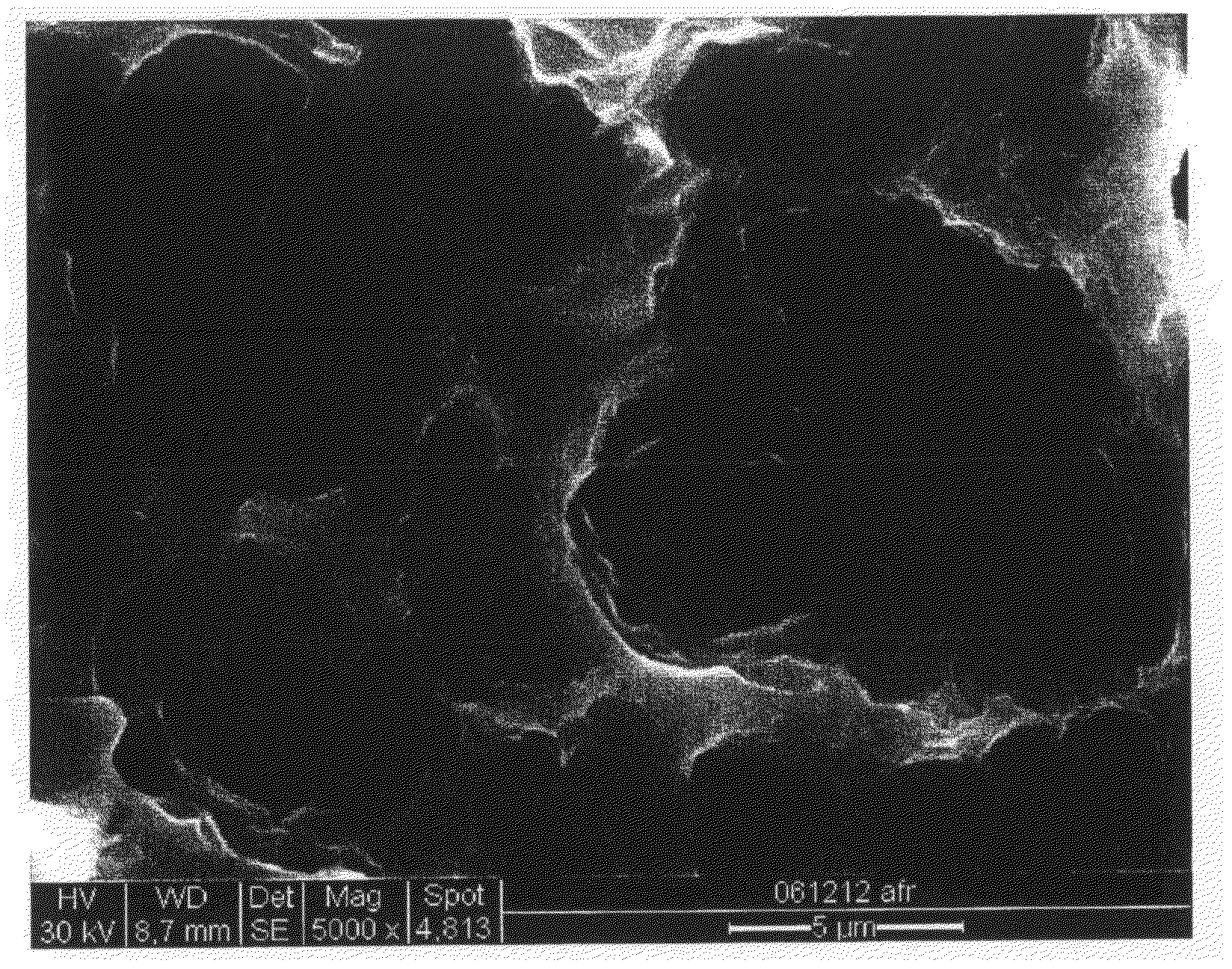
[0138]Titanium samples having the shape of a coin (machine worked and blasted, respectively), a fixture (blasted) and an abutment (machine worked) were cleaned by a conventional chemical treatment. The samples were immersed in an 1 M aqueous solution of oxalic acid and left at 80° C. for 30 minutes under vigorous agitation. After 30 minutes the samples were removed from the oxalic acid solution and rinsed in water followed by rinsing in water in an ultrasonic bath for 2 minutes. Approximately 10 minutes after rinsing, the samples were immersed in 0.1 M aqueous solution of hydrofluoric acid (HF) at room temperature and agitation until the start of active dissolution, followed by an additional active treatment time of 40 seconds. Next, the samples were removed from the HF solution and rinsed in water followed by rinsing in water in an ultrasonic bath for 5 minutes. The samples were dried in air at room temperature for about 60 minutes before s...
example 2
Cell Proliferation and Activity
[0146]Cell proliferation and production of alkaline phosphatase (ALP) and prostaglandin E2 (PGE2), respectively, was investigated for human osteoblast cells grown in vitro on titanium surfaces according to the invention in comparison to cells grown on a commercial implant surface (OsseoSpeed™; Asta Tech AB, Sweden).
(i) Cell Cultivation
[0147]MG-63 is a human cell line conventionally used for in vitro studies of osteoblasts. In this study, MG-63 cells (MG-63, ATCC No CRL-1427, U.S.) were grown in 300 ml Falcon cell culture flasks (BD, WWR, Sweden) in Dulbecco's Minimun Essential Medium (D-MEM) (Gibco, UK) containing 5% fetal calf serum (FCS; Gibco, UK) and 1% penicillin-streptomycin (PEST; Gibco, UK) from second passage from an ampulla of frozen cells. When adherent cells had grown to confluency, they were passaged using 0.05% Trypsin-EDTA (Gibco, UK) for 3 passages. Cell viability was high (>98%) as counted using light microscopy.
(ii) Cell Morphology (S...
example 3
Implantation
[0155]The integration of implants according to the invention was tested in a rabbit model. The objective was to qualitatively and quantitatively study the in vivo bone tissue response to two implant surface modifications according to the invention compared to the response to commercially available reference implants.
[0156](i) Implants for Removal Torque Study
[0157]Titanium torque fixtures (square headed removal torque design, 3.5×8.2 mm) prepared by immersion in oxalic acid and subsequently in HF as described in Example 1 (i.e., including steps b and c) were used (referred to as Test implant 2). Also, torque fixtures (3.5×8.2 mm) were used which were prepared by immersion in oxalic acid according to Example 1 (i.e., step c was omitted) (referred to as Test implant 1). Further, torque fixtures (3.5×8.2 mm) representing the commercially available OsseoSpeed™ oral implant were used as reference fixtures.
[0158](ii) Implants for Histological and Histomorphometrical Study
[0159...
PUM
| Property | Measurement | Unit |
|---|---|---|
| temperature | aaaaa | aaaaa |
| temperature | aaaaa | aaaaa |
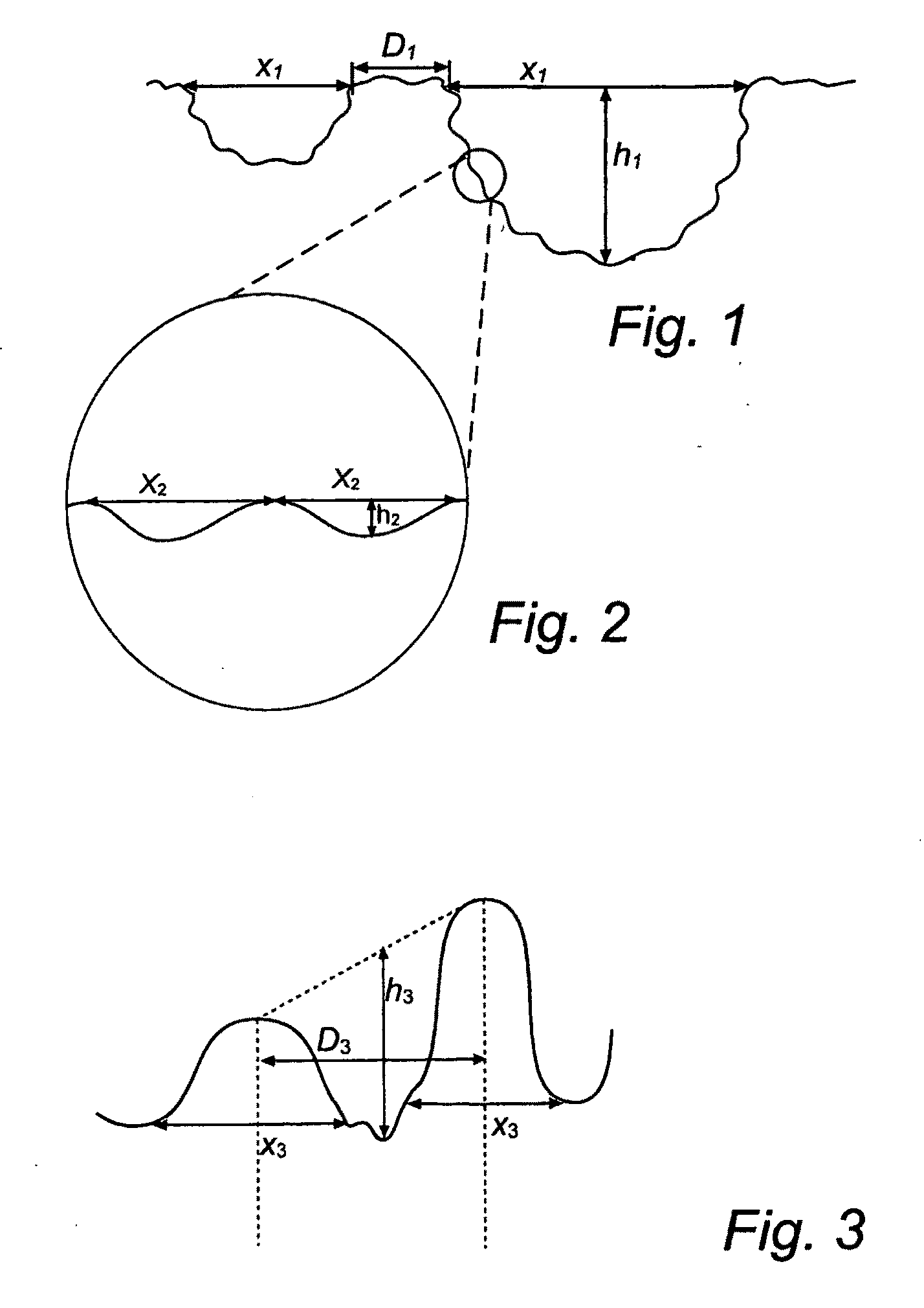
| pit diameter | aaaaa | aaaaa |
Abstract
Description
Claims
Application Information