In situ-forming hydrogel for tissue adhesives and biomedical use thereof

Active Publication Date: 2012-06-21
AJOU UNIV IND ACADEMIC COOP FOUND
View PDF1 Cites 76 Cited by
  • Summary
  • Abstract
  • Description
  • Claims
  • Application Information

AI Technical Summary

Benefits of technology

[0025]The present invention provides an in situ-forming bioadhesive hydrogel which has excellent biocompatibility and mechanical strength and excellent tissue adhesiveness thanks to modification with/without dopa derivatives. Particularly, the hydrogel according to the present invention finds

Problems solved by technology

Conventional surgical techniques, such as suturing, guarantee strong tensile strength, but have the disadvantages of pain and the need for the threads to be postoperatively removed.
On the other hand, tissue adhesives enjoy the advantages of a short adhesion time, simple usage, no requirements for postoperative removal, etc., but are problematic in that they exhibit low adhesiveness, poor biocompatibility and tensile strength and a remarkably decreased adhesiveness particularly in the presence of moisture.
These cyanoacrylate adhesives, consisting of a solitary material, can solidify in a short period of time at room temperature just by using water without the aid of initiators and exhibit a transparent appearance and strong adhesive strength, but low resistance to both impact and heat.
Moreover, their use is now restricted due to the high toxicity and fragility thereof although cyanoacrylate

Method used


Image

  • In situ-forming hydrogel for tissue adhesives and biomedical use thereof
  • In situ-forming hydrogel for tissue adhesives and biomedical use thereof
  • In situ-forming hydrogel for tissue adhesives and biomedical use thereof

Examples


Example

PREPARATION EXAMPLE 1

Synthesis of Tetronic-Tyramine / Dopamine (Tet-TA / DA)

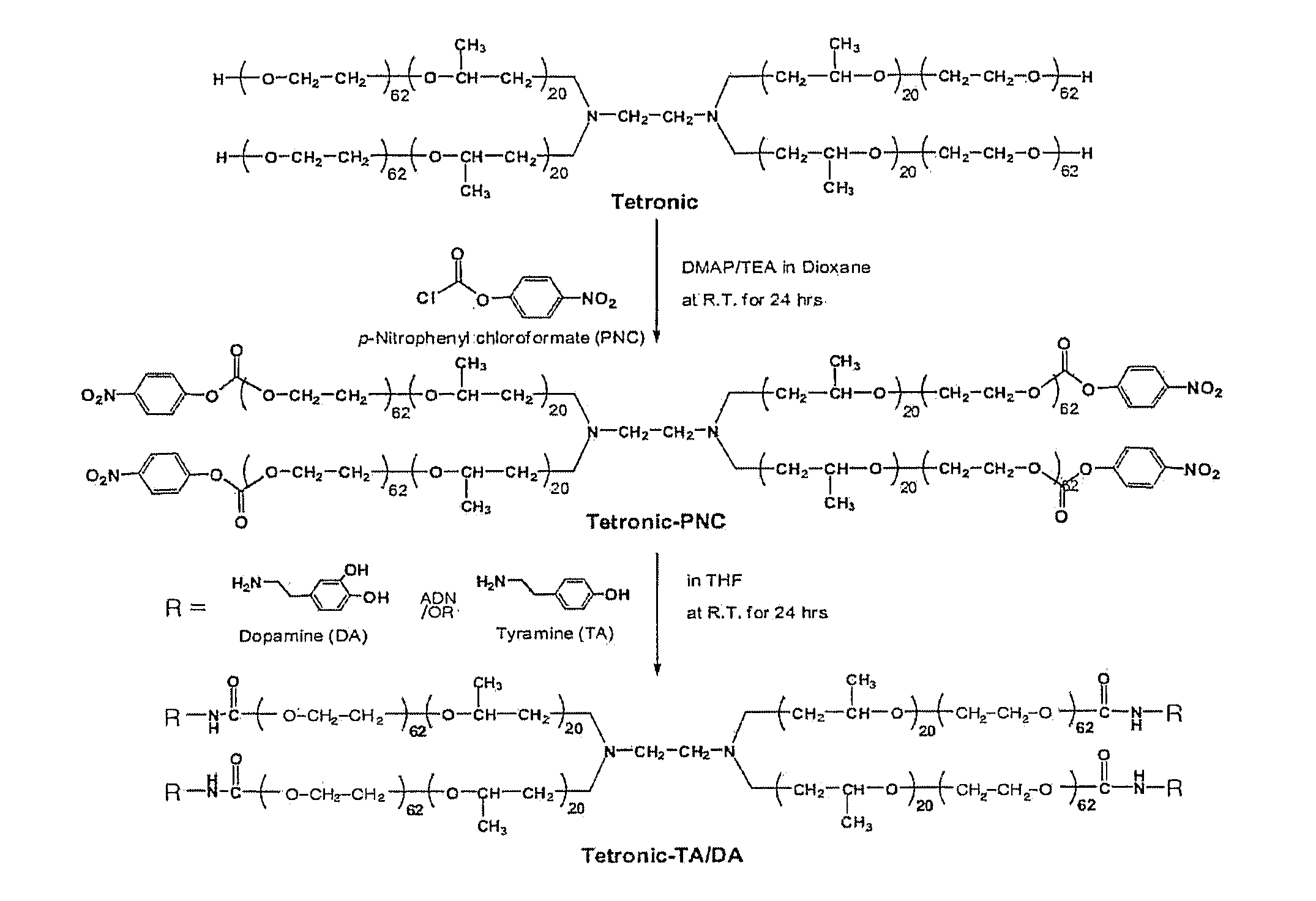
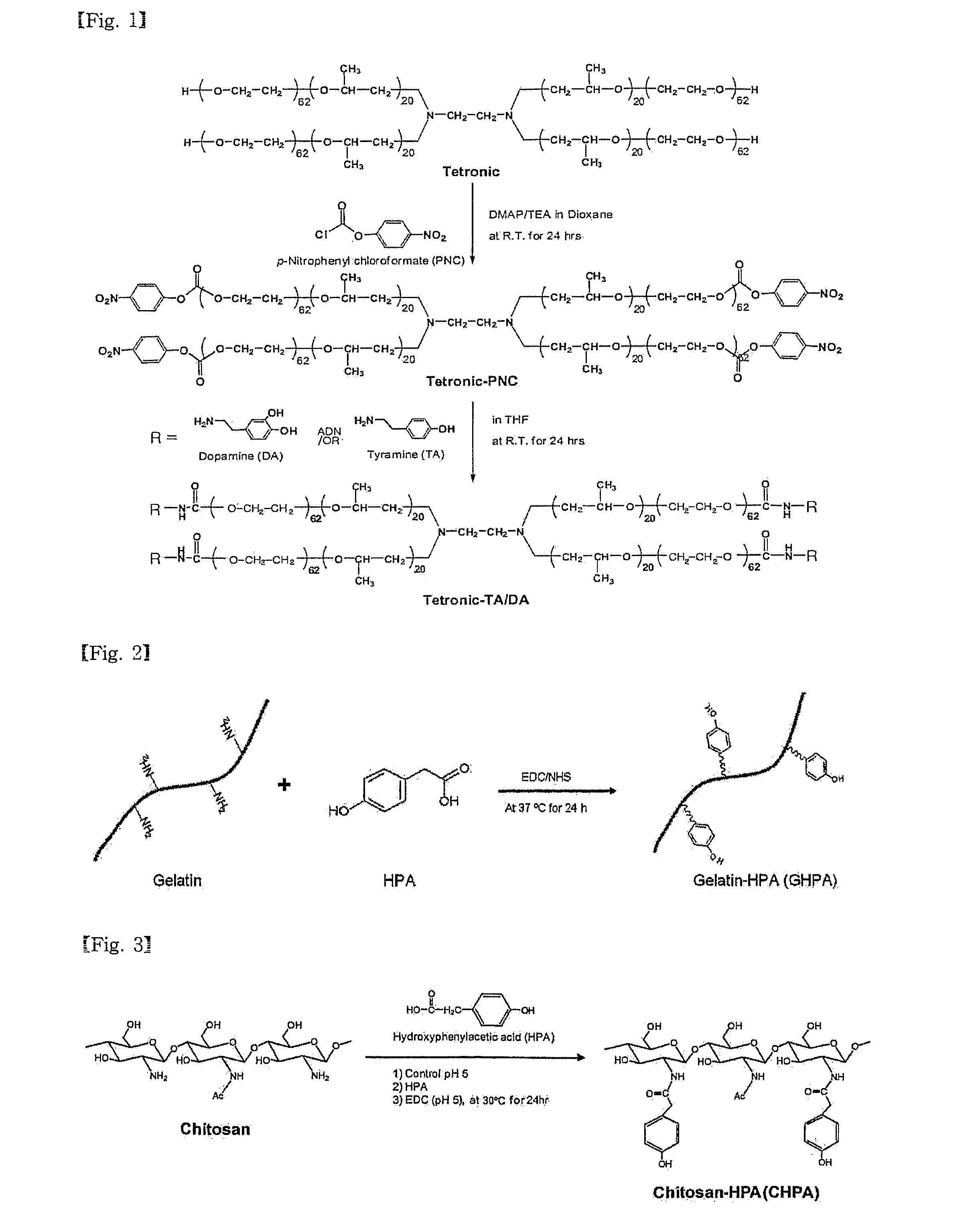
[0091]FIG. 1 is a reaction scheme for synthesizing Tet-TA / DA.

[0092]1. Synthesis of Tetronic-(p-nitrophenyl chloroformate) ┌Tet-PNC┐

[0093]A solution of 30 g (1.67 mmol) of tetronic in 300 mL of dioxin was mixed sequentially with a solution of 1.018 g (8.33 mmol) of 4-dimethylaminopyridine (DMAP) and 0.843 g (8.33 mmol) of triethylamine (TEA) in 40 mL of dioxane and a solution of 1.679g (8.33 mmol) of p-nitrophenyl chloroformate (PNC) in 50 mL of dioxane. The molar ratio of tetronic:PNC:DMAP:TEA was 1:5:5:5. The reaction was performed at 30° C. for 24 hrs in a nitrogen atmosphere.

[0094]After completion of the reaction, remaining reagents were removed through a filter and the reaction product was concentrated using a rotary evaporator. The concentrated solution was dropwise added to 1600 mL of chilled ether to form precipitates which were then filtered.

[0095]The filtered powder was left for 24 hrs in a vacuum oven ...

Example

PREPARATION EXAMPLE 2

Synthesis of Gelatin-Hydroxyphenylacetic Acid (GHPA)

[0101]FIG. 2 is a reaction scheme for the synthesis of GHPA.

[0102]10 Grams of gelatin was dissolved in 200 mL of 0.1 M 2-(N-morpholino)ethanesulfonic acid (MES) to prepare a solution A. Separately, 0.609 g (4 mmol) of 4-hydroxyphenylacetic acid (HPA) was dissolved in 50 mL of 0.1 M MES to give a solution B. 0.92 Grams (4.8 mmol) of 1-ethyl-3-(3-dimethylaminopropyl)-carbodiimide (EDC) and 0.276 g (2.4 mmol) of N-hydroxysuccinimide (NHS) were dissolved in respective 5 mL of 0.1 M MES. The EDC solution and the NHS solution were sequentially at intervals of 15 min to the solution B. 15 min after addition of the NHS solution, the solution B containing EDC / NHS was mixed with the solution A. The reaction was performed at 40° C. for 24 hrs.

[0103]After completion of the reaction, the reaction mixture was filtered through a syringe filter (450 nm). Then, the filtrate was subjected to membrane dialysis against distilled w...

Example

PREPARATION EXAMPLE 3

Synthesis of Chitosan-Hydroxyphenylacetic Acid (CHPA)

[0104]FIG. 3 is a reaction scheme for the synthesis of CHPA.

[0105]0.644 Grams of low molecular weight chitosan with a degree of deacetylation of 75˜85% was added to 80 mL of distilled water and its acidity was adjusted to a pH of 3 with 1 N HCl to form a solution. After 0.404 g (2.6 mmol) of HPA was added to the solution, its acidity was increased to a pH of 5 with 0.1 M NaOH. After the addition of 0.768 g (4 mmol) of EDC, the reaction was performed at 30° C. for 24 hrs.

[0106]When the reaction was completed, the reaction mixture was subjected to membrane dialysis against distilled water (molecular weight cutoff 3500 Da) to remove unreacted HPA. The dialyzed solution was lyophilized to produce CHPA in the form of semi-transparent non-woven fabric.


PUM

PropertyMeasurementUnit
Fractionaaaaaaaaaa
Pressureaaaaaaaaaa
Pressureaaaaaaaaaa

Abstract

Disclosed herein are an in situ-forming, bioadhesive hydrogel and the medical uses thereof. Being formed by in situ crosslinking through an enzymatic reaction, the hydrogel has an advantage over conventional bioadhesive hydrogels in terms of biocompatibility. In addition, the in situ-forming bioadhesive hydrogel has excellent biocompatibility and mechanical strength and has excellent tissue adhesiveness thanks to modification with/without dopa derivatives. The hydrogel finds a variety of applications in the biomedical field, including bioadhesives or hemostats, implant substances for tissue regeneration and augmentation, carriers for delivering biologically active materials or drugs, etc.

Description

TECHNICAL FIELD[0001]The present invention relates to in situ-forming bioadhesive hydrogel which is excellent in biocompatibility and mechanical strength and shows excellent bioadhesiveness, with a dopa derivative bonded thereto, and the biomedical uses thereof.BACKGROUND ART[0002]The market for wound care, the major proportion of which is for sealants and hemostats, has grown rapidly alongside great advances in the research and development of tissue adhesives. Triggered by the FDA granting permission to a fibrin sealant in 1998, a burst of novel tissue adhesives has been appearing in the market each year. Attention is now being focused on these tissue adhesives as alternatives to those used in conventional surgical or internal operations, such as suturing, clipping, cautery, etc.[0003]Conventional surgical techniques, such as suturing, guarantee strong tensile strength, but have the disadvantages of pain and the need for the threads to be postoperatively removed. On the other hand,...

Claims


Application Information

Patent Timeline
IPC IPC(8): A61L24/04A61L24/08A61K31/785A61K38/39A61K31/722A61K31/765A61K38/02A61K38/18A61K38/27A61K38/19A61K38/20A61K38/21A61K38/23A61K38/25A61K38/22A61K38/28A61K38/31A61K38/26A61K39/395A61K39/00A61K33/24A61P31/04A61P35/00A61P29/00A61P7/04A61P37/04A61P41/00A61P19/04A61P19/08A61P17/00A61P9/00A61P27/02A61P25/00A61P13/00A61Q19/00A61P43/00A61L24/10
CPCA61L24/0015A61L24/0031A61L2300/602A61L2300/43A61L2300/426A61L24/0042A61L24/046A61L27/18A61L27/52A61L27/54A61L27/58A61L2300/252A61L2300/256A61L2300/406A61L2300/41A61L2300/414A61L2300/416C08L71/02A61P7/04A61P9/00A61P13/00A61P17/00A61P19/04A61P19/08A61P25/00A61P27/02A61P29/00A61P31/04A61P35/00A61P37/04A61P41/00A61P43/00A61F2/02A61L27/14
Inventor PARK, KI-DONGJOUNG, YOON-KIPARK, KYUNG-MINLIH, EU-GENE
Owner AJOU UNIV IND ACADEMIC COOP FOUND
Why Eureka
Try Eureka