

Implantable particles for urinary incontinence
- Summary
- Abstract
- Description
- Claims
- Application Information
AI Technical Summary
Benefits of technology
Problems solved by technology
Method used
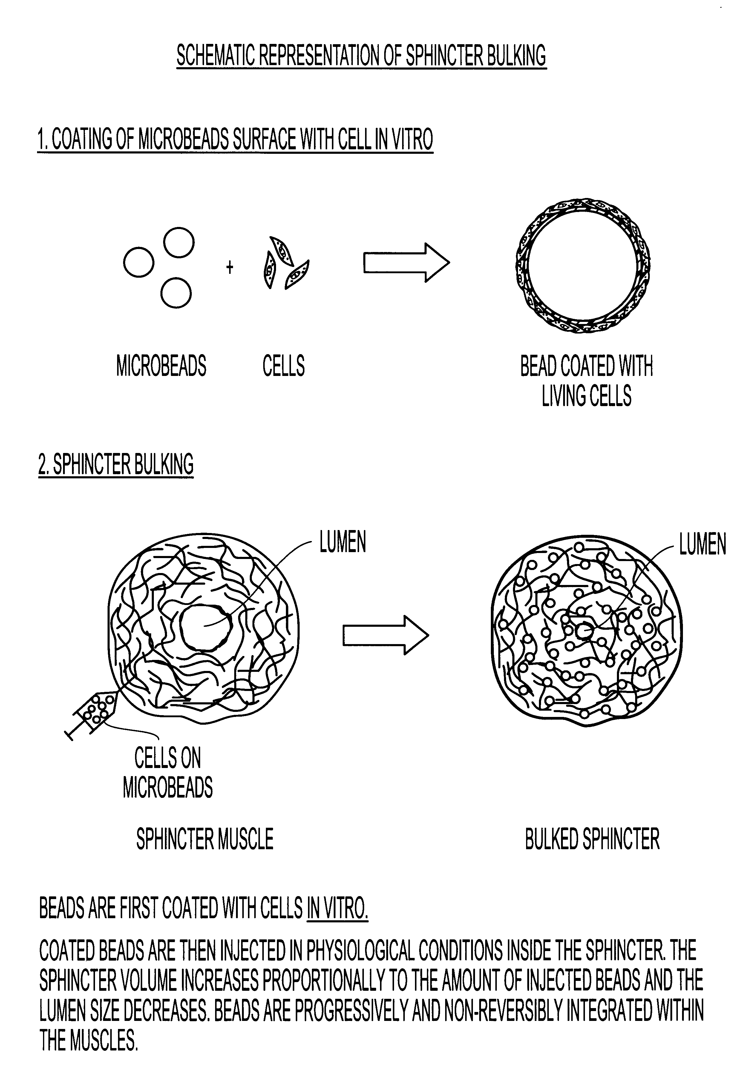
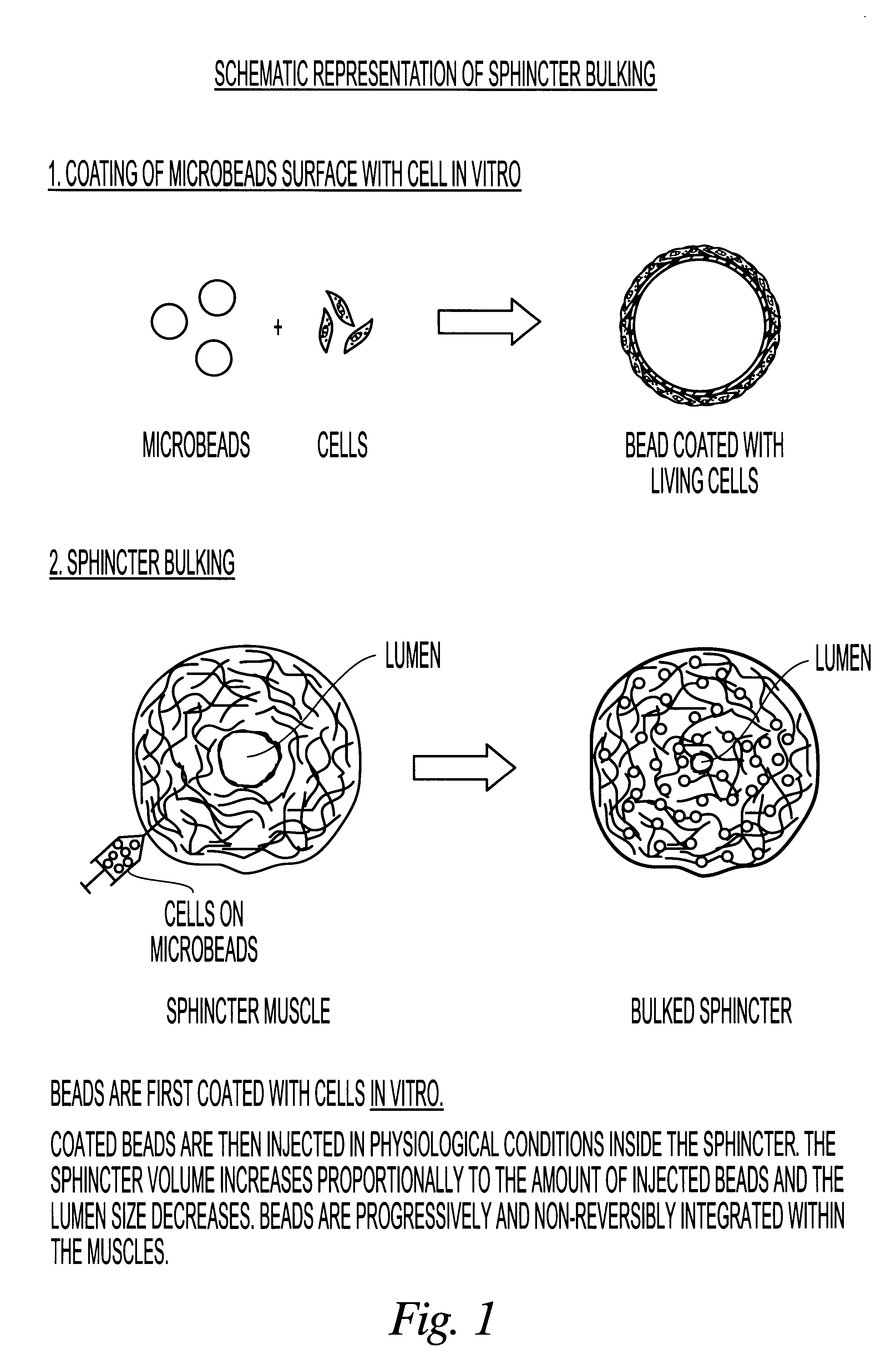
Image

Examples
example 1
6.1 Example 1
Preparation of Irregular Hydrogel Particles with Chemotactic Properties
58 grams of sodium chloride and 27 grams of sodium acetate were dissolved at room temperature in 100 ml of demineralized water. To this solution 400 ml of glycerol were added, the pH was adjusted to 6.0 and monomers were then dissolved. More specifically to this solution 90 gram of methylolacrylamide, 2 g of methacrylamidopropyl-trimethyl-ammonium-chloride hydrochloride and 10 gram of N,N'-methylene-bis-acrylamide were added and the mixture was agitated until complete solubilization. The solution was heated at about 70.degree. C. and 100 ml of a solution of gelatin at a concentration of 500 mg / ml was added. The total volume of the mixture was then adjusted to 1000 ml by addition of demineralized water. Finally 20 ml of 70 mg / ml ammonium persulfate aqueous solution and 4 ml of N,N,N',N'-tetramethyl-ethylene-diamine was added. The obtained mixture was stored at 70.degree. C. for about 3 hours until for...
example 2
6.2 Example 2
Preparation of Spherical Polyacrylic Hydrogel Gel Particles with Chemotactic Properties
The solution of monomers prepared as described in Example 1 above was poured slowly into 1500 ml of stirred and hot paraffin oil (50-70.degree. C.). After a few minutes a suspension / emulsion of liquids was obtained (the aqueous monomer solution was dispersed into oil and forms very small spherical droplets) and the polymerization occurred in suspension. The microdroplets were transformed into microbeads. The solid microbeads were recovered by centrifugation and suspended in 1 liter of physiological buffer containing 5% (w / v) glutaraldehyde and shaken for two hours. Finally the particles were extensively washed with water to eliminate completely the oil traces. Organic solvent extraction can be used for a more effective oil removal or an extensive washing in the presence of traces of nonionic detergents. The obtained microbeads are calibrated if necessary by sieving through a nylon net...
example 3
6.3 Example 3
Preparation of Hydrophilic Spherical Polystyrene Copolymer Particles Useful for Tissue Bulking
10 gram of styrene is mixed with 60 ml of toluene. 1 gram of divinylbenzene, 1 gram of dimethyl-aminoethyl-methacrylate and 1 gram of dimethyl-acrylamide are added to the resulting solution. After complete solubilization the monomer solution is mixed with 1% of AIBN (2,2'-azobisisobutyronitrile) as a polymerization catalyst and with 40 ml of paraffin oil as a viscosity inducer agent. The mixture is poured in an agitated water solution containing 0.5% Tween 80. In this situation there is formation of droplet suspension which turns into solid microbeads when the temperature is raised to 80-90.degree. C. for three to five hours. The resulting beads are dried and organic solvents extracted. They are then swollen in an aqueous solution of collagen in phosphate buffer at neutral pH. Embedded collagen is then crosslinked with glutaraldehyde as described in Examples 1 and 2. The result...
PUM
| Property | Measurement | Unit |
|---|---|---|
| Percent by mass | aaaaa | aaaaa |
| Percent by mass | aaaaa | aaaaa |
| Percent by mass | aaaaa | aaaaa |
Abstract
Description
Claims
Application Information