Method of detecting vulnerable atherosclerotic plaque

a technology of atherosclerotic plaque and detection method, which is applied in the field of infrared sensing, can solve the problems of plaque rupture, restnosis or transplant vasculopathy, and the sensitivity and/or toxicity of the device that cannot be detected by the fiberoptic catheter of this patent, so as to reduce the incidence of restnosis, reduce the incidence of vasculopathy, and reduce morbidity and mortality

Inactive Publication Date: 2006-01-31
BOARD OF RGT THE UNIV OF TEXAS SYST
View PDF74 Cites 35 Cited by
  • Summary
  • Abstract
  • Description
  • Claims
  • Application Information

AI Technical Summary

Benefits of technology

[0064]The invention's methods and devices will have a number of utilities. Each will reduce morbidity and mortality from coronary and carotid artery atherosclerosis. Each will reduce the incidence of restenosis and thus the need for repeated angioplasties or atherectomies. Each will also reduce the incidence of vasculopathy in organ-transplant patients. In turn, these outcomes will produce the benefits of better health care, improved public health, and reduced health care costs. These and other uses of the present invention will become clearer with the detailed description to follow.

Problems solved by technology

However, the fiberoptic catheter of this patent does not permit detection of heat.
The sensitivity and / or toxicity of these devices is unknown.
Moreover, no technique is described by this patent for predicting plaque rupture, restenosis or transplant vasculopathy.
Thus, it is not likely that such a device could be used to predict plaque rupture.
Indeed, recent studies have found that intravascular ultrasound cannot identify which plaques are at risk of rupturing. de Feytia Circulation 92:1408–13 (1995).
However, this device does not distinguish between plaque on the basis of heat differential.
Prior art approaches to intravascular arterial diagnosis and repair have been numerous yet have failed to provide certain capabilities.
In particular, such prior art catheters and methods have failed to provide means for detecting and treating atherosclerotic plaque since they have not been able to differentiate between those plaques at risk of rupturing and occluding and those that are not presently at such risk even if they are capable of determining the presence or absence of calcification of the plaque.
Similarly, prior art approaches have not provided effective means for identifying specific arterial sites at risk for arterial restenosis after angioplasty or atherectomy.
Prior art approaches have also failed to provide practical and effective means for detecting transplant vasculopathy.

Method used


Image

  • Method of detecting vulnerable atherosclerotic plaque
  • Method of detecting vulnerable atherosclerotic plaque
  • Method of detecting vulnerable atherosclerotic plaque

Examples


example i

Methods

[0087]Fifty carotid endarterectomy specimens were studied in the living state after gross inspection by a pathologist. Visible thrombi, noted in about 30% of the specimens, were typically removed by gentle irrigation, suggesting that they were surgical artifacts. The indications for surgery were generally a carotid stenosis and transient ischemic attack or stroke.

[0088]Twenty-four specimens from 22 patients were examined at room temperature (20° C.). Another 26 specimens from 26 patients were examined in a humidified incubator at 37° C.

[0089]Within 15 minutes after removal of a specimen, a Cole-Parmer model 8402–20 thermistor with a 24-gauge needle tip (accuracy, 0.1° C.: time constant, 0.15) was used to measure the temperature of the luminal surface in 20 locations. Temperatures were reproducible (±0.1° C.), and most measurements were found to be within 0.2° C. of each other and thus were designated as the background temperature.

[0090]In most plaques, several locations with ...

example ii

Limitations of the Study

[0103]A potential confounder identified by the inventors is plaque angiogenesis (neovascularization). The inventors studied living plaques ex vivo. In vivo, the presence and tone of the vasae vasorum might influence the temperature. However, since plaque angiogenesis correlates with inflammation, Nikkari et al., Circulation 92:1393–1398 (1995) and both are considered risk factors for plaque rupture, it is likely that temperature will still be predictive in vivo.

[0104]The inventors also believe that one must consider that what is true for atherosclerotic plaque in the carotid arteries may not be true in other sites, for example, the coronary arteries. The pathology of the plaque is somewhat different in the two locations. (Van Damme, et al., Cardiovasc Pathol 3:9–17 (1994)) and the risk factors are also different. Kannel, J Cardiovasc Risk 1:333–339 (1994); Sharrett, et al., Arterioscler Thromb 14:1098–1104 (1994).

example iii

Potential of Spectroscopy, Tomography, and Interferometry

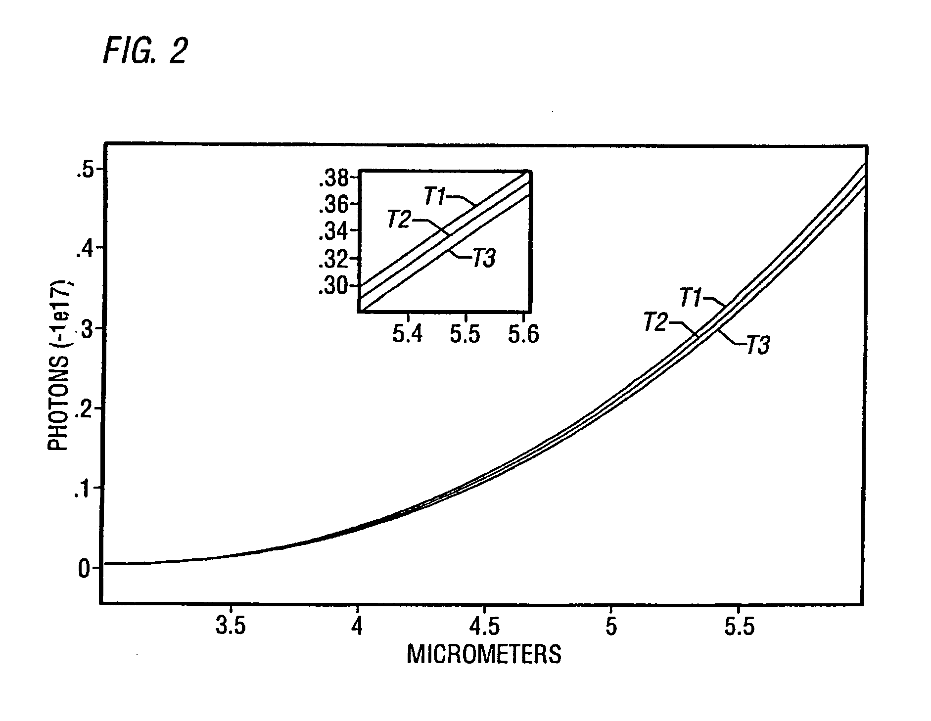
[0105]Infrared spectroscopy could prove useful in several ways. First, it should be able to corroborate the location of macrophages by the massive amounts of nitric oxide they produce, since nitric oxide has a characteristic near-infrared spectrum. Ohdan, et al., Transplantation 57:1674–1677 (1994). Near-infrared imaging of cholesterol has already been demonstrated. Cassis, et al., Anal Chem 65:1247–1256 (1993). Second, since infrared and near-infrared wavelengths penetrate tissue more deeply as wavelength increases, longer wavelengths should reveal metabolic activity in deeper (0.1- to 1-mm) regions.

[0106]This phenomenon could be used to develop computed infrared tomography, possibly in conjunction with interferometry, in which an incident beam is split by a moving mirror to produce a reference beam and a beam that is variably scattered and absorbed by the tissue. The nonsynchronous reflected wavelengths are reconstituted to ...


PUM


Abstract

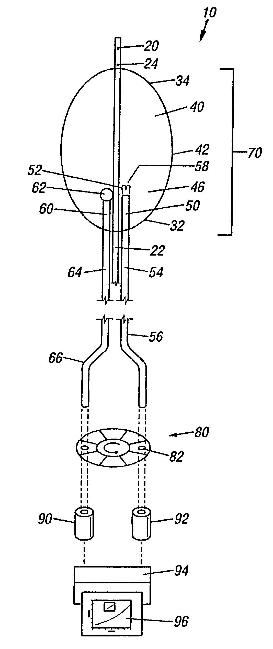
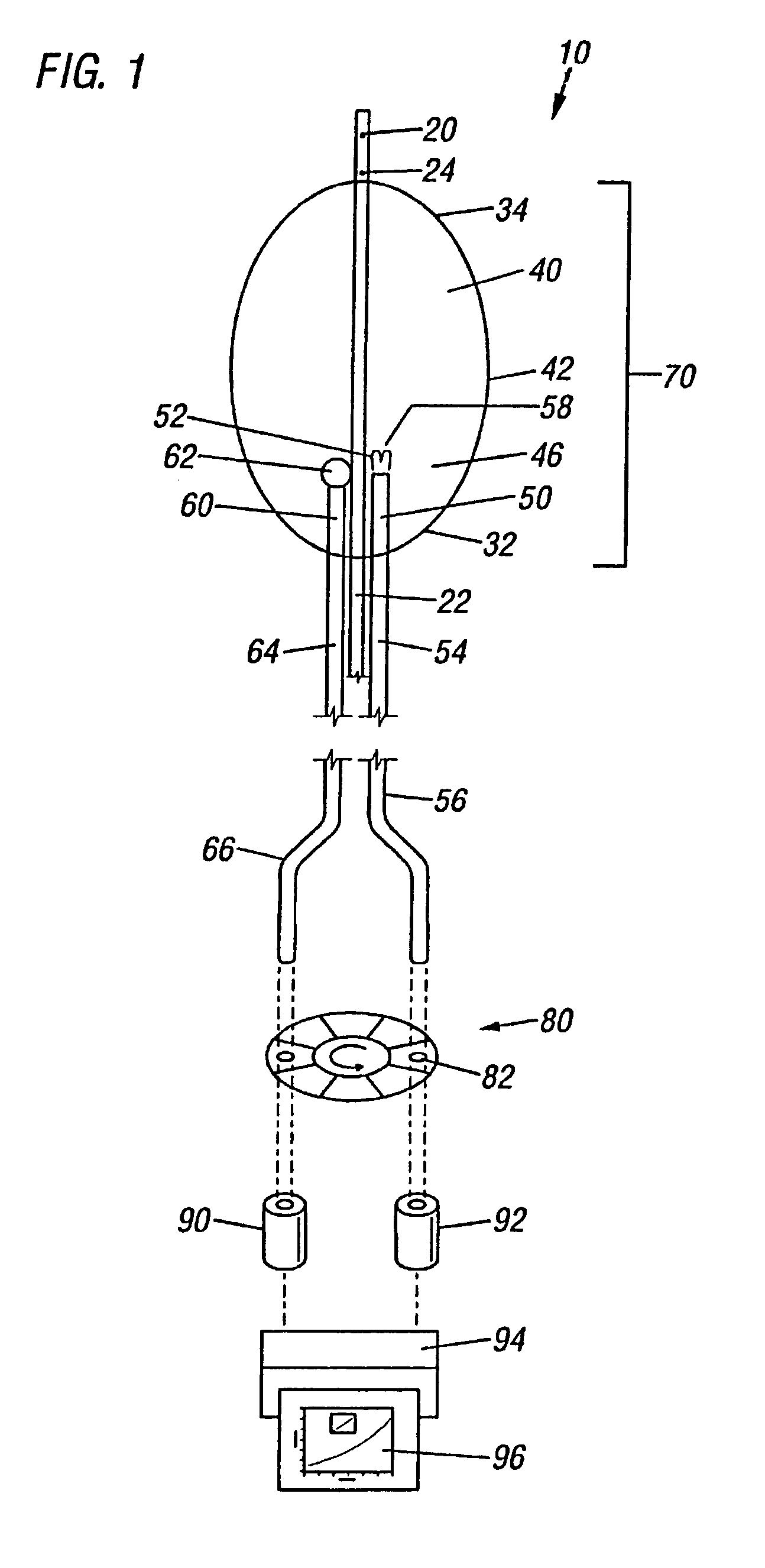
A method of detecting vulnerable arterial plaque formed on the vessel wall in vivo is disclosed. The method comprises obtaining an image of the arterial plaque, measuring the temperature of the arterial plaque, and determining whether the plaque exhibits an elevated temperature compared to the temperature of at least one adjacent vessel wall site. The method may include determining whether the plaque exhibits an elevated temperature of between 0.4 to 4° C. above at least one adjacent vessel wall site.

Description

CROSS-REFERENCE TO RELATED APPLICATIONS[0001]The present application is a continuation application of Ser. No. 09 / 268,609 filed Mar. 15, 1999 now U.S. Pat. No. 6,475,159, which is a divisional application of U.S. patent application Ser. No. 08 / 717,449, filed on Sep. 20, 1996, now U.S. Pat. No. 5,935,075, which claims the benefit of 35 USC 111(b) provisional application Ser. No. 60 / 004,061, filed Sep. 20, 1995, and entitled “Catheters and Methods Detecting Thermal Discrepancies in Blood Vessels.BACKGROUND OF THE INVENTION[0002]This invention was made with government support under Grant No. 91HL07 awarded by the National Heart Lung and Blood Institute, giving the federal government certain rights in the invention. In addition, the invention described herein was made in the performance of work under a NASA contract and is subject to the provisions of Section 305 of the National Aeronautics and Space Act of 1958, Public Law 85-568 (72 Stat. 435; 42 U.S.C. 2457).[0003]A. Field of the Inv...

Claims


Application Information

Patent Timeline
Patent Type & Authority Patents(United States)
IPC IPC(8): A61B5/04A61B5/01A61B5/00A61B17/00A61B17/22A61M25/00
CPCA61B5/0086A61B5/01A61B5/02007A61B5/0507A61B5/413A61B5/6853A61B5/015A61B5/0066A61B5/0071A61B5/0073A61B5/0075A61B2017/22001A61B2017/00084
Inventor CASSCELLS, S. WARDWILLERSON, JAMES T.BEARMAN, GREGORY H.EASTWOOD, MICHAEL L.KRABACH, TIMOTHY N.
Owner BOARD OF RGT THE UNIV OF TEXAS SYST
Why Eureka
Try Eureka