Three-dimensional porous chitosan/gelatine microsphere, preparation method thereof and application in liver cell culture

A three-dimensional porous and chitosan technology, which is applied in the preparation of microspheres, tumor/cancer cells, animal cells, etc., can solve the problems of increasing the risk factor, not keeping the shape well, and the surface and the interior are not connected, so as to increase Effects of specific surface area, improved exchange, and increased density

Inactive Publication Date: 2011-09-07
RUIJIN HOSPITAL AFFILIATED TO SHANGHAI JIAO TONG UNIV SCHOOL OF MEDICINE
View PDF3 Cites 29 Cited by
  • Summary
  • Abstract
  • Description
  • Claims
  • Application Information

AI Technical Summary

Problems solved by technology

First of all, in terms of material selection, chitosan and gelatin were selected as the main materials for the preparation of microspheres. Although there were also articles published on the preparation of microspheres with chitosan or (and) gelatin, the method used was mainly the emulsification cross-linking method. (Use liquid paraffin or edible oil as the oil phase, chitosan or (and) gelatin or sodium alginate as the water phase, stir, control the size of the liquid droplets to form an oil-in-water or water-in-oil suspension, add pentadiene The aldehyde-based cross-linking agent is reacted into spheres, washed, and dried to obtain microspheres. Most of the obtained microspheres are solid microspheres. The microspheres obtained after adding porogens are not really porous structures, but internally independent. The pores are not connected to the outside world, and the microspheres obtained by the emulsification method are easy to stick, and the cross-linking agent glutaraldehyde itself is toxic, which also increases a certain risk factor. At present, the microspheres prepared by the emulsification method are mainly used for microsphere drug delay Research on the release) and coagulation / precipitation method (using the characteristics of chitosan insoluble in alkaline solution or (and) the amphoteric properties of gelatin, drop chitosan or chitosan / gelatin solution into alkaline solution, such as NaOH, NaOH-methanol, etc., the microspheres are separated and purified by centrifugation and filtration, and the microspheres are obtained after washing, but the surface of the microspheres obtained by this method is only shrunk to form grooves, and the surface is not connected to the interior, that is, closed pores)
Although there are also literature reports on the freeze-drying method, most of them are directly mixed with the solution and poured into a mold to directly freeze-dry to prepare a film or cylindrical scaffold; or the freeze-dried scaffold is cross-linked with NaOH, glutaraldehyde, etc. , but because the spatial structure of chitosan or (and) gelatin material changes after freeze-drying, it is very soluble in water, and the original shape cannot be well maintained after cross-linking, so it is generally used to prepare membrane-like scaffolds for skin Tissue engineering or the preparation of mold scaffolds for bone tissue engineering, no three-dimensional porous microspheres with a spherical shape, a diameter of 300-800um, a surface pore size of 50-200um, the surface and the internal pore size, and the closer to the microsphere, the larger the internal pore size. ball coverage

Method used


Image

  • Three-dimensional porous chitosan/gelatine microsphere, preparation method thereof and application in liver cell culture
  • Three-dimensional porous chitosan/gelatine microsphere, preparation method thereof and application in liver cell culture
  • Three-dimensional porous chitosan/gelatine microsphere, preparation method thereof and application in liver cell culture

Examples


Embodiment 1

[0059] Preparation of a Three-dimensional Porous Chitosan / Gelatin Microspheres

[0060] (1) Preparation of primary freeze-dried microspheres

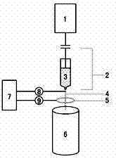
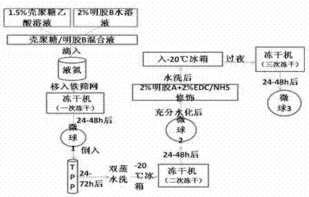
[0061] Prepare 1.5% chitosan (prepared with 1% acetic acid) and 2% gelatin B (prepared with double distilled water) respectively. : 1 phase mixed, after removing air bubbles, inhale into a 5ml syringe with a needle inner diameter of 0.7mm, and connect the microcapsule generator-electrostatic device (such as figure 1 Shown), in a high-voltage electrostatic environment (voltage U=60, frequency F=90, pulse width PW=6: high-voltage pulse microcapsule forming instrument, University of Shanghai for Science and Technology) drop into liquid nitrogen, wait for the microspheres to sink to the liquid nitrogen tank After bottoming, remove the microspheres and put them on an iron screen soaked in liquid nitrogen, put them on the freeze dryer, control the freeze drying temperature at -60°C to -80°C, turn off the freeze dryer after 24-48 hours and t...


PUM

PropertyMeasurementUnit
diameteraaaaaaaaaa
pore sizeaaaaaaaaaa

Abstract

The invention discloses a three-dimensional porous chitosan/gelatine microsphere, a preparation method thereof and an application thereof in liver cell culture. The preparation method thereof comprises the following steps of: rapidly freezing mixture into spheres by dropping liquid nitrogen by virtue of a high-pressure pulse microcapsule moulding instrument after chitosan and gelatine B are mixed, carrying out secondary freeze-drying after microspheres subjected to primary freeze-drying are crosslinked and cured with saturated tripolyphosphate-85% ethanol solution, fully hydrating the microspheres obtained by the secondary freeze-drying, adding cross linking agent carbodiimide/N-hydroxy succinyl, modifying the microspheres with gelatine A, removing unreacted carbodiimide/N-hydroxy succinyl and gelatine A by washing after water bath and reaction in dark place are carried out, and carrying out tertiary freeze-drying, thus the three-dimensional porous chitosan/gelatine microspheres with the diameter of 300-800Mum and the surface aperture of 50-200mu m are obtained, and the surface of each microsphere is connected with the aperture of the interior of the microsphere. The three-dimensional porous chitosan/gelatine microspheres obtained by the invention can be used for in vitro culture of high-density and high-activity liver cells.

Description

technical field [0001] The invention relates to a three-dimensional porous chitosan / gelatin microsphere, a preparation method thereof and an application in hepatocyte culture. More specifically, it involves the use of the macroporous properties of gelatin and the properties of gelatin as a good extracellular matrix, combined with microcapsule generator-electrostatic method, ion coacervation method and three-time freeze-drying method to prepare a three-dimensional porous chitosan / gelatin microparticles. Spheroids and their application in hepatocyte culture. Background technique [0002] Chitosan is a biopolymer obtained by deacetylation of chitin. It is the only positively charged alkaline polysaccharide found in nature so far. It is non-toxic, has good biocompatibility and biodegradability , in food, environmental protection, agriculture, medicine and other aspects have a wide range of applications. Collagen is the most abundant protein in animals. Gelatin, as a hydrolyze...

Claims


Application Information

Patent Timeline
Patent Type & Authority Applications(China)
IPC IPC(8): B01J13/02C12N5/071C12N5/09
Inventor 彭承宏黄芳韩宝三吴旭波董亚东崔龙
Owner RUIJIN HOSPITAL AFFILIATED TO SHANGHAI JIAO TONG UNIV SCHOOL OF MEDICINE
Why Eureka
Try Eureka