
Method for preparing small-diameter artificial blood vessels on basis of nanotechnologies

A technology of artificial blood vessels and nanotechnology, applied in medical science, fiber processing, textiles and papermaking, etc., can solve the problems of poor shape retention, low strength, slow degradation rate, etc., and achieve improved mechanical stability, high porosity, high The effect of histocompatibility

Inactive Publication Date: 2015-11-25
AFFILIATED HUSN HOSPITAL OF FUDAN UNIV
View PDF2 Cites 16 Cited by
  • Summary
  • Abstract
  • Description
  • Claims
  • Application Information

AI Technical Summary

Problems solved by technology

[0004] 1) Polyester artificial blood vessel is the earliest vascular material used, and has been successfully used for large blood vessel replacement for a long time due to its high patency rate, but it cannot fully meet the manufacturing requirements of small-diameter artificial blood vessels
[0005] 2) Silk artificial blood vessels are not stable enough due to their helical shrinkage, which can easily cause blood vessel suction and collapse, and have poor shape retention and low strength, which limits clinical application
[0006] 3) The artificial vascular material is expanded polytetrafluoroethylene, which has good biocompatibility and anticoagulant properties, but poor compliance, and the patency rate of the graft is only 30%, especially for ePTFE artificial vessels with a diameter less than 6mm. The above-mentioned shortcomings of blood vessels are more obvious, and the long-term patency rate is extremely poor
However, it has been reported in the literature that simple mixed PLA and PLLA materials were electrospun to make vascular stents due to inconsistent degradation rates and tissue generation replacement rates. After long-term observation, stent ruptures or aneurysms were found. The nanofibers made of molecular polymer blends are too densely arranged and the degradation rate is slow. After transplantation in rats, few smooth muscle cells enter the inner layer of the scaffold to participate in the reconstruction, so it is difficult to achieve satisfactory material degradation-tissue rebalance

Method used


Image

  • Method for preparing small-diameter artificial blood vessels on basis of nanotechnologies

Examples


Embodiment 1

[0044]Example 1 Making silk fibroin / PCL small-diameter nanofiber blood vessels with nanostructures

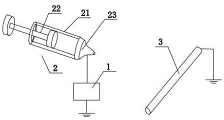
[0045] 1) Polycaprolactone and silk fibroin are dissolved in hexafluoroisopropanol (HFIP) to make a spinning solution for electrospun nanofiber blood vessels. The quality of polycaprolactone and silk fibroin in the spinning solution The percentage concentrations are 5% and 8%, respectively. The electrospinning equipment includes a DC high voltage generator, a microinjector, a cylindrical rod-shaped receiving screen and a power device, wherein the power device drives the cylindrical rod-shaped receiving screen to rotate, and the needle of the micro-injector Connect to the positive power supply of the DC high-voltage generator, the ground wire of the cylindrical rod-shaped receiving screen, and the ground wire of the cylindrical rod-shaped receiving screen. The diameter of the cylindrical rod-shaped receiving screen is 1-1.2mm. Put the spinning solution into the micro-injector, s...

Embodiment 2

[0048] Example 2 Heparinized treatment of nanofibrous vessels

[0049] By covalently combining heparin with polyethylene glycol diamine, the heparin is combined on the nanofibrils of the nanofibrous blood vessel to realize the heparinization treatment of the nanofibrous blood vessel.

[0050] The specific operation steps are as follows:

[0051] 1) Place the nanofiber blood vessel in a centrifuge tube, add 2ml of 70% deionized water and alcohol, and shake it on the machine at room temperature for 5 minutes;

[0052] 2) Discard the previous solution, add 2ml of deionized water to the centrifuge tube, and shake for 5 minutes x 4 times;

[0053] 3) Discard the former solution, add 2ml of sodium hydroxide (NaOH) with an equivalent concentration of 0.01N into the centrifuge tube, and shake it for 10 minutes to increase the amount of carboxyl groups on the surface of the nanofiber vascular material;

[0054] 4) Discard the previous solution, add 2ml of phosphate buffer solution in...

Embodiment 3

[0063] Example 3 Preparation of heparinized nanofibrous vessels compounded with mechanical growth factors

[0064] The aforementioned preliminarily heparinized nanofibrous blood vessels are covalently bound to the mechanical growth factor and heparin through a surface covalent binding method.

[0065] The specific operation steps are as follows:

[0066] 1) Place nanofibrous blood vessels in a centrifuge tube, add mechanical growth factor-phosphate buffer solution mixed solution with a concentration of mechanical growth factor of 20ng / ml, and shake the mixture at 4°C for 24 hours.

[0067] 2) Discard the previous solution, add 2ml of phosphate buffer solution into the centrifuge tube, and shake for 5 minutes x 3 times. Phosphate buffer was kept at 4°C until the mechanical growth factorization was completed at this step.

[0068] In this example, the preparation process of EDH-suflo-N-hydroxysuccinimide solution is as follows:

[0069] Use 0.5mmol / L 2-(N-morpholine)ethanesul...


PUM

PropertyMeasurementUnit
Diameteraaaaaaaaaa
Tube wall thicknessaaaaaaaaaa
Concentrationaaaaaaaaaa

Abstract

The invention belongs to the field of medicine and high-polymer materials, and discloses a method for preparing small-diameter artificial blood vessels on the basis of nanotechnologies. The method includes steps of dissolving, by weight, 8% of fibroin and 5% of polycaprolactone in hexafluoroisopropanol to obtain spinning liquor; manufacturing the nano-fiber blood vessels with the wall thicknesses of 130-170 micrometers on cylindrical rod-shaped receiving screens with the diameters of 1-1.2mm by means of electrospinning by the aid of electrospinning technologies. Compared with the prior art, the method has the advantages that requirements of tissue engineering on high tissue compatibility, high porosity, plasticity and degradability can be met owing to the electrospinning technologies, vascular stents can be modified by cell activity factors (mechanical growth factors) and medicine (heparin) with anticoagulant activity, and accordingly shortcomings of technological complexity and poor tissue compatibility of existing cell-modified vascular stents can be overcome by the aid of the method.

Description

technical field [0001] The invention relates to a preparation method of a small-diameter artificial blood vessel based on nanotechnology. Background technique [0002] Cardiovascular and cerebrovascular diseases have become the number one killer threatening national health in our country. Among the elderly in China, the incidence rate of cardiovascular and cerebrovascular diseases is as high as 30%. The relevant institutions in my country have reported that the death due to cardiovascular and cerebrovascular accidents accounts for more than 50% of the total deaths. With the continuous surge of the aging population in our country, the number of patients is still increasing The increase will bring a heavy burden to our national economic construction. Vascular replacement surgery is widely used in the treatment of arterial stenosis, aneurysm, atherosclerosis and other vascular diseases. However, autologous blood vessel transplantation has many defects such as difficulty in obta...

Claims


Application Information

Patent Timeline
IPC IPC(8): A61L27/18A61L27/56A61L27/20D01D5/00
Inventor 虞剑孙一睿黄冰清祝翊倩吴惺胡锦
Owner AFFILIATED HUSN HOSPITAL OF FUDAN UNIV
Learn More