
Preparation method and applications of 3D printing composite magnetic metal scaffold

A composite magnetic and 3D printing technology, applied in the field of biomedical materials, can solve the problems of lack of biological activity, single component, lack of cell recognition sites, etc., achieve good hydrophilicity and biocompatibility, and the preparation method is simple and easy Effects on manipulation, adhesion promotion and proliferation

Inactive Publication Date: 2018-02-23
PEKING UNION MEDICAL COLLEGE HOSPITAL CHINESE ACAD OF MEDICAL SCI
View PDF9 Cites 12 Cited by
  • Summary
  • Abstract
  • Description
  • Claims
  • Application Information

AI Technical Summary

Problems solved by technology

Natural polymer materials such as collagen, chitosan, and alginate have good biological activity, can promote cell adhesion and proliferation, and have good degradability at the same time, but the problem is that the processing and shaping performance is poor , and the mechanical strength of its preparations is often insufficient, which limits its clinical use
Artificially synthesized polymer materials is a field that has developed rapidly in recent years. Its composition is single, its properties are controllable, and its preparations have certain mechanical strength. It can also make various complex porous structures that meet the requirements by means of rapid prototyping technology. It has become a biological material. The main force of research in the field, but its problems are generally lack of biological activity, lack of sites for cell recognition, and its degradation metabolites often have a negative impact on local cells
Some scholars have found that materials such as poly-L-lactic acid and polyglycolic acid will produce acidic substances after degradation, which will reduce the pH value of the local environment and lead to cell and tissue necrosis.
The third category: metal materials, including stainless steel, titanium alloys, cobalt-chromium-molybdenum alloys, tantalum metals, magnesium alloys, etc., their advantages are that they can provide sufficient mechanical strength and have good biocompatibility, but the disadvantage is that they have no biological activity and often cannot be degraded and absorbed
However, titanium alloy is an inert metal, which has no biological activity and no sites that cells can recognize. Therefore, how to modify titanium alloy implants with osteogenesis-promoting modification technology is the focus of current research.
However, because the porous structure of titanium alloy scaffolds brings difficulties to the application of various modification technologies such as plasma spraying, surface active coatings, and loaded growth factors, how to comprehensively promote osteogenesis of porous metal scaffolds Modification is a research hotspot

Method used


Image

  • Preparation method and applications of 3D printing composite magnetic metal scaffold
  • Preparation method and applications of 3D printing composite magnetic metal scaffold
  • Preparation method and applications of 3D printing composite magnetic metal scaffold

Examples


Embodiment 1

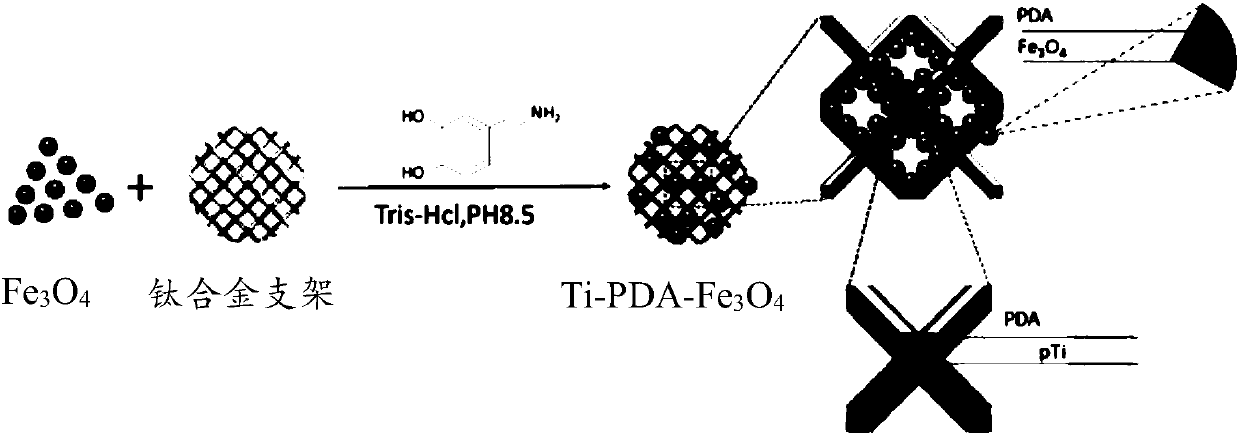
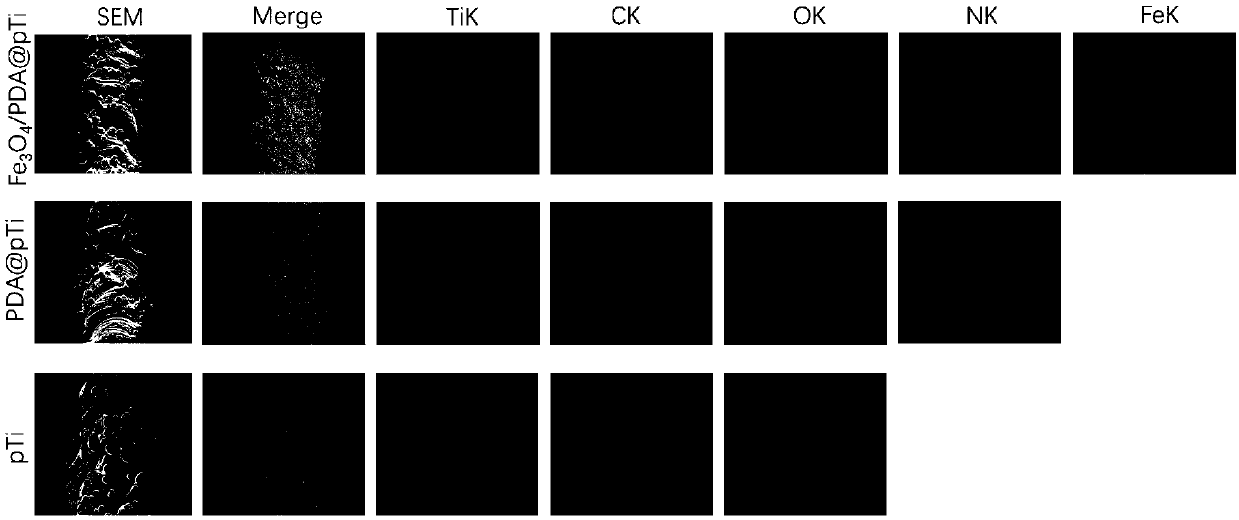
[0035] Example 1 Fe 3 o 4 @PDA Composite Magnetic Titanium Alloy Stent (Ti-PDA-Fe 3 o 4 ) preparation

[0036] 1. Use CAD software to construct a 3D digital model with a dodecahedron as the basic unit, a pore diameter of 300 μm, and a pore column of 200 μm.

[0037] 2. Input the above preparation parameters into the laser 3D printing equipment (ConceptLaser Germany), the maximum scanning speed is 7m / s, and the construction speed is 1-5cm 3 / h. Set the printing layer thickness, melting speed, scanning direction, laser spot interval, powder spreading speed, melt the powder, build up layers, and then use wire cutting to separate samples and heat treatment to release stress.

[0038] 3. Soak in 10% dilute hydrochloric acid overnight, and ultrasonically clean to remove residual titanium alloy powder to obtain a three-dimensional porous titanium alloy stent.

[0039] 4. Fe 3 o 4 The magnetic nanoparticles and the three-dimensional porous titanium alloy scaffold are immersed ...

Embodiment 2

[0041] Example 2 Fe 3 o 4 @PDA Composite Magnetic Titanium Alloy Stent (Ti-PDA-Fe 3 o 4 ) preparation

[0042] 1. Use CAD software to construct a 3D digital model with a dodecahedron as the basic unit, a pore diameter of 600 μm, and a pore column of 300 μm.

[0043] 2. Input the above preparation parameters into the laser 3D printing equipment (ConceptLaser Germany), the maximum scanning speed of this equipment is 7m / s, and the construction speed is 1-5cm 3 / h. Set the printing layer thickness, melting speed, scanning direction, laser spot interval, powder spreading speed, melt the powder, build up layers, and then use wire cutting to separate samples and heat treatment to release stress.

[0044] 3. Soak in 10% dilute hydrochloric acid overnight, and ultrasonically clean to remove residual titanium alloy powder to obtain a three-dimensional porous titanium alloy stent.

[0045] 4. Fe 3 o 4 The magnetic nanoparticles and the three-dimensional porous titanium alloy scaf...

Embodiment 3

[0047] Example 3 Fe 3 o 4 @PDA Composite Magnetic Titanium Alloy Stent (Ti-PDA-Fe 3 o 4 ) preparation

[0048] 1. Use CAD software to construct a 3D digital model with a dodecahedron as the basic unit, a pore diameter of 800 μm, and a pore column of 300 μm.

[0049] 2. Input the above preparation parameters into the laser 3D printing equipment (ConceptLaser Germany), the maximum scanning speed is 7m / s, and the construction speed is 1-5cm 3 / h. Set the printing layer thickness, melting speed, scanning direction, laser spot interval, powder spreading speed, melt the powder, build up layers, and then use wire cutting to separate samples and heat treatment to release stress.

[0050] 3. Soak in 10% dilute hydrochloric acid overnight, and ultrasonically clean to remove residual titanium alloy powder to obtain a three-dimensional porous titanium alloy stent.

[0051] 4. Fe 3 o 4 The magnetic nanoparticles and the three-dimensional porous titanium alloy scaffold are immersed ...


PUM

PropertyMeasurementUnit
Apertureaaaaaaaaaa
Elastic modulusaaaaaaaaaa

Abstract

The present invention discloses a 3D printing composite magnetic metal scaffold preparation method, which comprises: establishing a three-dimensional model with a porous structure, printing a three-dimensionally-penetrating porous metal scaffold by using a 3D printing technology, and modifying the porous metal scaffold with poly dopamine and Fe3O4@PDA composite magnetic nanoparticles through a one-step method so as to obtain the 3D printing composite magnetic metal scaffold. According to the present invention, the 3D printing composite magnetic metal scaffold can meet the requirement of the bone repair material on the mechanical strength, and further has advantages of advantages of good biocompatibility, good hydrophilicity and reduced cytotoxicity, wherein the coated material has weak magnetism, such that the osteoblast differentiation can be easily achieved, and the osteogenesis is easily achieved; and the preparation method has advantages of simple and easy operation, rapidness, high efficiency and easy production.

Description

technical field [0001] The invention relates to the technical field of biomedical materials, in particular to a preparation method and application of a 3D printed composite magnetic metal stent. Background technique [0002] The treatment of bone defects has always been a clinical problem. In vivo experiments show that 6×6×10mm 3 Bone defects need to be repaired by bone grafting. It has been reported that delayed union or nonunion occurs in up to 13% of tibial fractures. In addition, plastic surgery, oral and maxillofacial surgery, and large bone defects caused by severe trauma, infection, bone tumors, and skeletal deformities all require Repaired by bone grafting. [0003] Clinically, graft materials for bone defects include autologous bone and allogeneic bone. Autologous bone transplantation has always been the "gold standard" for the treatment of bone defects, but autologous bone is generally taken from the patient's ilium and fibula, not only the amount of bone taken...

Claims


Application Information

Patent Timeline
IPC IPC(8): A61L27/56A61L27/34A61L27/30A61L27/06A61L27/04A61L27/50B22F3/105B33Y70/00B33Y80/00
CPCA61L27/042A61L27/06A61L27/306A61L27/34A61L27/50A61L27/56B33Y70/00B33Y80/00A61L2300/412A61L2300/606A61L2400/18A61L2430/02A61L2300/102B22F10/00B22F10/62B22F10/68B22F10/28B22F10/80C08L79/04Y02P10/25
Inventor 黄振飞于凌佳贺宇王海吴志宏
Owner PEKING UNION MEDICAL COLLEGE HOSPITAL CHINESE ACAD OF MEDICAL SCI
Learn More