Anti-adhesion tendon material and preparation method thereof

An anti-adhesion and tendon technology, applied in pharmaceutical formula, medical science, prosthesis, etc., can solve the problems of unfavorable tendon tissue to obtain nutrients, prolong the regeneration and reconstruction process of tendon tissue, and lack of targeting of immune regulation, reaching the shelf Long effective period, shorten the time of regeneration and reconstruction, easy to obtain the effect

Active Publication Date: 2022-06-03
BEIJING XINKANGCHEN MEDICAL SCI & TECH DEV
View PDF8 Cites 0 Cited by
  • Summary
  • Abstract
  • Description
  • Claims
  • Application Information

AI Technical Summary

Problems solved by technology

[0003] At present, physical isolation is one of the most commonly used clinical methods to reduce tendon adhesions. The most common ones on the market are silica gel membrane, hyaluronic acid membrane, and chitin membrane , polylactic acid membrane, biological amniotic membrane, autologous or allogeneic fascia, etc. (Zhou Xiujun. Evaluation of different biomaterials for preventing adhesion after tendon injury[J]. Chinese Tissue Engineering Research and Clinical Rehabilitation, 2010, 14(21): 3915-3918 Kang Dong. The anti-adhesion effect of absorbable biomaterials in tendon injury reconstruction [J]. Chinese Tissue Engineering Research, 2016, 20(21): 3185-3192.), as disclosed in patent 202010574129.9 is chitosan modified Collagen film, patent 202110906851.2 discloses that fresh amniotic membrane is loaded with the drug ecoxib, and patent 202110264223.9 is a layered composite film of polylactic acid and type I collagen in a certain proportion. These film products have a certain effect of preventing tendon adhesion. However, the adhesion between the membrane and the body is poor, sutures are often required during surgery, and the fixing and wrapping operation is cumbersome; at the same time, these membranes mainly isolate the fibrin, multinucleated cells, and inflammatory cells exuded around the tendon from the tendon through the physical barrier function, hindering It prevents the fibroblasts secreting collagen fibers from the tendon and plays an anti-adhesion role. In add

Method used


Image

  • Anti-adhesion tendon material and preparation method thereof
  • Anti-adhesion tendon material and preparation method thereof
  • Anti-adhesion tendon material and preparation method thereof

Examples


Embodiment 1

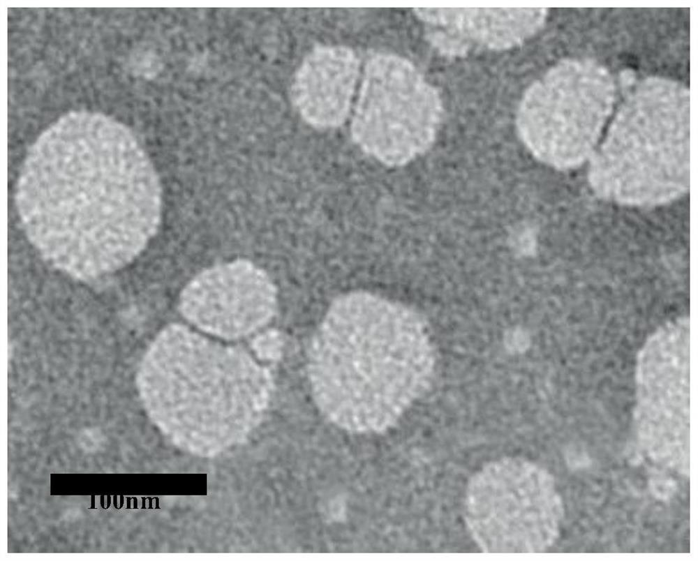
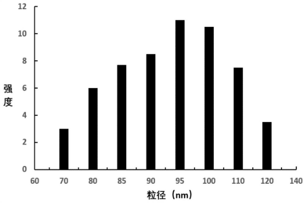
[0028] Example 1 Preparation of Treg cell exosomes

[0029] (1) Primary culture of induced cells: aseptically collect human peripheral blood, and separate mononuclear cells from peripheral blood by density gradient centrifugation. 6 The density of / ml was added to the culture vessel pre-coated with anti-CD3 monoclonal antibody and anti-CD28 monoclonal antibody, and then the medium containing TGF-β, IL-2 and 10% serum replacement was added, and the temperature was 37 °C, 5% CO. 2 Cultured in an incubator to induce Treg cell expansion.

[0030] (2) Cell passage expansion: supplement the medium containing TGF-β and IL-2 every 2 days, and after culturing for 1 week, change the medium to only add IL-2 at 37°C, 5% CO 2 The culture was continued in the incubator for a total of 3 weeks.

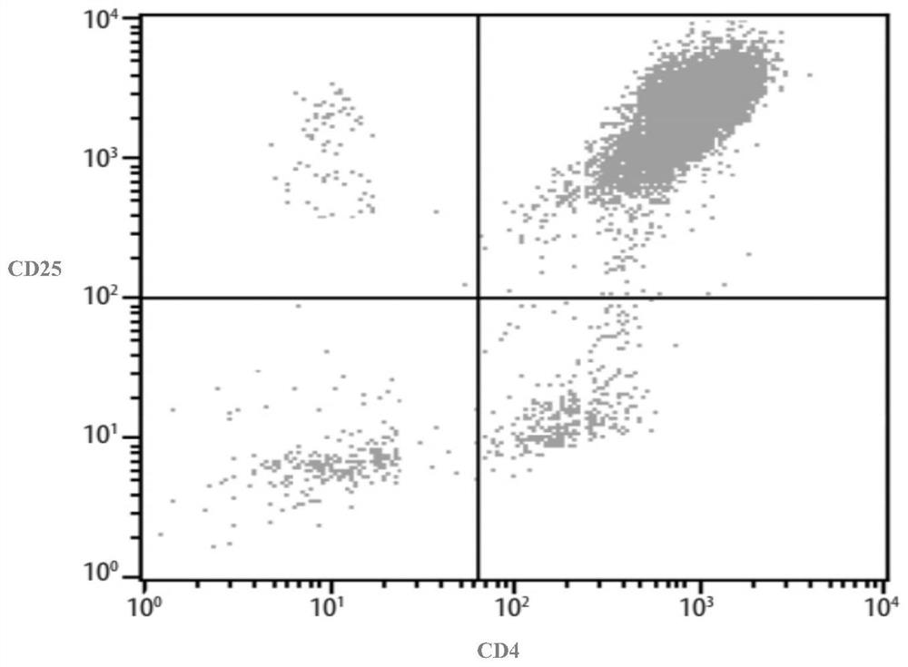
[0031] (3) Cell phenotype detection: collect Treg cells, pipette 2 × 10 6 cells were added with anti-human CD4-FITC and anti-human CD25-PE flow antibodies, incubated for 30 min, washed and detecte...

Embodiment 2

[0036] Embodiment 2 The compound method of exosome and tendon

[0037] (1) The tendon raw material is physically removed from fat and fascia, sterilized with 75% alcohol, and then frozen at low temperature (-40°C) for 4 weeks;

[0038] (2) The frozen tendon is placed in a freeze dryer, and freeze-dried for 48h;

[0039] (3) The extracted Treg cell exosomes were suspended in sterile saline to prepare a Treg cell exosome suspension, which was compounded by 1 g of tendon at 1 × 10 12 The Treg cell exosome suspension was evenly applied to the freeze-dried tendon, let stand at 18°C ​​for 3h, and then cryogenically frozen (-40°C) for 32h;

[0040] (4) Finally, the tendons of the composite Treg cell exosomes were placed in a freeze dryer and freeze-dried for 48 hours;

[0041] (5) The tendon is taken out from the freeze dryer to obtain the tendon that can prevent adhesion.

Embodiment 3

[0042] Example 3 Anti-adhesion test of tendon combined with Treg cell exosome composite material for repairing chicken tendon sheath defect

[0043] Diazepam injection was intramuscularly injected into the middle and upper part of chicken thighs, routinely sterilized, and a longitudinal 2.5 cm incision was made at the proximal interphalangeal joints on the metatarsal side of the bilateral third toes. The subcutaneous tissue was separated, and the tendon sheath was exposed for 1.5 cm. cm and superficial flexor tendon, transversely cut off 1 / 2 of the deep flexor tendon, use non-invasive suture for central suture with modified Kessler method, and use non-invasive suture for peripheral suture. Treg-Exo group: Treg cell exosomes combined with tendon to cover the tendon sheath defect, and non-invasive sutures were used for end-to-end suture fixation; MSC-Exo group: MSC exosomes combined with tendon to cover the tendon sheath defect, non-invasive sutures were used to cover the tendon ...


PUM

PropertyMeasurementUnit
Particle sizeaaaaaaaaaa

Abstract

The invention discloses an anti-adhesion tendon material and a preparation method thereof. The tendon material is composed of regulatory T lymphocyte exosomes and tendons, and is prepared by compounding the T lymphocyte exosomes and the tendons and then freeze-drying the T lymphocyte exosomes and the tendons. The tendon tissue of the prepared composite Treg cell exosome is exposed in body fluid, nutrient substances can be fully obtained, the growth of implanted tendon cells into the tendon tissue can be accelerated, the regeneration and reconstruction time of the tendon tissue can be shortened, the Treg cells can be cultured on a large scale, the Treg cell exosome is easy to obtain, and after the Treg cell exosome and the tendon material are freeze-dried, the preparation process is simple, and the preparation cost is low. The tendon material can be stored for a long time under the refrigeration condition or the room temperature condition, the shelf life is long, and the prepared tendon material is convenient to use and does not need physical isolation.

Description

technical field [0001] The invention belongs to the technical field of tissue engineering regenerative medical products, in particular to an anti-adhesion tendon material and a preparation method thereof. Background technique [0002] Tendon injuries are a common sports injury that usually requires surgical repair or suturing. Due to the poor blood supply of the tendon, it is difficult for the tendon to heal itself after injury. About 40% of the patients will experience different degrees of tendon adhesion after surgery, which seriously affects the functional recovery of the patients. Current research believes that tendon adhesion is a kind of aseptic fibroproliferative inflammatory reaction of the body. The body releases vasoactive substances and cytokines such as histamine and bradykinin under the stimulation of various physical, chemical and biological factors, which makes the local Increased capillary permeability, leading to congestion, edema, and the formation of infl...

Claims


Application Information

Patent Timeline
IPC IPC(8): A61L27/36A61L27/38A61L27/50
CPCA61L27/386A61L27/3662A61L27/3804A61L27/3691A61L27/50A61L2430/10A61L2400/18
Inventor 刘永进韩丽伟胡先同郝丽芳赵子腾李金衷鸿宾
Owner BEIJING XINKANGCHEN MEDICAL SCI & TECH DEV
Why Eureka
Try Eureka