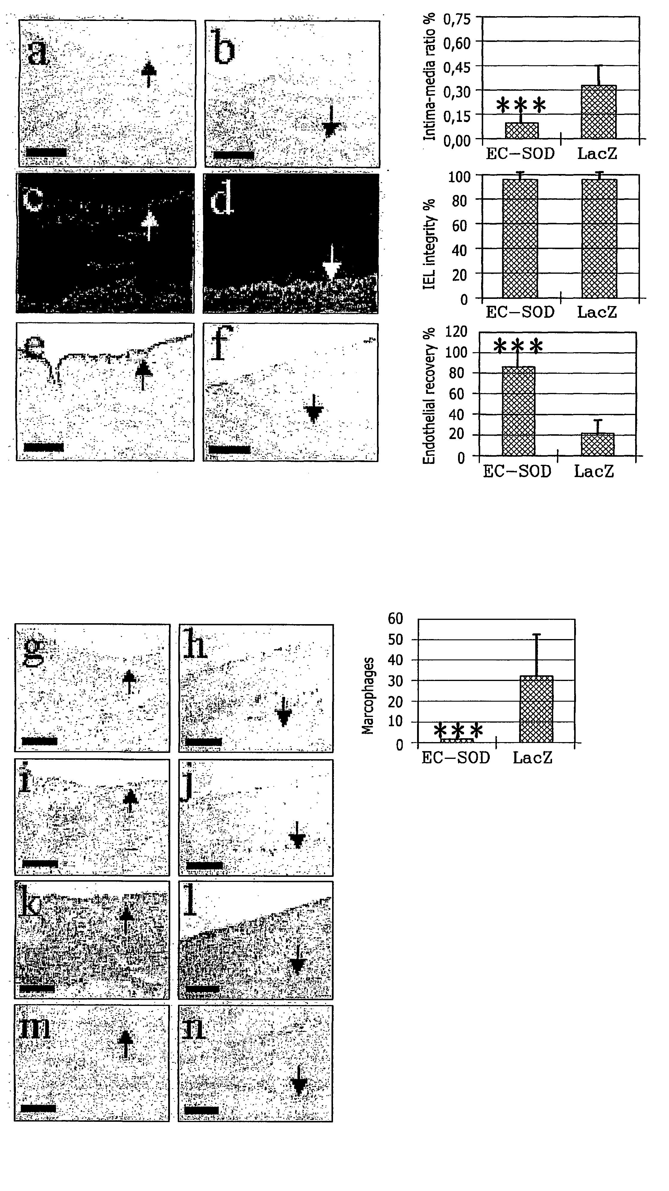
These reactive changes in tissues are difficult to control and cause complications.
All these procedures suffer from reaction to trauma caused by the intervention with following
scar tissue formation or fibrotic reaction.
Replacement of the body part with native structures is usually preferred method but suffers from tissue reaction in the connection site.
Tissue transplantation is costly, and suffers from significant failure rates because of acute inflammatory and long-term fibrotic reactions.
Use of artificial or synthetic medical
implant devices has been subject of considerable attention, but also this technology suffers from
foreign body reaction against the implants with following increase in connective tissue or
fibrosis.
Although
implant devices can be used in some instances as an alternative to donor-based transplants, they too often produce unsatisfactory results because of the tissue response to trauma,
implant's incompatibility with the body, induction of
foreign body inflammatory reaction and induction of connective
tissue formation with following geometrical changes.
Also, lack of
cell lining of the cardiovascular device synthetic surface sets up conditions, which increases the risk of
thrombosis and other medical / surgical procedural complications.
Cardiovascular diseases affect a large segment of the human
population, and are a cause for significant morbidity, costs and mortality in the society.
Cerebral
vascular disease, strokes and bleedings also cause significant morbidity, costs and mortality.
Native blood vessels used as grafts suffer from increased connective
tissue formation and accelerated atherosclerosis.
This causes subsequently narrowing of the vessel lumen.
However, stents actually increase the amount of late luminal narrowing due to
intimal hyperplasia, and the overall rate of
stent restenosis remains unacceptably high (Kuntz et al.
These devices suffers from both
thrombosis until they are covered with endothelial cells and bleeding complications post-operatively.
All endovascular and surgical devices are complicated by same problems—lack of endothelial surfaces on the synthetic surface, formation of excessive connective tissue and inflammatory reaction.
Currently there is no strategy in the market to reduce restenosis after simple
angioplasty without a device inserted.
When using
stent devices after angioplasty procedure pharmaceutically coated stents have not been evaluated for long term effects and there is no verified method in humans which would securely reduce long term restenosis rate in stents or
stent grafts.
The main strategy has been use of various
pharmaceutical substances with stents to reduce
hyperplasia following the trauma to the tissue such as rapamycin,
sirolimus,
paclitaxel,
tacrolimus,
dexamethasone, cytochalasine D and Actinomycin C. One drawback with the current pharmacologically coated devices is the possible disappearance of the effect after the substance has been released from the device surface.
Furthermore, the nature of compounds eluting at high local concentration into the vessel wall and downstream vasculature or tissue is an issue of concern.
Another drawback is that none of these substances is naturally occurring in the body and thereafter fail to promote natural healing of the
foreign body surface.
In native vessel graft the main problem has been
intimal hyperplasia both in the site for connection of the graft and the vessel in the specific body location and the
intimal hyperplasia in the graft vessel lumen.
In humans, Dacron and ePTFE vascular prostheses have met certain clinical success in large and middle-sized arterial reconstructions, but are yet not ideal.
However, the success is limited for vessel substitutes smaller than 6 mm in
diameter, due to anastomotic
hyperplasia i.e., the propensity to develop excessive connective tissue growth in the area where either two native blood vessels or an synthetic artificial
blood vessel and an autologous vessel are connected or due to thrombosis (i.e. propensity to develop clots) in the open thrombogenic surface (Nojiri, Artif.
The major drawback in these systems is the cumbersome use of the sleeve and the used substances are growth factors or encoding for a
growth factor.
This period without endothelial surface may result in undesired effects and problems due to e.g.
thrombogenicity of the surface.
The lack of endothelial surface has led to inferior performance of synthetic grafts compared to autologous grafts (Nojiri, Artif.
Autologous grafts, on the other hand, comprise a step for the harvesting thereof, which leads to longer operation times and also possible complications in the harvesting area.
Transposition of omentum with uncompromised vasculature around a porous carotid
artery PTFE graft has been demonstrated to increase endothelial
cell coverage in the graft lumen in dogs (Hazama, J. of Surg. Res. 1999; 81; 174-180), however, entailing problems with a cumbersome and complex procedure, such as discussed above.
Accordingly, a substantial drawback with these methods is that they are
time consuming and cumbersome in practice, and they also require a specific expertise in the area as well as the suitable equipment.
In addition to the drawbacks discussed above, this method is even more cumbersome and therefore costly to be useful in practice.
However, the problems are still as mentioned above.
In
cell sodding, endothelial cells are administered directly on the polymeric graft surface after harvesting, whereby the graft is implanted, but this technique also includes several steps as mentioned above, which makes it cumbersome as well.
Also,
tissue engineering, which is also a complex and therefore costly procedure, has been used in order to construct vascular tissues for implantation.
Drawback here is that the use of
growth factor stimulating effect of growth factors
on cells could cause uncontrollably connective tissue growth.
Arterial homografts have been described, but they give rise to problems regarding arterial preservation and
antigenicity.
These interventions cause trauma and tissue injury to the vessel wall with disruption of the endothelial cell lining.
After the vessel to dilatation stents have been associated with subacute thrombosis and neointimal thickening leading to obstruction.
Endothelial
cell seeding on the stent has been used as a method to deliver recombinant
protein to the
vascular wall, in order to overcome thrombosis, but as mentioned above, this technology is cumbersome and therefore costly.
In humans, the main problem with stent grafts is the formation of neointimal thickening and lack of complete endotheliahisation leading to
occlusion, as discussed above in relation to grafts.
However, hitherto, no such method that works in practice has yet been developed.
Some studies have been made on endothelial seeding in this context, but it is clinically cumbersome due to the many steps required, as described above.
However, when foreign biomaterials are implanted, an inflammatory foreign body reaction starts, which in the end encapsulates the device, and inhibits
diffusion of nutritive substances to the cells inside the
semipermeable membrane.
The lack of vascularity is an obstacle for
diffusion of substances.
It decreases long-term viability of the encapsulated endocrine tissue, and it also makes vascular implants more susceptible to infections.
The fibrotic
capsule without vascularity can also limit the device performance.
Some other materials used in the procedure of implanted devices also encounter similar problems as the ones discussed above.
To summarise, the major drawbacks in this field is the development of excessive connective tissue after endovascular and
surgical procedures.
For example, after
balloon dilatation procedure the result is excessive connective tissue growth with following complications.
Also, in the autologous vascular grafts excessive connective growth causes narrowing.
In vascular
surgical procedures with or without implanted devices the excessive connective
tissue formation in the anastomotic areas leads to narrowing of the vessel connection with limitation to the flow and following dysfunction in the organ supplied by that vessel.
In vascular implants, when
synthetic materials are used, problems also arise due to open thrombotic surfaces where the implant is performed, which in turn generate
blood clotting and inferior performance.
In synthetic tissue implants, the consequence is a nonvascularised fibrotic non-nutritive zone, which leads to dysfunction of the implant.
This together with the inflammatory reaction causes that
biocompatibility of the mammalian body, especially the
human body, with implanted medical devices cannot be achieved in any satisfactory degree using the prior art methods.


