
Fragment complementation assays for G-protein-coupled receptors and their signaling pathways

a gprotein-coupled receptor and fragment complementation technology, applied in the field of molecular biology, chemistry and biochemistry, can solve the problems of difficult to express the protein of interest and/or obtain a sufficient quantity, take 6 months or longer to develop, and in vitro assay, so as to reduce the screening time, improve the sensitivity, and facilitate the construction of screening assays.

Inactive Publication Date: 2005-08-18
ODYSSEY THERA INC
View PDF4 Cites 26 Cited by
  • Summary
  • Abstract
  • Description
  • Claims
  • Application Information

AI Technical Summary

Benefits of technology

[0030] It is an object of the present invention to provide methods for functional annotation and screening of GPCRs and GPCR-dependent pathways on a large scale. It is an object of this invention to allow the rapid construction of screening assays for GPCRs, starting with genes encoding the receptors or their downstream signaling elements. Another object of this invention is to enable either high-throughput assays or high-content assays for GPCRs and GPCR-dependent pathways. A further object of this invention is to provide compositions useful for such assays, including suitable signaling proteins that can be used in assay construction. It is also an object of this invention to provide for a variety of detection options, including fluorescent, luminescent, phosphorescent or colorimetric readouts. Advantages of the invention include the ability to screen for agonists, antagonists and inhibitors for any GPCR or GPCR-dependent pathway, in any cell type of interest. Another advantage of the invention is the ability to de-orphanize GPCRs for drug discovery. A further advantage is the ability to construct assays suitable for a wide range of detection methods, laboratory instrumentation and automation. An additional advantage is the ability to construct assays for any assay format, either in vitro or in vivo, and in any cell type.
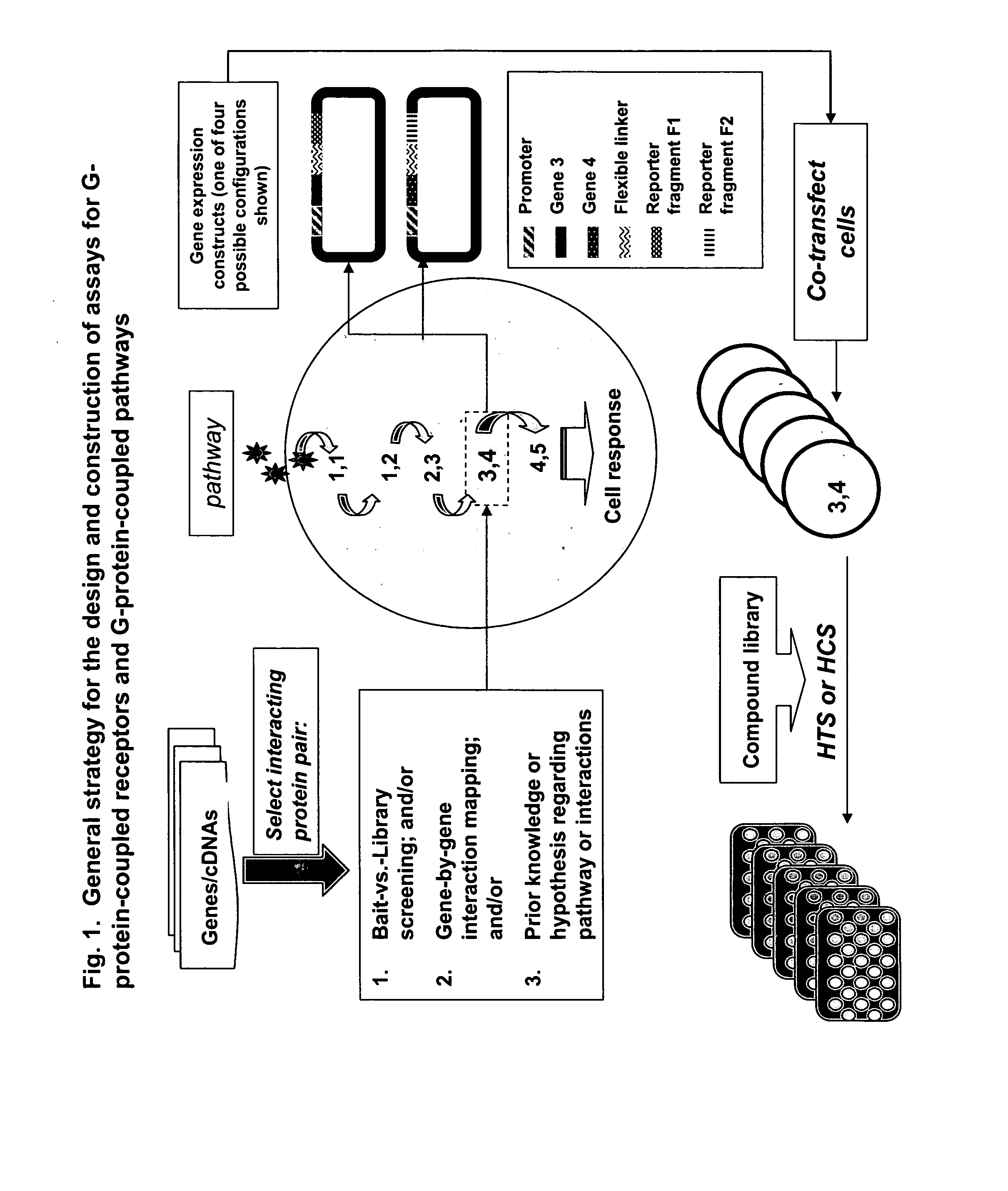
[0031] The present invention seeks to provide the above-mentioned needs for drug discovery. The present invention provides a general strategy for constructing and employing high-throughput assays for GPCRs and GPCR-dependent pathways based on reporter complementation strategies, including protein-fragment complementation assays (PCAs) and enzyme-fragment complementation assays. The present invention also teaches how such assays can be applied to de-orphanization of receptors, mapping of pathways, and screening of compound libraries in order to identify natural products, small molecules, peptides, nucleic acids, or other pharmacologically active agents that can inhibit or activate specific biochemical pathways in live cells.
[0032] The present application teaches general approaches to developing a GPCR assay, including methods for identifying and selecting an interacting protein pair for the construction of assays for GPCRs and G-protein-coupled signaling pathways. Multiple methods for identifying or selecting an interacting protein pair are described, including cDNA library screening, gene-by-gene interaction mapping. These methods can be used to map the intracellular signaling elements linked to a specific GPCR, thereby generating additional assays for drug screening. Prior knowledge or hypothesis regarding a pathway or a protein-protein interaction can also be used to design and construct such assays.
[0033] Methods and compositions are provided both for high-throughput screens (HTS) and for high-content screens (HCS). These assays can be used for the screening of compound libraries to identify compounds of potential therapeutic value, and for the screening of biological compounds or extracts for natural ligands. These assays can be used to identify the native ligand of a GPCR (de-orphanization) and to identify the signaling pathway(s) for ‘orphan’ GPCRs. Signals that are optically detectable in live cell assays, such as fluorescence, luminescence or phosphorescence, can be generated. Cell lysates and fixed cells can also be used in the present invention. Cell fixation offers advantages over live cell assays for purposes of laboratory automation, since entire assay plates can be fixed at a specific time-point after cell treatment, loaded into a plate stacker or carousel, and read at a later time. Alternatively, in vitro assays can be constructed using the strategies and methods described herein. While in vitro assays have the disadvantage of being de-coupled from live-cell signaling events, they may offer certain advantages for ultra high-throughput, low-cost screening.
[0034] In the case of purely quantitative assays, the signal generated in the assay is quantified with a microtiter fluorescence plate reader, flow cytometer, fluorimeter, microfluidic device, or similar devices. The intensity is a measure of the quantity of the protein-protein complexes formed and allows for the detection of changes in protein-protein complex formation in live cells in response to agonists, antagonists and inhibitors. In the case of high-content assays, cells are imaged by automated microscopy, confocal, laser-based, or other suitable high-resolution imaging systems. The total fluorescence / cell as well as the sub-cellular location of the signal (membrane, cytosol, nucleus, endosomes, etc.) can be detected. The choice of HTS or HCS formats is determined by the biology and biochemistry of the signaling event and the functions of the proteins being screened. It will be understood by a person skilled in the art that the HTS and HCS assays that are the subject of the present invention can be performed in conjunction with any instrument that is suitable for detection of the signal that is generated by the chosen reporter.

Problems solved by technology

Although such assays are inexpensive to perform, they can take 6 months or longer to develop.
A major problem is that the development of an in vitro assay requires specific reagents for every target of interest, including purified protein for the target against which the screen is to be run.
Often it is difficult to express the protein of interest and / or to obtain a sufficient quantity of the protein in pure form.
Moreover, although in vitro assays are the gold standard for pharmacology and studies of structure activity relationships (SAR) it is not possible to perform target validation with an in vitro assay, in vivo assays are necessary in order to obtain information about the biological availability and cellular activity of the screening hit.
Transcription reporter assays are highly sensitive screening tools; however, they do not provide information on the mechanism of action of the compound, enable mapping of the components of the pathway leading to transcription, or enable studies of individual steps within signaling cascades.
Quantifying FRET or BRET can be technically challenging its use in imaging protein-protein interactions is limited by the very weak FRET signal.
However, the prior art is silent on the use of either protein-fragment or enzyme-fragment complementation assays for G-protein-coupled receptors or G-protein-coupled signaling pathways.

Method used


Image

  • Fragment complementation assays for G-protein-coupled receptors and their signaling pathways
  • Fragment complementation assays for G-protein-coupled receptors and their signaling pathways
  • Fragment complementation assays for G-protein-coupled receptors and their signaling pathways

Examples


example 1

Dimerization of GPCRs

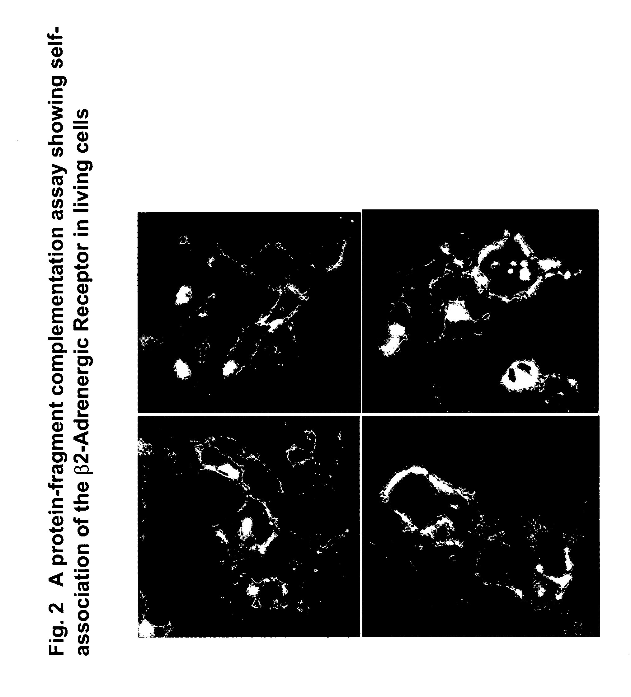
[0071] A growing number of studies have shown that GPCRs are capable of forming homodimers or heterodimers (S. Angers et al., 2002, Dimerization: an emerging concept for GPCR ontogeny and function, Annu. Rev. Pharmacol. Toxicol. 42: 409-435; M K Dean et al., 2001, Dimerization of G-protein-coupled receptors, J. Med. Chem. 44: 4594-4614). Receptor self-association and subsequent changes in receptor activity have been reported for the beta2-adrenergic receptor (T. E. Hebert, 1996, A peptide derived from a beta2-adrenergic receptor transmembrane domain inhibits both receptor dimerization and activation, J. Biol. Chem. 271: 16384-16392), in addition to the delta-opioid receptors, the dopamine receptors, and other GPCRs. It has been shown that agonists can stabilize the dimeric forms of different GPCRs (U. Gether et al., 2000, Uncovering molecular mechanisms involved in activation of G-protein-coupled receptors, Endocrine Reviews 21:90-113), suggesting that homodime...

example 2

Associations of GPCRs with Subunits of Guanine Nucleotide Binding Proteins (G-Proteins)

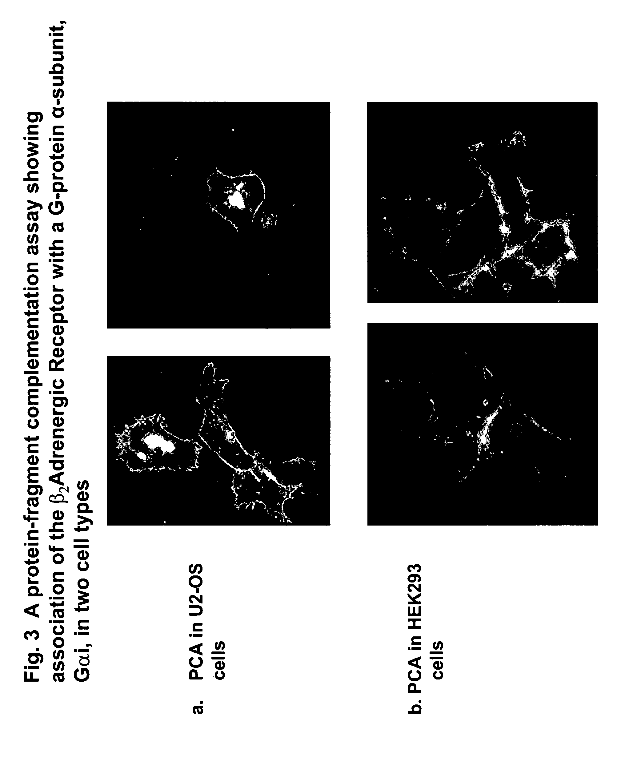
[0074] GPCRs are coupled to their second messenger systems by heterotrimeric guanine nucleotide-binding proteins (G-proteins) comprised of subunits Galpha, Gbeta and Ggamma (C C Malbon & A J Morris, 1999, Physiological regulation of G protein-linked signaling, Physiol. Rev. 79: 1373-1430; T Gudermann et al., 1996, Diversity and selectivity of receptor-G-protein interaction, Annu. Rev. Pharmacol. Toxicol. 36: 429-459). When an extracellular ligand or agonist binds to a GPCR, the receptor exerts guanine nucleotide exchange factor activity, promoting the replacement of bound guanosine diphosphate (GDP) for guanosine triphosphate (GTP) on the Ggammasubunit. Upon binding GTP, conformational changes within the three flexible ‘switch’ regions of Galphaallow the release of Gbeta-gammaand the subsequent engagement of downstream effectors that are specific to each Galphasubtype. The intrinsic GTPase activi...

example 3

Intracellular Events Involving Beta-Arrestin

[0076] Beta-arrestins are adapter proteins that form complexes with most GPCRs and play a central role in receptor desensitization, sequestration and downregulation (for a review see Luttrell and Lefkowitz, J. Cell Science 115 (3): 455-465, 2002). Beta-arrestin binding to GPCRs both uncouples receptors from their cognate G-proteins and targets the receptors to clathrin-coated pits for endocytosis. Beta-arrestins may also function as GPCR signal transducers. They can form complexes with other signaling proteins, including Src framily tyrosine kinases and components of the ERK1 / 2 and JNK3 MAP kinase cascades. Beta-arrestin / Src complexes have been proposed to modulate receptor endocytosis and to act as as scaffolds for several kinase cascades. Beta-arrestin movement from the plasma membrane to intracellular vesicles has been visualized by tagging beta-arrestin with GFP and monitoring the subcellular distribution of the fluorescence in living...


PUM

PropertyMeasurementUnit
Fluorescenceaaaaaaaaaa
Chemiluminescenceaaaaaaaaaa
Phosphorescence quantum yieldaaaaaaaaaa

Abstract

This invention relates generally to the fields of biology, molecular biology, chemistry and biochemistry. The invention is directed to a large number of novel assays for G-protein-coupled receptors (GPCRs) and their signaling pathways. Methods are described for constructing such assays for one or more steps in a GPCR pathway. The invention can be used for functional characterization of GPCRs, target validation, de-orphanization of receptors, high-throughput screening, high-content screening, pharmacological profiling, and other drug discovery applications. The assays can be used directly to assess whether a compound library or a biological extract contains an agonist or antagonist of a receptor. Assay compositions are also provided. The development of such assays is shown to be straightforward, providing for a broad, flexible and biologically relevant platform for the discovery of novel drugs and natural ligands that act on GPCRs or their cognate pathways.

Description

[0001] This application claims the priority benefit under 35 U.S.C. section 119 of U.S. Provisional Patent Application No. 60 / 505,447 entitled “Fragment Complementation Assays For G-Protein-Coupled Receptors And Their Signaling Pathways”, filed Sep. 25, 2003, which is in its entirety herein incorporated by reference.FIELD OF THE INVENTION [0002] This invention relates generally to the fields of biology, molecular biology, chemistry and biochemistry. The invention is directed to a large number of novel assays for G-protein-coupled receptors (GPCRs) and their signaling pathways. The invention also relates to methods for constructing such assays for one or more steps in a GPCR pathway. The invention can be used for functional characterization of GPCRs, target validation, de-orphanization of receptors, high-throughput screening, high-content screening, pharmacological profiling, and other drug discovery applications. The assays can be used directly to assess whether a compound library o...

Claims


Application Information

Patent Timeline
IPC IPC(8): C07K16/28C12P21/02C12Q1/00C12Q1/68G01NG01N33/50G01N33/53G01N33/543G01N33/74
CPCC12Q1/00G01N33/5041G01N2500/00G01N2333/726G01N33/74
Inventor WESTWICK, JOHNKEON, BRIGITTEMACDONALD, MARNIE
Owner ODYSSEY THERA INC
Learn More