
Sensors for fluorescence and magnetic resonance imaging

Active Publication Date: 2008-06-12
MASSACHUSETTS INST OF TECH
View PDF58 Cites 12 Cited by
  • Summary
  • Abstract
  • Description
  • Claims
  • Application Information

AI Technical Summary

Benefits of technology

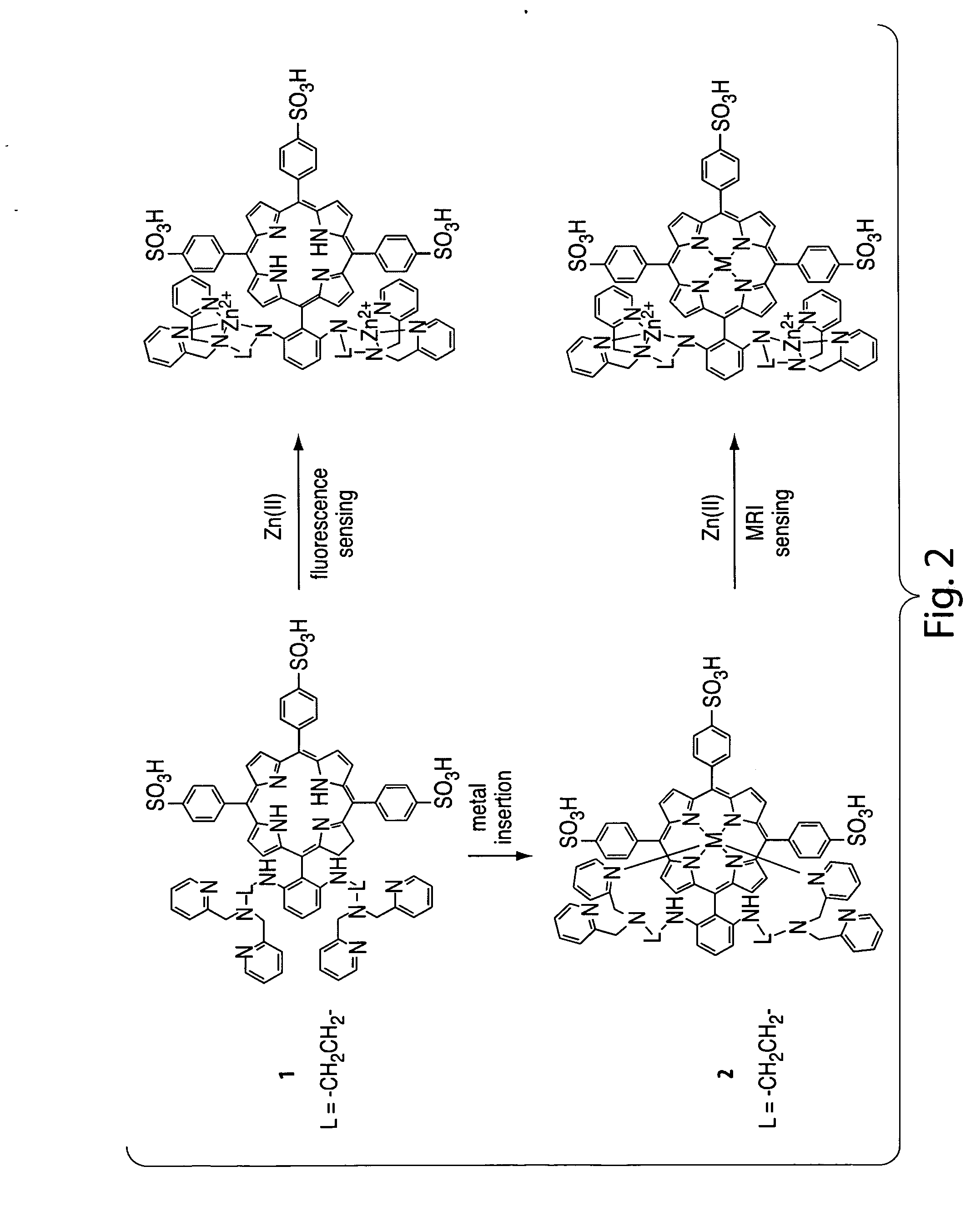
[0033]Another advantage of the present invention relates to the ability to synthesize a wide variety of agents or compositions having different detection mechanisms (e.g., MRI-based detection, fluorescence-based detection, etc.) and tailored to specifically interact with a particular target analyte, using one or more common synthetic intermediates. For example, the invention may involve the synthesis of a molecular platform that may be easily modified to suit several applications. In some cases, the molecular platform may be bound to a metal ion which may establish the mechanism or property by which the agent determines an analyte. In an illustrative embodiment, the invention may comprise the synthesis of a molecular platform which may be useful as an optical-based (e.g., fluorescence-based) agent. Subsequent binding of the same molecular platform to a paramagnetic metal ion may then produce an MRI-based agent. The molecular platform may also comprise one or more attachment sites for various binding moieties selected to specifically interact with a target analyte.
[0034]In some embodiments, the present invention provides methods for determination (e.g., detection) of an analyte, such as zinc ions, calcium ions, nitric oxide, and / or other biological analytes. The agent or composition may comprise a chelator group having an MRI or optical signal and a binding moiety (e.g., for binding an analyte) covalently attached to the chelator group. Determination of the analyte may comprise exposing the agent or composition to a sample suspected of containing an analyte. If present, the analyte can interact with the agent or composition to produce a change in the optical signal or MRI signal of the agent or composition. The shift in the MRI signal or optical signal of the agent may then be determined, thereby determining the analyte. As used herein, the term “determining” generally refers to the analysis of a species or signal, for example, quantitatively or qualitatively, and / or the detection of the presence or absence of the species or signals. “Determining” may also refer to the analysis of an interaction between two or more species or signals, for example, quantitatively or qualitatively, and / or by detecting the presence or absence of the interaction.
[0035]For example, the agent may be an MRI-based agent having a first relaxivity upon exposure to magnetic resonance imaging conditions. Exposure of the agent to a sample suspected of containing an analyte, wherein the species interacts with (e.g., binds) the analyte, if present, may then generate a second relaxivity of the analyte-bound species upon exposure to said magnetic resonance imaging conditions. Determination of a change (e.g., shift), or lack thereof, between the first and second relaxivities may then determine the presence and / or amount of the analyte in the biological sample. In some embodiments, the shift in the magnetic resonance imaging signal comprises a shift in T1 relaxivity. In some embodiments, the shift in the magnetic resonance imaging signal comprises a shift in T2 relaxivity.
[0036]In another embodiment, the agent may have a luminescence absorption or emission, wherein upon interaction with an analyte, the agent may undergo a change in a characteristic of the luminescence absorption or emission. In some cases, the change comprises a decrease in luminescence intensity of the optical signal. In some cases, the change comprises an increase in luminescence intensity of an optical signal. In some cases, the change comprises a change in the wavelength of the luminescence absorption or emission, either alone or in combination with a change in the luminescence intensity of the optical signal.
[0037]In some embodiments, the invention comprises the use of agents (e.g., compounds) that are capable of being inserted into a cell or a portion of a cell, i.e., are cell permeable. As used herein, a species “capable of being inserted into a cell or a portion of a cell” refers to a species which may penetrate the cell membrane and / or interior portions of the cell (e.g., cytoplasm or the nucleus) without substantially damaging the cell or causing cell death. The agent may comprise one or more functional groups that may facilitate interaction between the agent and the cell membrane, transporters positioned within or adjacent the cell membrane, and / or other components associated with the cell that may be involved with cellular uptake of the agent. Such functional groups can include ionic groups, hydrophobic groups, hydrophilic or water-soluble groups, or the like, so long at they, in combination with other features of the species, render the species able to be inserted into a cell, or portion thereof. For example, the agent may comprise one of more sulfonate groups or N-alkyl pyridinium groups.
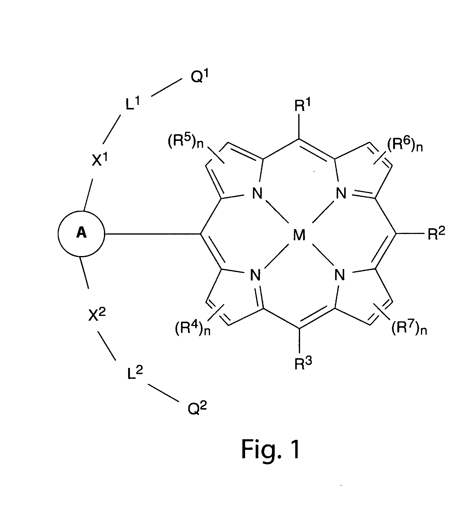
[0038]In one set of embodiments, agents capable of being inserted into a cell or a portion of a cell are those that include one or more functional groups comprising conjugated pi-electron systems (e.g., one or more aromatic groups). Without wishing to be bound by theory, transfer of such an agent across the cell membrane may be facilitated by the delocalization of partial charges in a conjugated pi-electron systems. The aromatic group may be an aryl group, a heteroaryl group (e.g., an aromatic heterocycle), substituted derivatives thereof, and the like. Examples of aromatic groups include phenyl, pyrrole, pyridine, porphyrin, expanded porphyrin groups, or the like, optionally substituted. FIG. 1 shows an illustrative embodiment wherein an agent comprises a porphyrin or metalloporphyrin group.

Problems solved by technology

However, detailed studies of the molecular mechanisms of intracellular Zn2+ accumulation, trafficking, and function, or mechanisms for other metal ions or biological species, have been limited due to a lack of suitable methods for detection in living biological systems.
Previous methods have involved the irreversible precipitation of metal complexes, which are typically restricted to use in postmortem samples.
Fluorescent metal ion sensors have been investigated, but have been limited due to photobleaching and high background signals caused by light scattering.
Additionally, as with other optical methods, fluorescence imaging techniques have limited penetration depth and lateral range, making them unsuitable for global analysis of relatively large and opaque specimens, such as live animals.
However, for in vivo applications, many MRI techniques have been limited due to insufficient cell membrane permeability.
Moreover, some MRI agents comprise toxic metal ions, which have been shown to dissociate from the MRI agent in physiological conditions due to insufficient stability of the metal complex.

Method used


Image

  • Sensors for fluorescence and magnetic resonance imaging
  • Sensors for fluorescence and magnetic resonance imaging
  • Sensors for fluorescence and magnetic resonance imaging

Examples


example 1

[0111]The following example describes the synthesis of 2,2′-(phenylmethylene)bis(1H-pyrrole) (compound 3). A solution containing 200 mL of H2O and 50 mL of pyrrole (0.72 mol) was prepared and bubbled with argon for 15 min. A 1 -mL portion of HCl (37% aq.) was added, followed by benzaldehyde (14.2 mL, 0.14 mol) in a dropwise manner over 15 min. The solution turned brown and was maintained under argon for an additional 2 h, after which it was carefully neutralized with NH4OH and then diluted with 100 mL of H2O and extracted with CH2Cl2 (3×150 mL). The combined organic phases were dried over Na2SO4 and allowed to evaporate to remove solvent and excess pyrrole. The resulting solid mixture was dissolved in 100 mL of toluene at 80° C. and slowly cooled to room temperature to yield a first crop of product (11.0 g white powder) by filtration. The mother liquor was further concentrated to yield two more batches of product with acceptable purity. A total of 17.1 g of pure material was isolate...

example 2

[0112]The following example describes the synthesis of 5-(2,6-N-dinitrophenyl)-10,15,20-trisphenylporphyrin (2NO2-TPP) (compound 4). (Phenylmethylene)bis(1H-pyrrole) (3, 3.112 g, 14.0 mmol), freshly distilled benzaldehyde (712 μL, 7.0 mmol), and 2,6-dinitrobenzaldehyde (1.575 g, 8.0 mmol) were dissolved in a liter of dry CH2Cl2 under argon. A 1-mL portion of BF3·Et2O was injected into the reaction solution, which was then stirred at room temperature for 2 h in the dark. 2,3-Dichloro-5,6-dicyano-1,4-benzoquinone (DDQ) (6.8 g, 30 mmol) was added and the reaction was allowed to proceed under argon in the dark overnight. The solvent was removed and the resulting solid residue was dissolved in CH2Cl2 / MeOH (3:1) and then mixed with aluminum oxide (˜80 g). The mixture was evaporated to dryness. Flash chromatography using an aluminum oxide column, followed by passage through a second, silica gel column afforded 569.0 mg of a purple solid. Yield: 11.5%. 1H NMR (CD2Cl2, 400 MHz) δ:2.73 (2H, s...

example 3

[0113]The following example describes the synthesis of 5-(2,6-diaminophenyl)-10,15,20-trisphenylporphyrin (2NH2-TPP) (compound 5). A mixture of 2NO2-TPP, (4, 98.4 mg) and SnCl2 (390.1 mg) was stirred in 5 mL of 1 N aqueous HCl under argon in the dark at room temperature for 12 h and cooled in an ice bath. The solution was then carefully titrated with NH4OH until the color of mixture changed from green to brown at pH 10. The mixture was diluted with 50 mL of water and extracted with CH2Cl2 (3×50 mL). The organic layer was washed with water and brine and then dried over Na2SO4. The solvent was removed and the residue was purified by silica gel column chromatography using a gradient eluent from CH2Cl2 / hexan (4:1) to CH2Cl2 / ethyl-acetate (6:1); 77.0 mg of product was obtained as a purple solid (85.5%). 1H NMR (CD2Cl2, 400 MHz) δ: 2.75 (2H, s), 3.39 (4H, s, br), 6.64 (2H, d, J=8.1 Hz), 7.43 (1H, t, J=8.1 Hz), 7.83 (9H, m), 8.27 (6H, dd, J1=7.5 Hz, J2=1.5 Hz) 8.90 (4H, s), 8.92 (2H, d, J=...


PUM

PropertyMeasurementUnit
Wavelengthaaaaaaaaaa
Optical propertiesaaaaaaaaaa
Paramagnetismaaaaaaaaaa

Abstract

The present invention generally relates to agents and compositions having MRI and / or optical signals, and methods for their use in the determination of an analyte. In some cases, an optical, MRI, or other signal produced by the agent or composition may be affected by the presence of an analyte. Some embodiments of the present invention may provide agents or compositions which are cell permeable. Examples of analytes that may be determined by the present invention include zinc ions, calcium ions, and other biological analytes.

Description

RELATED APPLICATIONS [0001]This application claims priority under 35 U.S.C. § 119(e) to co-pending U.S. Provisional Application Ser. No. 60 / 844,956, filed Sep. 15, 2006, the contents of which are incorporated herein by reference.STATEMENT REGARDING FEDERALLY SPONSORED RESEARCH OR DEVELOPMENT [0002]This invention was made with the support under the following government contract: 5-R01-GM065519-06 awarded by the National Institutes of Health. The government has certain rights in the invention.FIELD OF THE INVENTION [0003]The present invention generally relates to agents and compositions having a magnetic resonance imaging (MRI) signal or an optical signal, and related methods.BACKGROUND OF THE INVENTION [0004]Sensors for metal ions and other species involved in biological signaling pathways and other biological functions can provide valuable information regarding the physiological roles of the metal ions and / or species and can serve as a tool in the diagnosis of a variety of pathologi...

Claims


Application Information

Patent Timeline
IPC IPC(8): A61K49/06
CPCA61K49/0002A61K49/0036A61K49/106A61K49/085A61K49/10A61K49/0052
Inventor ZHANG, XIAO-ANJASANOFF, ALAN PRADIPLIPPARD, STEPHEN J.
Owner MASSACHUSETTS INST OF TECH
Learn More