Devices and methods for imaging particular cells including eosinophils

a technology for eosinophils and apparatuses, applied in the field of apparatus and methods for imaging particular cells, can solve the problems of putting a substantial financial burden on our health care system, time-consuming and frustrating for patients and their families, and eosinophilic inflammation may lead to significant and permanent damage, etc., to facilitate the placement of rcm probe optics, facilitate the scan, and facilitate the effect of rcm

Inactive Publication Date: 2011-06-09
THE GENERAL HOSPITAL CORP
View PDF118 Cites 25 Cited by
  • Summary
  • Abstract
  • Description
  • Claims
  • Application Information

AI Technical Summary

Benefits of technology

[0027]For example, a transnasal or transoral RCM probe can be designed and fabricated based on the nasogastric (NG) tube, e.g., a predicate device. A pressure sensor can be utilized to facilitate placement of the RCM probe's optics near the gastroesophageal junction. The probe's optics can scan from the distal to proximal esophagus, creating a three-dimensional RCM image that covers a surface area of a surface area of the esophagus that may range from 0-1000 mm2. In addition, a faster and higher resolution RCM system can be provided to facilitate the scan to be completed in a few minutes.
[0028]Thus, in accordance with the exemplary embodiments of the present disclosure, it is possible to provide devices and methods which can measure esophageal eosinophils directly, and without the cost and complexities of endoscopic biopsy. The exemplary embodiments can use reflectance confocal microscopy techniques, e.g., implemented through a flexible, small-diameter (approximately 3 mm) transnasal probe. The exemplary device can be configured or structured to be deployed without sedation in the outpatient setting and operated by a technician. Once inserted, images from the entire length of the esophagus can be automatically obtained and analyzed by a computer to provide eosinophil counts. The exemplary embodiments of the present disclosure can provide a cost-effective and less invasive tissue image-based biomarker for EoE that can be used to follow these patients during their therapeutic course. Additionally, the exemplary device can be utilized to study this disease to answer questions ab

Problems solved by technology

It is generally thought that if left untreated, the eosinophilic inflammation may lead to significant and permanent damage, including fibrosis of the lamina propria, strictures, and perforations.
This process is time consuming and frustrating for patients and their families.
Because endoscopic biopsy requires conscious sedation, it can also be costly, placing a substantial financial burden on our health care system.
Patients with EoE can experience a variety of unpleasant symptoms such as chest pain, nausea, vomiting, dysphagia, food impaction, and failure to thrive.
While the natural history of this disease is not fully understood; experts fear that longstanding eosinophilic inflammation may give rise to permanent esophageal damage, leading to fibrosis of the lamina propria, esophageal narrowing, stricture, and perforation.
The main limitation of corticosteroids is that EoE universally recurs when they are withdrawn, and as a result, if corticosteroids are the only line of therapy, they must be continued throughout the duration of the disease.
However, the elemental diet is very restrictive, poorly tolerated, and may have to be administered by a feeding tube.
Methods for finding the culprit dietary antigen, such as skin prick and allergy patch testing (APT) have been proposed, but are not yet accurate enough to be recommended for managing the diets of EoE patients.
Dietary elimination is very difficult for patients to tolerate and as a result, foods are gradually reintroduced into the diet until symptoms and/or esophageal eosinophils recur.
This process is inconvenient and taxing to patients and their families.
It is also associated with a small but real risk of complications associated with endoscopy.
In addition, at an average of currently about $1000 per procedure, endoscopic biopsy for EoE monitoring can be a significan

Method used


Image

  • Devices and methods for imaging particular cells including eosinophils
  • Devices and methods for imaging particular cells including eosinophils
  • Devices and methods for imaging particular cells including eosinophils

Examples


Embodiment Construction

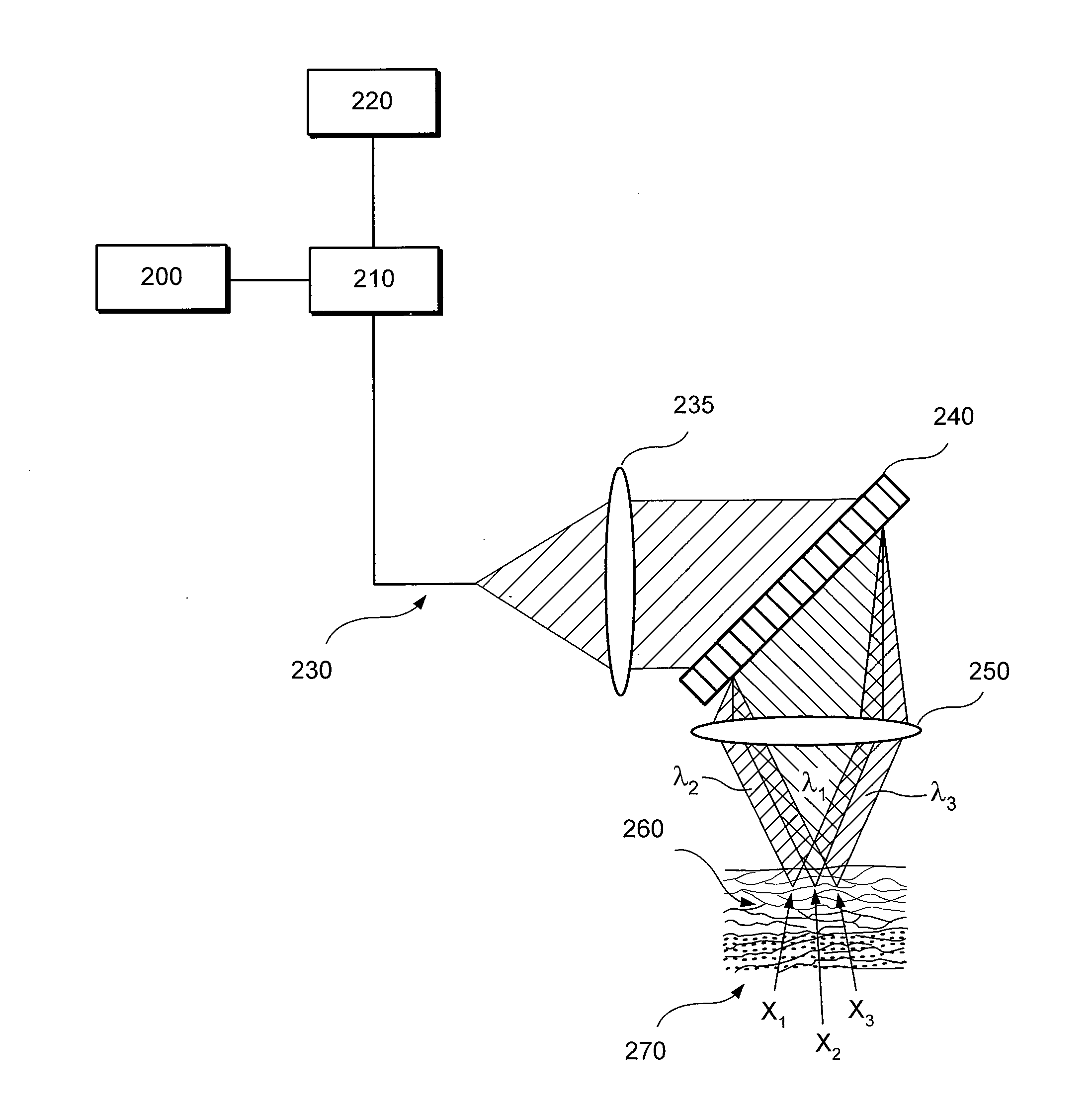
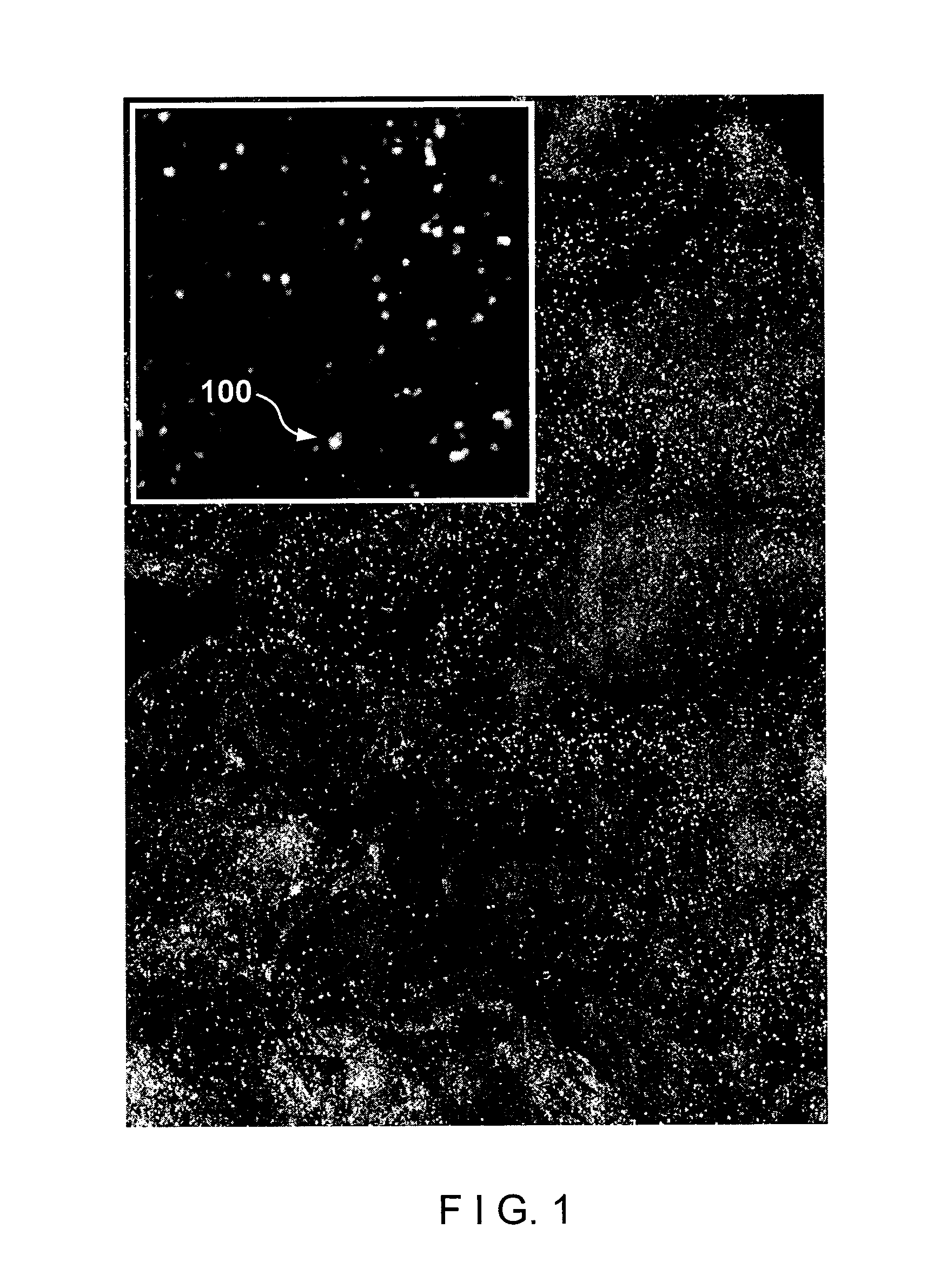
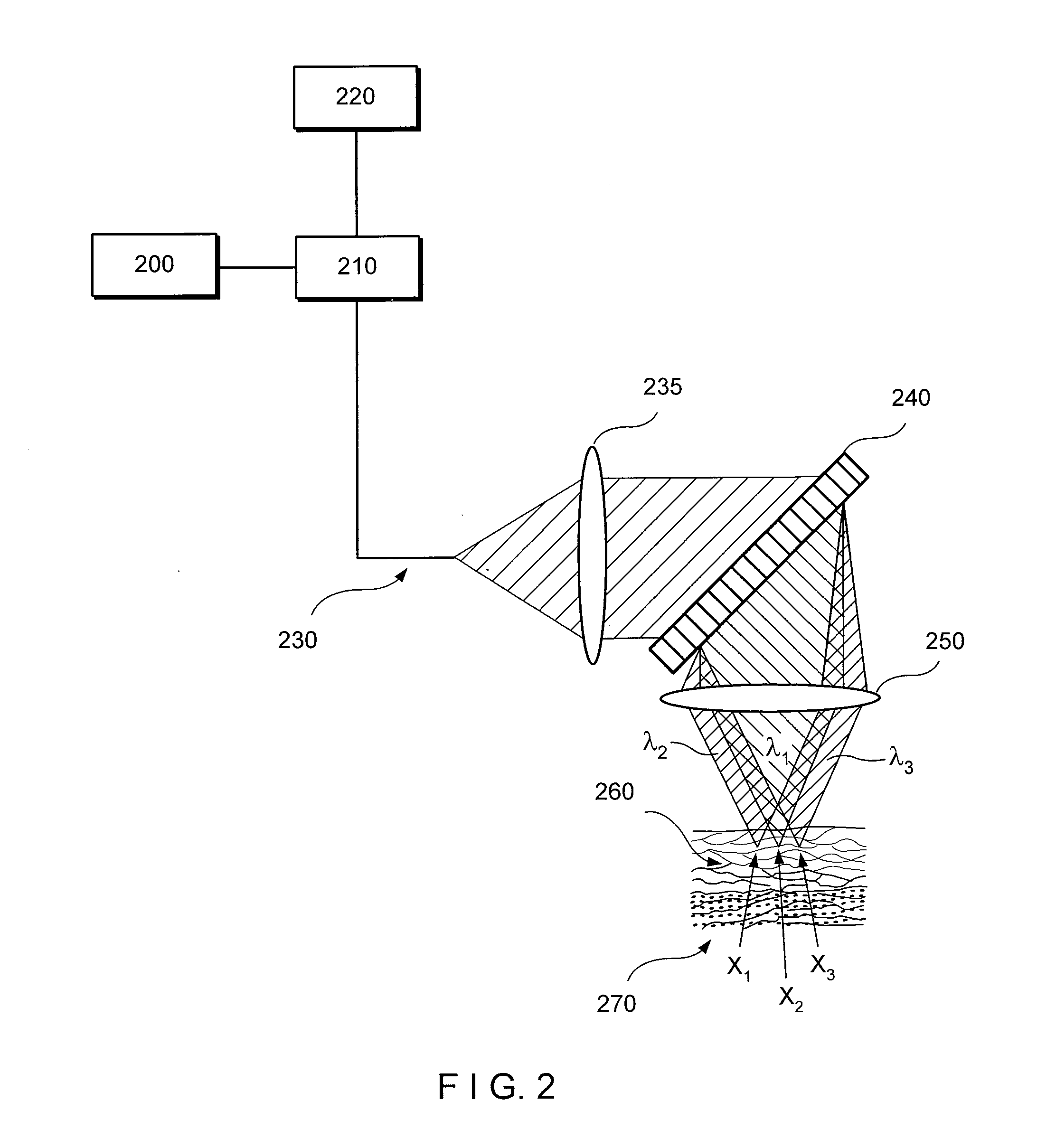
[0024]According to one exemplary embodiment of the present disclosure, exemplary RCM techniques, methods and apparatus according to the present disclosure can be configured to identify esophageal eosinophils.

[0025]One of the objects of exemplary embodiments of the present disclosure is to determine the presence or absence of esophageal eosinophils in human patients. Another object of the present disclosure is to count, in a manual, semi-automatic, or computer processing manner, the number of eosinophils in human patients. A further object of the present disclosure is to obtain cross-sectional images of tissue at the cellular level to enable the counting of eosinophils. Another object of the exemplary embodiments of the present disclosure present disclosure is to identify other cells, such as leukocytes, including mast cells, lymphocytes, basophils, and neutrophils, that may also be elevated in EoE. A further object of the exemplary embodiments of the present disclosure present discl...


PUM


Abstract

An exemplary embodiment of apparatus and method according to the present disclosure can be provided. For example, using at least one first arrangement, it is possible to direct at least one first electro-magnetic radiation to at least one portion of tissue within a body. Using at least one second arrangement, it is possible to receive at least one second electro-magnetic radiation provided from the portion, which is based on the first electro-magnetic radiation. Further, using at least one third arrangement, it is possible to differentiate at least one particular cell which is eosinophil, mast cell, basophil, monocyte and/or nutrophil from other cells in the portion based on the second electro-magnetic radiation.

Description

CROSS-REFERENCE TO RELATED APPLICATION(S)[0001]This application is based upon and claims the benefit of priority from U.S. Patent Application Ser. No. 61 / 249,207, filed on Oct. 6, 2009, the entire disclosure of which is incorporated herein by reference.FIELD OF THE DISCLOSURE[0002]The present disclosure relates to exemplary embodiments of apparatus and methods for imaging particular cells, including but not limited to eosinophils, and more particularly to such exemplary apparatus and methods which can use, e.g., confocal microscopy to image particular cells including eosinophils.BACKGROUND INFORMATIONOverview[0003]Eosinophilic esophagitis (EoE) is a disease that afflicts both children and adults, thought to be caused by food allergy, and characterized by the presence of eosinophils within the esophageal squamous epithelium. EoE is now known to be common and rapidly increasing in incidence. EoE patients can experience symptoms ranging from nausea and vomiting to dysphagia and food im...

Claims


Application Information

Patent Timeline
IPC IPC(8): A61B6/00
CPCA61B5/0062A61B5/0068A61B5/0075A61B5/0084A61B5/0086G02B21/0076A61B5/411A61B5/68A61B5/6852A61B5/6853G02B21/0064A61B5/037A61B5/0071A61B5/4233
Inventor TEARNEY, GUILLERMO J.BOUMA, BRETT E.KANG, DONGKYUNYOO, HONGKI
Owner THE GENERAL HOSPITAL CORP
Why Eureka
Try Eureka