Activation and monitoring of cellular transmembrane potentials

a transmembrane potential and activation technology, applied in the field of monitoring and manipulating cellular transmembrane potentials, can solve the problems of affecting the well-being of living organisms, affecting the quantum yield of materials, etc., and achieves high quantum yield, large stokes shift, and different optical properties.

Active Publication Date: 2012-02-09
LIFE TECH CORP
View PDF7 Cites 28 Cited by
  • Summary
  • Abstract
  • Description
  • Claims
  • Application Information

AI Technical Summary

Benefits of technology

[0019]In one aspect, a light-controlled activation platform is provided that includes one or more type of nanomaterials (e.g., semiconductor nanocrystals). In certain embodiments, the nanomaterials are capable of functioning as optical voltage sensors. In other embodiments, the nanomaterials are capable of activating changes in cellular membrane potential. In certain embodiments, the nanomaterials comprise nanocrystals (e.g., semiconductor nanocrystals).
[0020]In another aspect, compositions are provided for monitoring and manipulating cellular transmembrane potentials, comprising: a substrate and an activation platform, wherein the activation platform is disposed on a surface of the substrate, wherein the activation platform comprises one or more layers of immobilized nanocrystals. In certain aspects, the activation platform comprises two or more types of nanocrystals, wherein the two or more types have different optical properties. The nanocrystals can be semiconductor nanocrystals. Each nanocrystal can comprise a semiconductor core. The semiconductor core can further comprise a coating on the core. The coating can comprise a hydrophilic compound. Each nanocrystal can further comprise a semiconductor shell between the core and the coating, such as a positively charged compound or a negatively charged compound. The nanocrystal can comprise a surface coating, wherein the coating comprises a material having one or more thiol, sulfonate, or carboxylate groups. The coating can comprise 1-thioglycerol, thioglycolic acid, 2-mercaptoethane sulfonate, poly(acrylic acid) or a derivative thereof, lipoic acid, dihydrolipoic acid, polyethylenimine, cysteamine, polyallylamine, histidine, polyhistidine, lysine, or polylysine. The activation platform can further comprise an adhesion substrate, and the adhesion substrate can comprise poly-L-lysine, fibronectin, collagen, elastin, hyaluronic acid, laminin, matrigel, lectins, antibodies to cellular membrane proteins, or RGD peptides and their progeny. The one or more layers of nanocrystals can further comprise a polymer, such as agarose, polymethyl methacylate, or polyacrylamide. The activation platform can comprise two or more layers of immobilized nanocrystals (e.g., about 2 to about 15 layers or about 5 to about 10 layers). The composition can further include one or more layers of an organic material, such as a polymer (e.g., a synthetic polymer). The polymer can be PDDA. In certain embodiments, the nanocrystals are water-dispersible.
[0021]In yet another aspect, a method of making an activation platform is provided, comprising providing a substrate; applying one or more layers of nanocrystals onto a surface of the substrate; and applying a cell adhesion layer onto the one or more layers of nanocrystals. The one or more layers of nanocrystals can be applied onto the surface of the substrate by any method including spin casting, drop casting, roll coating, drop on demand inkjet printing, PDMS (polydimethylsiloxane) stamp printing, electrostatic layer by layer assembly.
[0022]In anothe

Problems solved by technology

Ion channels are involved in many vital functions, and a dysfunction of ion channels caused by changes in biochemical regulation, expression levels, or structural mutations can adversely impact the well-being of living organisms.
In humans, inherited or induced changes in ion channel function could result in serious complications to health.
Although this technique allows some detailed biophysical characterization of ion channel physiology, throughput is quite low and ease-of-use of patch-clamp instrumentation is generally unsatisfactory

Method used


Image

  • Activation and monitoring of cellular transmembrane potentials
  • Activation and monitoring of cellular transmembrane potentials
  • Activation and monitoring of cellular transmembrane potentials

Examples


example 1

Membrane Labeling with Quantum Dots

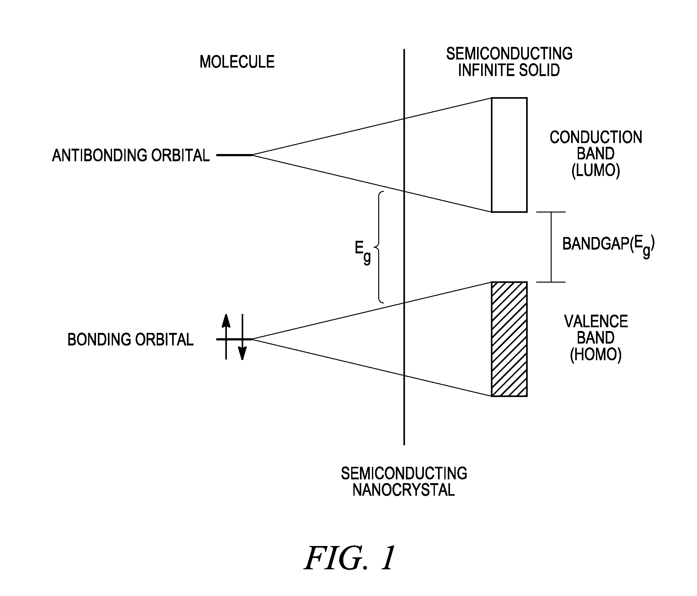
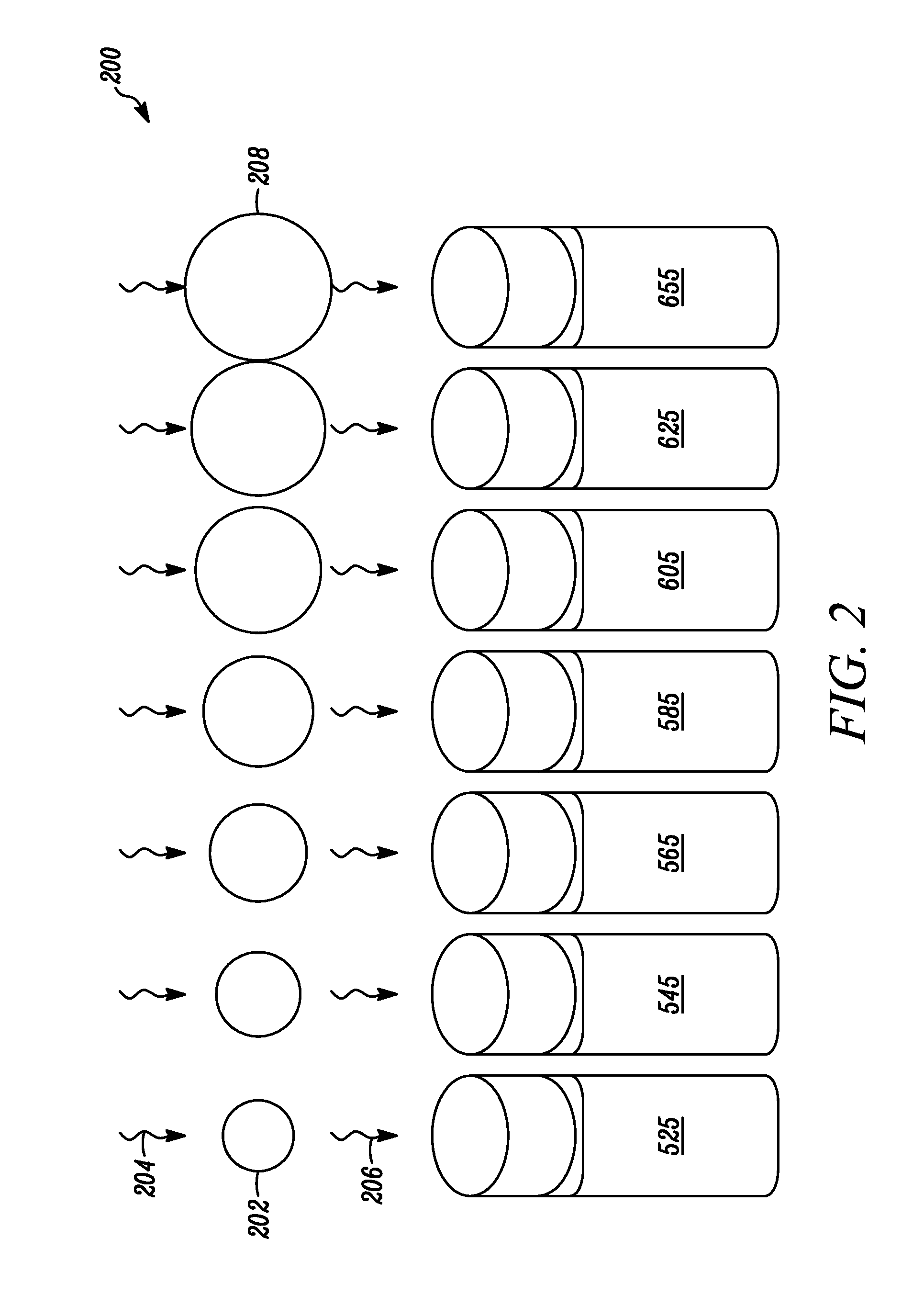
[0126]Quantum dots are ideal candidates for use as voltage-sensitive probes, as their physical size is comparable with the thickness of the cell membrane, and their electronic properties make them potentially tunable to the external electromagnetic field. Referring to FIG. 6A, the absorption of a photon 602 having a first wavelength and with energy higher than the band gap 604 between the HOMO valence band 606 and the LUMO conduction band 608 of a quantum dot forms electron-hole pairs than can radiatively recombine to emit a photon 610 having a second wavelength. An intense local electric field (e.g., due to a change in the membrane potential across the lipid bilayer of a cell) can interact with the free charge carriers generated by excited quantum dots. FIG. 6B illustrates schematically how an electrical field 612 can modulate the optical properties of the emitted light 614. For example, a membrane potential of 100 mV across the hydrophobic lipid ...

example 2

Screening Assays of Quantum Dots

[0128]QDOT nanocrystal-based optical voltage sensors are provided to monitor rapid changes in cell membrane potential. When localized inside the cellular membrane, the nanoparticles are sensitive to and detect changes in transmembrane voltage gradient and report it as the change in their emission properties. A series of quantum-dots were prepared and screened to monitor their optical properties in response to chemical and electrical stimulation of cells.

[0129]A. Effect of Chemical Stimulation

[0130]The effect of chemical stimulation on cells treated with quantum dots was monitored according to the following protocol. Cells were incubated with nanocrystal-based voltage sensors in 96-well plates for 1 hour, and then any excess nanoparticles were washed away. A population of labeled cells was depolarized using a chemical stimulus (100 mM KCl), appropriate for high-content imaging experiments. FIG. 10 shows the emission intensity (expressed as relative flu...

example 3

Quantum Dot-Based Activation Platforms

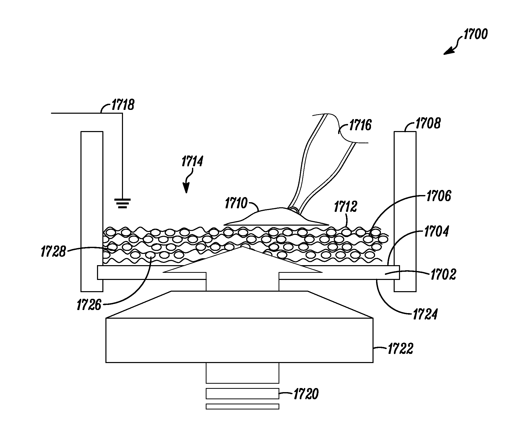
[0134]A light-controlled activation platform is described for remote reversible manipulation of the membrane potential. The described materials and methods provide an alternative to passive monitoring of the cell functional activity, and provide the ability to non-invasively manipulate the membrane potential of cells, which is vital for understanding their development, communications and fate. Traditional non-physiological methods rely most commonly on pharmacological intervention via addition of “high K+” solution or pharmacological channel openers or direct stimulation of cells with microelectrodes. These non-physiological methods, however, can have serious shortcomings, including the lack of control of the membrane potential, irreversibility of elicited changes, and low temporal resolution.

[0135]The alternative approach provided herein uses light to trigger remote reversible manipulation of the membrane potential in a temporally precise and s...


PUM


Abstract

The use of nanostructures to monitor or modulate changes in cellular membrane potentials is disclosed.

Description

CROSS REFERENCE TO RELATED APPLICATIONS[0001]This application claims priority to U.S. Provisional Application No. 61 / 059,155, filed Jun. 5, 2008, the disclosure of which is incorporated herein by reference in its entirety.FIELD OF THE INVENTION[0002]The invention relates to compositions and methods useful for monitoring and manipulating cellular transmembrane voltages. In particular, nanoparticles and their use in monitoring and manipulating transmembrane voltages are disclosed.DESCRIPTION OF RELATED ART[0003]Cells have phospholipid membranes that serve as bimolecular barriers that separate cell contents from the extracellular environment. The phospholipid membranes maintain the necessary compositional differences between cellular compartments and the extracellular milieu by regulating the passage of materials through the membrane as a function of intracellular signaling. The normally impermeable phospholipid membrane has a resting membrane potential originating from an unequal dist...

Claims


Application Information

Patent Timeline
IPC IPC(8): G01N33/566B05D5/00B05D1/36
CPCB82Y5/00B82Y15/00G01N2500/10G01N33/6872G01N33/588
Inventor IGNATIUS, MICHAEL J.MOLOKANOVA, ELENASAVTCHENKO, ALEXEIBARTEL, JOSEPH A.ZHAO, WEIWENTREADWAY, JOSEPH A.
Owner LIFE TECH CORP
Why Eureka
Try Eureka