Dendrimer-bioadhesive polymer hydrogel nanoglue and use thereof

a technology of bioadhesive and nano-glue, which is applied in the field of dendrimerbioadhesive polymer hydrogel nanoglue, wound repair and treatment of disorders, can solve the problems of requiring removal, uneven healing, and invasive procedures, and achieves the effects of preventing infection and scar tissue formation, reducing the time window of surgery, and reducing the risk of infection

Pending Publication Date: 2020-06-04
THE JOHN HOPKINS UNIV SCHOOL OF MEDICINE
View PDF0 Cites 3 Cited by
  • Summary
  • Abstract
  • Description
  • Claims
  • Application Information

AI Technical Summary

Benefits of technology

[0013]Dendrimer nanoglues have been developed which both seal and provide for controlled, sustained local release of therapeutic, prophylactic and / or diagnostic agent. The nanoglue has desirable mechanical properties to hold the tissue. It also can be used to deliver agents accelerating wound healing and delivering antibacterial and anti-inflammatory drugs to prevent infection and scar tissue formation. The formulation is surgeon friendly. It is a viscous liquid during application and polymerizes rapidly upon laser illumination, providing a longer time window for surgeons to work with. The strength of the nanoglue can be modulated by the duration of laser application or by altering the percentages of individual components of the nanoglue.
[0014]Other materials can be incorporated into the formulations to increase flexibility, strength or control drug delivery properties. An example is hyaluronic acid to increase strength and flexibility. Therapeutic, prophylactic and / or diagnostic agents can be incorporated directly, bound to the dendrimers, or formulated into particles, to enable sustained release of therapeutic agents such as steroids and antibacterial drugs (antibiotic drugs). This can be used to reduce eye drop usage, thereby reducing side-effects.
[0015]The formulation has multimodal applications and can be modified for administration for different locations or purposes, for example, as a looser gel for subconjunctival administration to increase drug availability and aid sustained drug release for various corneal and anterior segment diseases such as corneal inflammation, corneal neovascularization, corneal graft rejection, and possibly iritis and anterior uveitis. The percentage of individual components is modified with appropriate dilution to form transparent flexible drug depot gels that can be injected intravitreally for sustained release of drugs for a variety of posterior segment diseases such as diabetic retinopathy, choroidal neovascularization (CNV) secondary to age related macular degeneration (AMD) and other retinal pathologies. Using hyaluronic acid as the major component in the modified formulation integrates well with the vitreous gel, causing dendrimer with drugs to release from the gel and delivering drugs to targeted retinal cells for enhanced and long-term efficacy resulting in significant decreased frequency in intravitreal injections.
[0016]The formulation provides advantages for treatment of conditions requiring frequent eye drops that often cause ocular surface irritation and toxicity and may have a better safety profile in terms of intraocular pressure (TOP) and patient discomfort. The subconjunctival gels have reduced side effects as compared to eye drops. The photocrosslinkable dendrimer-hyaluronic acid based nanoglues are useful in sealing corneal incisions and simultaneously releasing antibiotic / steroids for prevention of infection and inflammation and to accelerate corneal wound healing. In addition to the benefits of being able to be applied in situ and photo-cured in a tailored manner to provide a high crosslink density through the use of the dendrimer; the inclusion of hyaluronic acid in the gel promotes wound healing and integrates into corneal stroma; while simultaneously releasing antibiotics / steroids to address infections / inflammation in a sustained manner. The formulation is also transparent, which is clearly beneficial in ocular applications.
[0017]In preferred embodiments, the nanoglue is administered to seal corneal tissue and / or administered to treat various corneal and anterior segment disorders such as corneal inflammation, corneal neovascularization, and complications with corneal graft procedures, iritis, and uveitis. The nanoglue improves the healing of corneal incision, compared to that treated with sutures or non-treated. It also withstands the high intraocular pressure of the wounded eye compartments, avoids leakage, adheres strongly to the cornea, and results in less corneal edema or fibrosis, compared to sutures. The healing of the cornea is more rapid and has less scarring, inflammation, and neovascularization compared to treatment with sutures.

Problems solved by technology

However, sutures have a number of disadvantages, including invasive procedures, uneven healing, propensity to become loose and / or broken, and often requiring removal by a skilled practitioner.
Furthermore, suturing may cause inflammation, increase the risk for infection and, in the case of corneal wounds, may lead to neovascularization and induce astigmatism.
Previous studies have explored hydrogel adhesives, but they are disfavored due to lack of biocompatibility, lack of tensile strength, and limited sites for application (Grinstaff M W, et al., Biomaterials, 28(35):5205-5214 (2007); Grinstaff M W, et al., Chemistry-A European Journal, 8(13):2838-2846 (2002)).
However, the drugs are often cleared through tear secretions, and therefore repeated dosing is required which may lead to patient non-compliance, discoloration of cornea, elevated intraocular pressure, and corneal irritation and toxicity (Gibson J M, et al., US Ophthalmic Review, 8(1):2-7 (2015); Mahajan H S, et al., Carbohydrate Polymers, 122:243-247 (2015)).

Method used


Image

  • Dendrimer-bioadhesive polymer hydrogel nanoglue and use thereof
  • Dendrimer-bioadhesive polymer hydrogel nanoglue and use thereof
  • Dendrimer-bioadhesive polymer hydrogel nanoglue and use thereof

Examples


example 1

on of Photocrosslinked, Dendrimer-Hyaluronic Acid Hydrogel with Covalently Attached Dendrimer-Drug Conjugates

[0116]Materials and Methods

[0117]Modification of Dendrimer with Methacrylate (D-MA)(Formulation 1)

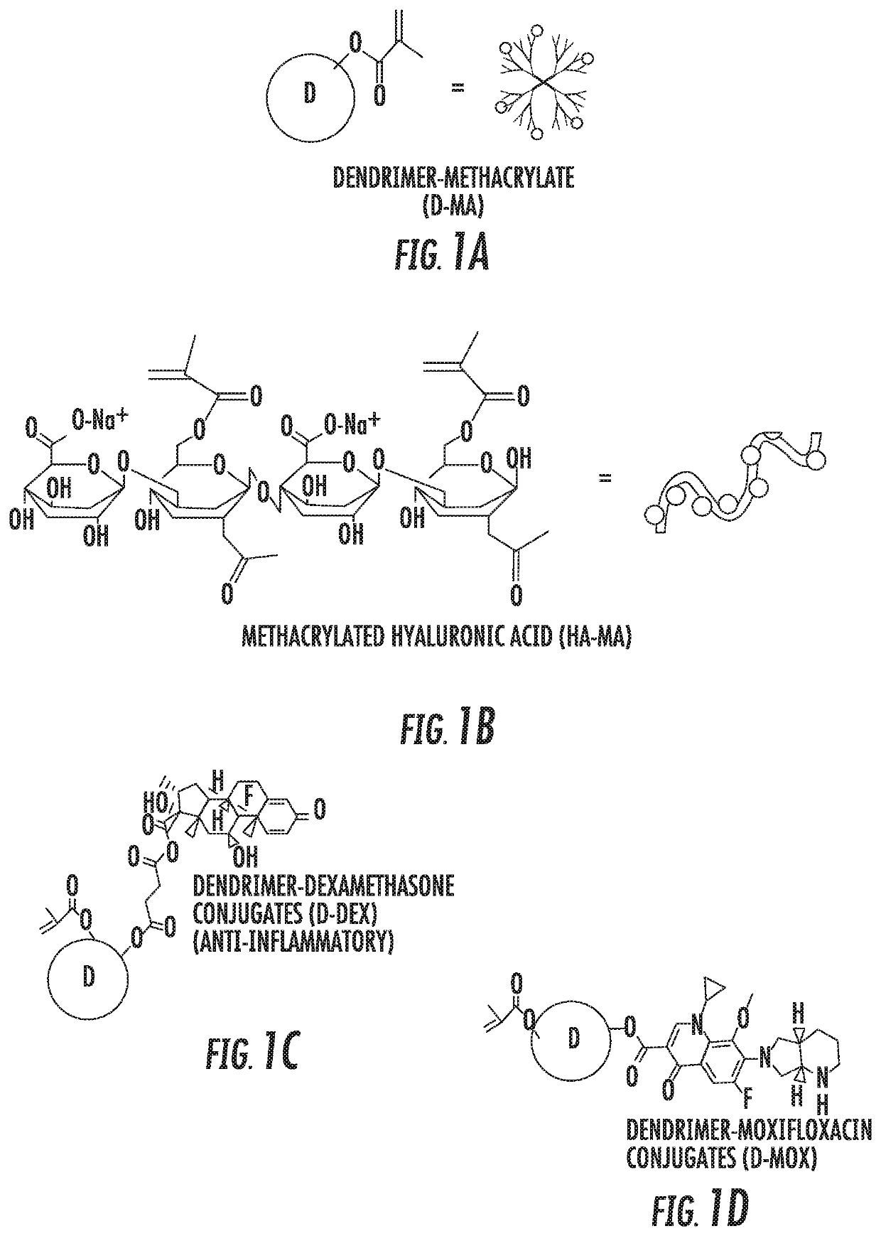
[0118]The surface of PAMAM G2 and G4 hydroxyl dendrimers are modified with methacrylate groups thereby enabling photo crosslinking (Component 1). Photo crosslinkable dendrimers (G2, G3, G4 and G6) are synthesized by adopting and optimizing previously established lab procedures. Briefly, methacrylic acid was covalently conjugated to the surface groups of the dendrimer using PyBOP / DIEA coupling reaction and the resultant product was purified and dialyzed using water. The formation of the product (D-MA) was confirmed using 1H NMR and HPLC (FIG. 1A).

[0119]Synthesis of Photo-Crosslinkable, Dendrimer-Drug Conjugate (Component 2)

[0120]Synthesized dendrimer dexamethasone conjugates with photocrosslinkable groups (MA-D-Dex) (G2-G6) were made that can enhance wound healing and prevent corn...

example 2

nd Mechanical Properties of Nanoglue Compared to Sutures in Ex Vivo Wounded Rabbit Eyeballs

[0132]Materials and Methods

[0133]Different types of corneal incisions were created on freshly enucleated mature rabbit eyeballs. We used different wound architecture and measured the burst pressure using custom designed manometer system with saline infusion. The nanoglue formulation with premixed formulations were applied on corneal incisions with different wound architecture and a argon green laser (325 nm) (for methacrylate system, formulation 1) or cobalt blue light (for thiol-ene click chemistry, formulation 2) was applied for 20-30 seconds resulting rapid photocrosslinked transparent ocular bandage that can withstand high intraocular pressures than compared to sutures. Briefly, a 3 mm linear incision was created in the central cornea resulting in a gaping wound. A tunnel incision was created. A 3-mm trephine central incision was created. The burst pressure with saline infusion was measure...

example 3

nd Mechanical Properties of Nanoglue Compared to Sutures in In Vivo Wounded Rat Cornea

[0136]Materials and Methods

[0137]In-vivo evaluation the rats were anesthetized with and pupils were dilated with tropicamide and 10% phenylephrine. A 2.75-mm keratome was used to create a full thickness central corneal incision. The blade was initially directed posteriorly, and after the penetration it was directed to the angle to avoid damaging the lens and its capsule. The incision was then either sutured (10-0 prolene or nylon) or glued with above mentioned nanoglue. Following the suturing / gluing, the corneal incision was confirmed to be closed with fluorescein and cobalt blue light.

[0138]Follow-up was performed 24, 72 hours, 7 days and 2 weeks following surgery. The rat corneas were imaged using optical coherence tomography (OCT) on day 7. The animals were clinically assessed for wound healing until day 14.

[0139]Results

[0140]Following the suturing / gluing with nanoglue, the corneal incision was ...


PUM

PropertyMeasurementUnit
pHaaaaaaaaaa
pHaaaaaaaaaa
temperatureaaaaaaaaaa

Abstract

A nanoglue is formed with one or more bioadhesive polymers, one or more dendrimers, and optionally one or more therapeutic, prophylactic, or diagnostic agents. The bioadhesive polymers and dendrimers are modified with functional groups to permit crosslinking upon one or more stimuli, e.g., ultraviolet irradiation, and form hydrogel in situ at tissue sites. In the repair of corneal wounds, the nanoglue leads to improved rate of healing with less scarring and less inflammation, compared to non-treated cornea or ones treated with sutures. Therapeutic agents can be covalently conjugated to the precursor components and be delivered to specific eye compartments, providing a more efficacious treatment formulation of ocular disorders than delivering drugs in their free forms. Methods of making and using the hydrogel and hydrogel precursor compositions are also provided.

Description

REFERENCE TO RELATED APPLICATIONS[0001]This application claims the benefit of U.S. Provisional Patent Application No. 62 / 292,741, filed on Feb. 8, 2016, which is hereby incorporated by reference for all purposes as if fully set forth herein.INCORPORATION-BY-REFERENCE OF MATERIAL SUBMITTED ELECTRONICALLY[0002]The instant application contains a Sequence Listing which has been submitted in ASCII format via EFS-Web and is hereby incorporated by reference in its entirety. Said ASCII copy, created on Feb. 8, 2017, is named P13910-02_ST25.txt and is 2,544 bytes in size.FIELD OF THE INVENTION[0003]The present invention relates to the field of hydrogel adhesives, and more particularly, to wound repair and treatment of disorders.BACKGROUND OF THE INVENTION[0004]Repair of wounds after traumatic or surgical injury is of significant clinical importance. These wounds are often sealed with sutures. However, sutures have a number of disadvantages, including invasive procedures, uneven healing, prop...

Claims


Application Information

Patent Timeline
Patent Type & Authority Applications(United States)
IPC IPC(8): A61L24/08A61L24/00C08G83/00A61K31/573A61K9/00A61K9/06
CPCA61K45/06A61L24/08A61K47/58C08G83/004A61K47/61A61K9/0048A61K9/0019A61K9/06B82Y30/00A61K31/573A61L24/0015A61L2300/222A61L24/0031A61L15/58A61L24/0042A61L2400/06A61L2400/12A61K47/6903C08J3/075C08J3/246C09J201/005C08L101/005C08J2300/202C08J2305/08A61P17/02A61P27/02A61P27/14C08L5/08
Inventor RANGARAMANUJAM, KANNANSTARK, WALTERKAMBHAMPATI, SIVA PRAMODHSOIBERMAN, URIYIU, SAMUELAL-TOWERKI, ABDUL-ELAH ABAD
Owner THE JOHN HOPKINS UNIV SCHOOL OF MEDICINE
Why Eureka
Try Eureka