Peptide-based scaffolds for cartilage regeneration and methods for their use

a cartilage regeneration and peptide-based technology, applied in the direction of peptides, drug compositions, peptide/protein ingredients, etc., can solve the problems of poor cell attachment, poor integration into the site where tissue engineered materials are utilized, and the preparation of any synthetic material with a nanoscale structure that mimics natural tissue is a challenging problem, so as to facilitate nanofiber assembly and growth factor binding, improve biocompatibility, and facilitate the effect of nanofiber assembly

Active Publication Date: 2013-05-28
NORTHWESTERN UNIV
View PDF138 Cites 18 Cited by
  • Summary
  • Abstract
  • Description
  • Claims
  • Application Information

AI Technical Summary

Benefits of technology

[0013]TGF-β1 has a significant role in the regulatory network of growth factors that maintains articular cartilage in the differentiated phenotype.41 Additionally, TGF-β1 is a necessary and critical factor for inducing chondrogenesis in marrow-derived MSCs.42 In articular cartilage tissue engineering, TGF-β1 has been shown to increase collagen and proteoglycan production and inhibit matrix breakdown.43 Though not intending to be bound by theory, the addition of a TGF-binding peptide to a peptide-amphiphile nanofiber scaffold can enhance the chondrogenic influence of the scaffold in vivo. As demonstrated herein, when a self-assembled peptide scaffold is applied to an articular cartilage defect after microfracture, which allows bone marrow containing MSCs to infiltrate the defect, the scaffold successfully promotes cartilage matrix production and the regeneration of hyaline-like cartilage.
[0017]It is, furthermore, an object of the present invention to provide a peptide-amphiphile (PA) molecule as described above, wherein the spacer segment includes the amino acid sequence “(Gly)mXaa(Xbb)n”, wherein m and n are integers that independently range between 0 and 5, more preferably between 1 and 3, wherein Xaa is any amino acid (serine in a preferred embodiment), and wherein Xbb is an amino acid residue selected from those with acidic side-chains, including, for example, glutamic acid (E) and aspartic acid (D). The use of this spacer peptide segment facilitates homogenous blending with other peptide amphiphiles containing amino acid residues with acidic side-chains, and provides adequate gelation strength under physiological conditions.
[0025]It is also an object of the present invention to provide mixtures of the TGF-binding peptide amphiphile and a non-specific ‘filler’ peptide amphiphile. Mixing in this fashion enables the concentration of the TGF-binding peptides to be varied independently and in a controlled manner from the overall concentration of peptide amphiphile nanofibers. While not intending to be bound by theory, mixing or ‘diluting’ the TGF-binding peptide with a filler PA may facilitate nanofiber assembly and growth factor binding to the self-assembled structure. Furthermore, appropriate selection of the ‘filler’ PA may lead to improved biocompatibility with chondrogenic cell types, more homogenous blending with the TGF-binding PA and improved gelation under physiological conditions; however, determining the optimum combination involves only routine experimentation and thus is well within ordinary skill. In a preferred embodiment, the filler PA has the following structure:H3C(CH2)nCO—(Xxx)x—(Xyy)y—(Xzz)z where Xxx and Xyy are amino acids with non-polar side-chains, Xzz is an amino acid with an acidic side-chain, n is an integer ranging from 4 to 20, and x, y, and z are integers that can range independently from 2 to 6. In a more preferred embodiment, Xxx is valine, Xyy is alanine, Xzz is glutamic acid and n=14. An even more preferred embodiment of the filler PA has the peptide sequence VVVAAAEEE (SEQ ID NO:7). When conjugated to a preferred lipophilic segment this peptide amphiphile has the following structure:

Problems solved by technology

However, most polymers used today to create these scaffolds, such as polylactic acid, polyorthoesters, and polyanhydrides, are difficult to control and result in, among other things, poor cell attachment and poor integration into the site where the tissue engineered material is utilized.
The preparation of any synthetic material with structure on the nanoscale that mimics natural tissue is a challenging problem.
Untreated articular cartilage lesions lead to pain, dysfunction, and accelerated osteoarthritis.
Focal articular cartilage lesions have limited regenerative potential.
The regenerative potential of untreated articular cartilage is limited to the formation of a fibrocartilage scar.
Clinical and histological outcome studies of these techniques have demonstrated varying, and often confounding clinical results.
The limited self-healing capability of articular cartilage is largely due to the nature of the tissue.
First, the avascularity of articular cartilage cannot support the formation of a fibrin clot.
Second, the dense extracellular matrix (ECM) of articular cartilage restricts chondrocyte migration to the defect space.
Third, chondrocytes have low mitotic activity, which results in insufficient cell proliferation and matrix synthesis for complete regeneration.
With this limited natural healing capability, clinical intervention is necessary to prevent further articular cartilage degradation and early progression of degenerative osteoarthritis.
Additionally, there may be a paucity in the quantity of the extracellular matrix and availability of endogenous growth factors to induce chondrocyte differentiation.
These techniques require a two-stage surgery, with a significantly higher cost and increased morbidity from the harvest site.
In a recent systematic review of treatment of focal knee articular cartilage defects Magnussen et al. conclude that there is not one technique that is superior to another in clinical or histological evaluation.38 Clearly no clinical solution has been found that can provide a large percentage of patients with excellent outcomes at long term follow-up.

Method used


Image

  • Peptide-based scaffolds for cartilage regeneration and methods for their use
  • Peptide-based scaffolds for cartilage regeneration and methods for their use
  • Peptide-based scaffolds for cartilage regeneration and methods for their use

Examples


example 1

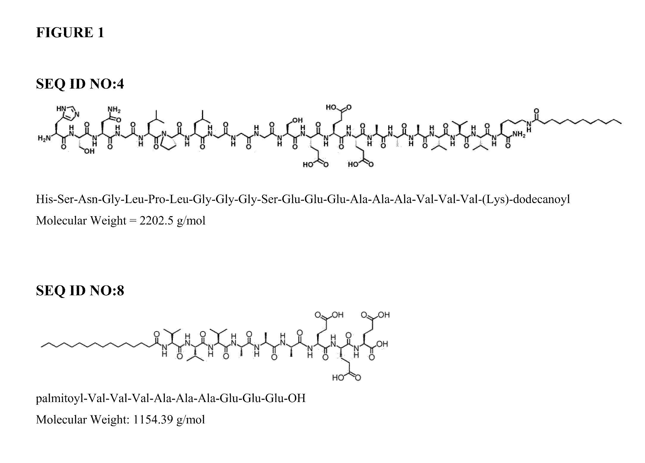
Automated Synthesis and Purification of Peptide Amphiphiles Containing the TGF-Binding Segment HSNGLPL (SEQ ID NO:1), Including SEQ ID NO:4

[0121]1.1 Reagents:

[0122]The following reagents, or equivalents, were used as received: HBTU (2-(1H-Benzotriazol-1-yl)-1,1,3,3-tetramethyluronium hexafluorophosphate), piperidine, DIEA (n,n,-diisopropylethlamine), DMF (n,n-dimethylformamide), DCM (dichloromethane), TFA (trifluoroacetic acid), TIS (triisopropylsilane). All water was purified by reverse osmosis and filtered using a Millipore™ system to a resistivity of 18.2 Mohm-cm. 9-Fluorenylmethoxycarbonyl (Fmoc) protected amino acids and orthogonally protected Fmoc-Lys(Mtt)-OH were purchased from EMD Biosciences (La Jolla, Calif.). Peptides were synthesized on Rink amide resin (loading 0.6-0.75 mmole / g).

[0123]1.2 Peptide Synthesis:

[0124]Peptides were synthesized via solid-phase methodology on an automated peptide synthesizer (CS Bio Co. model 136XT), using a 250 mL glass reaction vessel which w...

example 2

Synthesis of Filler Peptide Amphiphile C16H31O-VVVAAAEEE (SEQ. ID NO:8)

[0133]The filler PA was synthesized via automated solid phase peptide synthesis as described above, except a pre-loaded glutamic acid Wang resin was used. Additional amino acids were coupled as described, proceeding from C- to N-terminus. After coupling of the N-terminal valine, the peptide was capped with a palmitoyl moiety, which was accomplished as described in Example 1.2 by substituting palmitic acid for dodecanoic acid. Peptide cleavage from the resin, HPLC purification, aseptic filtration and lyophilization were performed as described in Examples 1.3-1.6.

example 3

Chemical Characterization of Peptide Amphiphiles

[0134]Peptide amphiphiles were characterized as to identity and purity using mass spectroscopy (MS), high performance liquid chromatography (HPLC), and amino acid analysis (AAA), as described below.

[0135]3.1 Mass Spectroscopy

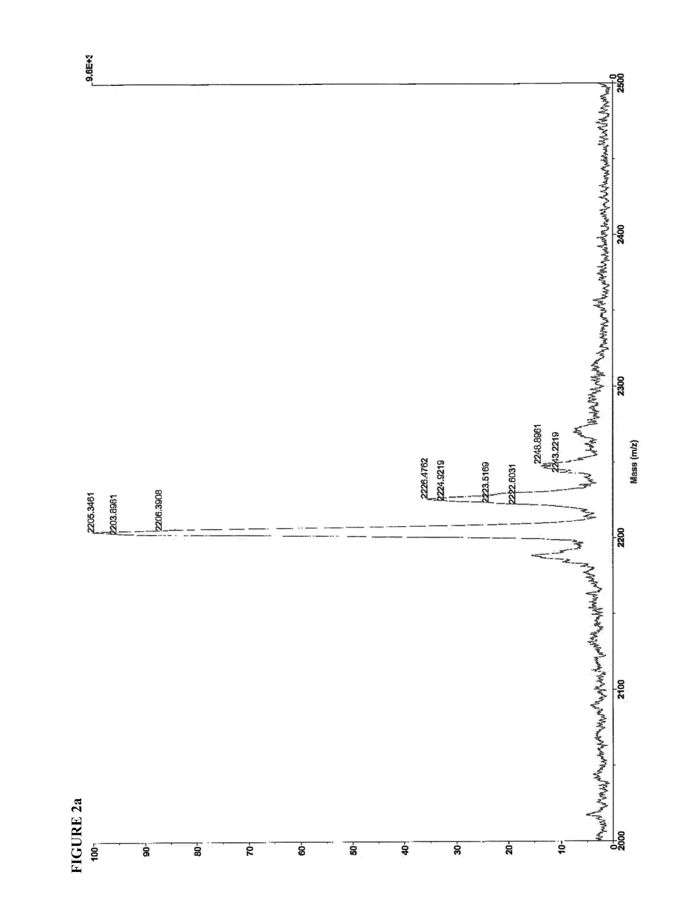
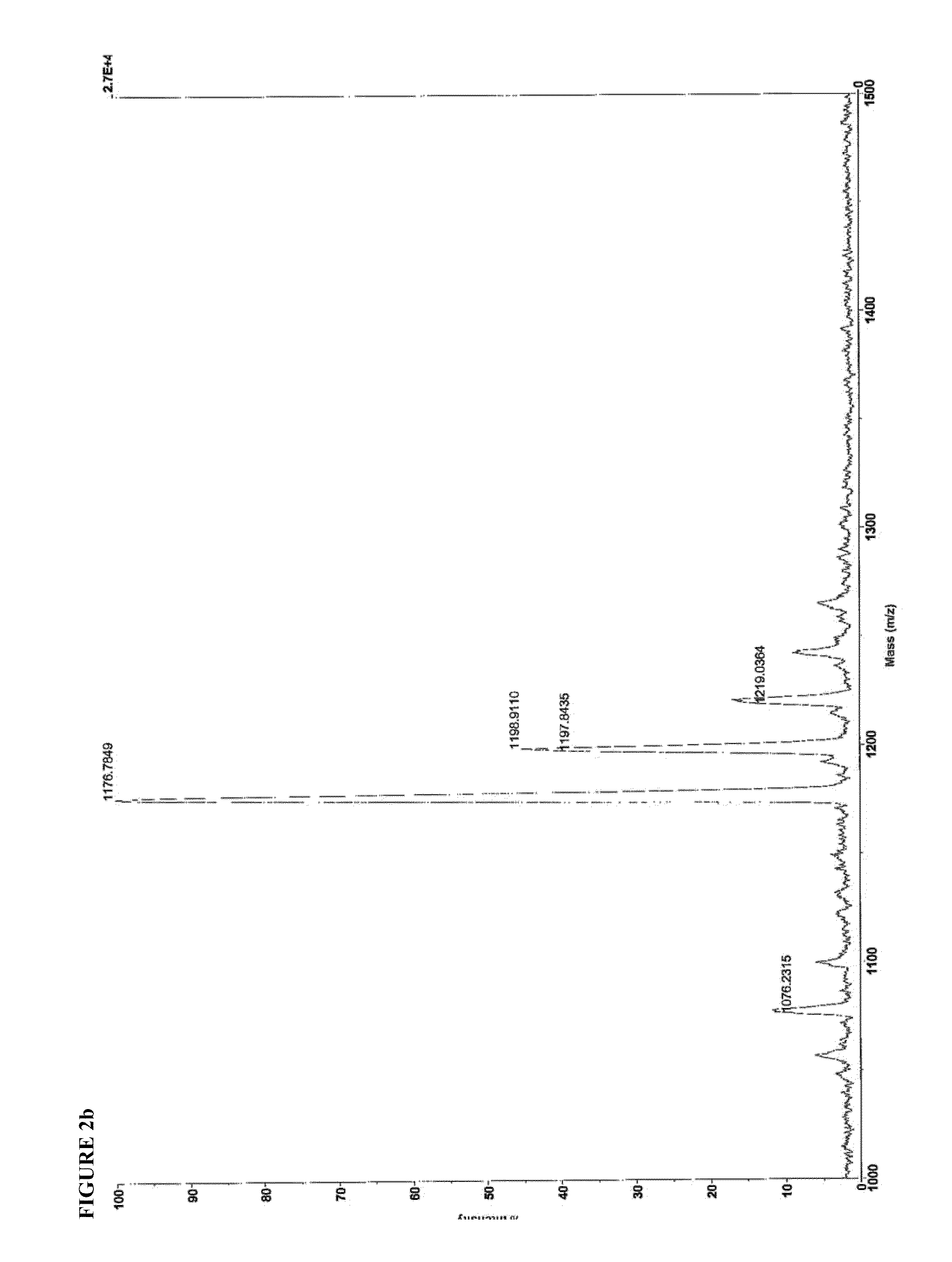
[0136]Identity of SEQ ID NO:4 and SEQ ID NO:8 were confirmed by matrix-assisted laser desorption-ionization time-of-flight mass spectroscopy (MALDI-TOF MS), using a Voyager DE-PRO instrument operating in positive ion mode, with CHCA matrix. 1 uL of a 1 mg / mL solution was spotted onto the MALDI plate. The mass spectra are shown in FIG. 2.

[0137]

SEQ ID. NO: 4Expected m / z = 2201.22Found m / z =2203.90[M + H+]2226.48[M − H + Na+]2243.22[M − H + K+]2248.90[M − H + 2Na+]SEQ ID. NO: 8Expected m / z = 1153.68Found m / z =1176.78[M − H + Na+]1198.91[M − H + 2Na+]1198.92[M − H + 3Na+]1076.23Valine deletion

[0138]3.2 Analytical HPLC

[0139]Analytical, reverse-phase HPLC was performed to determine the peptide purity of the molecules syn...


PUM

PropertyMeasurementUnit
diameteraaaaaaaaaa
pHaaaaaaaaaa
flow rateaaaaaaaaaa

Abstract

Disclosed herein are novel peptide amphiphile molecules and compositions composed of a peptide sequence that non-covalently binds the growth factor TGF-β1. Also disclosed are methods of using these peptide amphiphiles to create a gel scaffold in situ that enhances articular cartilage regeneration when used in combination with microfracture. Significant improvement in tissue quality and overall O'Driscoll histological scores were observed in rabbits with full thickness articular cartilage defects treated with the TGF-binding peptide amphiphile. The gel can further serve as a delivery vehicle for recombinant TGF-β1 protein growth factor. Scaffolds that localize and retain chondrogenic growth factors may synergistically enhance cartilage repair when combined with microfracture, by inducing bone marrow mesenchymal stem cells into chondrogenic differentiation. This invention represents a promising new biomimetic approach to enhance current techniques of articular cartilage regeneration in the clinical setting.

Description

RELATED APPLICATIONS[0001]This application claims priority from U.S. Provisional Application No. 61 / 168,894, filed Apr. 13, 2009, which is incorporated herein by reference in its entirety.FIELD OF THE INVENTION[0002]The present invention relates generally to new peptide-based gels and their therapeutic use to enhance articular cartilage regeneration. More particularly, the present invention relates to a mixture of self-assembling peptide amphiphiles (PA) having a peptide epitope sequence capable of non-covalently binding to transforming growth factor beta (TGF-β1). Hydrogel scaffolds composed of this peptide amphiphile and autologous cells are shown to enhance the regeneration of articular cartilage in an in vivo rabbit model. This is the first example of a self-assembling peptide amphiphile gel that can be used alone or as a therapy to augment current orthopedic strategies used clinically for cartilage defect repair, as in the context of microfracture or autologous chondrocyte tran...

Claims


Application Information

Patent Timeline
Patent Type & Authority Patents(United States)
IPC IPC(8): A61K38/18A61K38/00A61K38/04C07K14/495C07K5/00A61P19/00
CPCA61K38/1841C07K7/06C07K14/00A61K2300/00A61P19/00A61P19/02A61K38/04C07K7/08
Inventor SHAH, RAMILLE N.SHAH, NIRAV A.STUPP, SAMUEL I.
Owner NORTHWESTERN UNIV
Why Eureka
Try Eureka