
Stable and easy-to-store vaginal secretion microbial cell fluorescence detection dye liquor

A technology of vaginal secretions and microbial cells, applied in the field of medical diagnosis, can solve the problems of many interference factors, easy missed detection, complex background, etc.

Pending Publication Date: 2021-03-19
广州翰德泽信医药科技有限公司
View PDF0 Cites 3 Cited by
  • Summary
  • Abstract
  • Description
  • Claims
  • Application Information

AI Technical Summary

Problems solved by technology

[0010] (1) Traditional wet film method: smear observation after dilution with normal saline, the wet film method is convenient and economical, and can be completed within a few minutes, but the observation of clue cells requires experience, the background is complex, it is easy to miss, and the overall positive detection rate is low and no way to check for dead trichomonas
[0011] (2) Gram staining: cells and microorganisms can be colored, and the shape after staining is clearer than that of wet slides, but trichomonas will be broken, and the background is relatively complex, so it is easy to miss or misdetect
[0012] (3) Enzyme chemistry method: It can be equipped with fully automated equipment and has a high degree of automation, but there are many interference factors of enzymes, no international authoritative report support, low clinical compliance rate, high probability of false positive and false negative, and complicated operation, which takes 45 minutes. Incubate at 37°C, cold chain transport
[0014] (5) PCR method: It has the advantages of specificity, sensitivity, high yield, quickness, simplicity, good repeatability, and easy automation, etc., but the operation requirements are high, the steps are complicated, and special PCR laboratories and professional laboratory physicians are required, and the detection cost is low. high
[0015] The key to the treatment of vaginitis is to accurately determine the pathogen of vaginitis, and microscopic examination is the gold standard for the diagnosis of vaginitis, but there are relatively many limitations in the detection of wet slides or ordinary staining. However, the general fluorescent staining solution has problems such as poor dyeing effect, fast quenching of fluorescence effect after staining, and unstable storage of the staining solution for a long time, resulting in low fluorescence intensity after staining.

Method used


Image

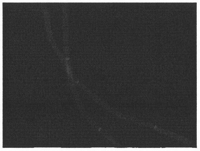
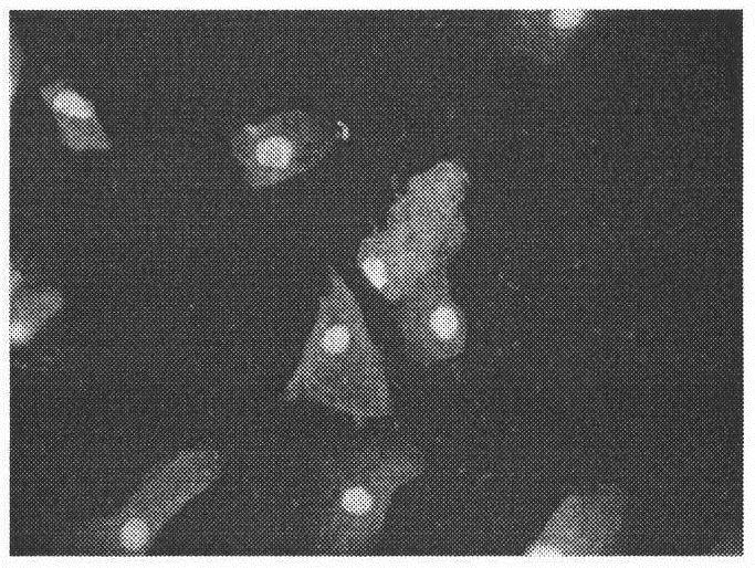
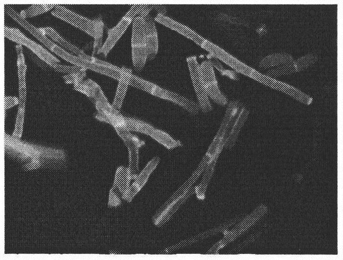
  • Stable and easy-to-store vaginal secretion microbial cell fluorescence detection dye liquor
  • Stable and easy-to-store vaginal secretion microbial cell fluorescence detection dye liquor
  • Stable and easy-to-store vaginal secretion microbial cell fluorescence detection dye liquor

Examples


Embodiment 1

[0071] Reagent 1 configuration

[0072] Liquid A:

[0073] (1) Liquid A is mixed according to the weight percentage of the following formula: 0.5 parts of fluorescent whitening agent-28, 15 parts of dimethyl sulfoxide, 0.05 parts of Evans blue, 20 parts of glycerol, 10 parts of potassium hydroxide, 54.45 parts of ionized water;

[0074] (2) Potassium hydroxide of corresponding weight is taken and dissolved in deionized water to form an aqueous solution, naturally cooled to room temperature, and set aside;

[0075] (3) Dissolve fluorescent whitening agent-28 of corresponding weight in deionized water to form an aqueous solution for use;

[0076] (4) The aqueous solution described in the above (2) (3) is mixed and set aside;

[0077] (5) take by weighing dimethyl sulfoxide and glycerin of corresponding weight and mix, stand-by;

[0078] (6) Add the mixed liquid described in (5) dropwise into the aqueous solution described in (4), stir while adding, add Evans blue after mixin...

Embodiment 2

[0085] Reagent 2 configuration

[0086] Liquid A:

[0087] (1) Liquid A is mixed according to the weight percentage of the following formula: 0.5 parts of fluorescent whitening agent-28, 15 parts of dimethyl sulfoxide, 0.05 parts of Evans blue, 2-hydroxypropane-1,2,3-tricarboxylate 5 parts of potassium hydroxide, 20 parts of glycerin, 10 parts of potassium hydroxide, 49.45 parts of deionized water;

[0088] (2) Potassium hydroxide of corresponding weight is taken and dissolved in deionized water to form an aqueous solution, naturally cooled to room temperature, and set aside;

[0089] (3) Dissolve fluorescent whitening agent-28 of corresponding weight in deionized water to form an aqueous solution for use;

[0090] (4) The aqueous solution described in the above (2) (3) is mixed and set aside;

[0091] (5) Dimethyl sulfoxide, 2-hydroxypropane-1,2,3-potassium tricarboxylate and glycerol are weighed and mixed for use;

[0092] (6) Add the mixed liquid described in (5) dropwi...

Embodiment 3

[0099] Embodiment 3 solution stability test

[0100] Reagents 1 and 2 were subjected to high temperature experiment and normal temperature experiment respectively.

[0101] (1) High temperature experiment

[0102] The test sample of the staining solution was placed at 80°C, protected from light, and taken out after one week; then the mixed sample of Candida, lactic acid, trichomonas and epithelial tissue was stained. The stability of the reagents was judged from the staining effect, and the results are shown in Table 1.

[0103] (2) Normal temperature experiment

[0104] The staining solution test samples were placed in an opaque plastic bottle and taken out after 6 months at room temperature; then the mixed samples of Candida, lactic acid, trichomonas and epithelial tissue were stained. The stability of the reagents was judged from the staining effect, and the results are shown in Table 1.

[0105] Table 1

[0106]

[0107]The results showed that after the high-temper...


PUM


Abstract

The invention discloses vaginal secretion microbial cell fluorescence detection dye liquor which is easy and convenient to operate, clear in dyeing effect, high in detection rate, not prone to precipitation, stable in system and easy to store. The vaginal secretion microbial cell fluorescence detection dye liquor comprises two reagents: a solution A and a solution B, fluorescein in the solution Acan be combined with beta-polysaccharide, such as chitin and cellulose, on fungal cell walls with high affinity, and fungal components existing in a sample are marked. The solution B is composed of water, fluorescein and a fluorescent auxiliary agent, and the fluorescein can fluorescently mark cells and pathogenic microorganisms in vaginal secretion. The anti-interference fluorescent auxiliary agents are respectively added into the reagents, so that the combination rate of fluorescent dyes, cells and microorganisms can be improved, the fluorescence intensity is enhanced, the cell and microorganism contours are clear and visible, the stability of a staining solution component system is improved, and the staining solution fluorescence quenching risk is reduced. The dye liquor is simple and rapid to operate, clean in background, clear in image and easy to judge, and the accuracy of vaginitis and other gynecological leucorrhea detection items can be greatly improved through clinical use.

Description

technical field [0001] The invention belongs to the technical field of medical diagnosis, and in particular relates to a dye solution for fluorescent detection of vaginal secretion microbial cells. [0002] technical background [0003] Vaginitis is a general term for a variety of vaginal mucosal inflammatory diseases caused by different etiologies. In normal physiological state, the tissue anatomy and biochemical characteristics of the vagina are sufficient to defend against the invasion of external microorganisms. If it is destroyed, pathogenic bacteria can take advantage of the opportunity to enter and cause vaginal inflammation through various factors. [0004] Vaginitis is the most common disease in obstetrics and gynecology patients. It can be divided into specific and non-specific vaginitis. Most of them are bacterial vaginitis, fungal vaginitis, and trichomonas vaginitis caused by Gardnerella, fungi, and trichomonas infection. . Bacterial vaginosis is one of the mo...

Claims


Application Information

Patent Timeline
IPC IPC(8): G01N1/30G01N21/64
CPCG01N1/30G01N21/6428G01N2001/302
Inventor 杨文虹李海丽
Owner 广州翰德泽信医药科技有限公司
Learn More