Sequence-based karyotyping

a karyotype and sequence-based technology, applied in the field of gene therapy, can solve the problems of limited ability to resolve detailed mutations (involving only a small part of a chromosome) and cannot be used to detect smaller alterations

Inactive Publication Date: 2005-10-06
454 CORP
View PDF5 Cites 252 Cited by
  • Summary
  • Abstract
  • Description
  • Claims
  • Application Information

AI Technical Summary

Benefits of technology

[0011] The plurality of DNA sequences obtained are mapped to a genomic scaffold to create a distribution of mapped sequence to a region of the genome. At least 1000, 10,000, 100,000, 1,000,000 or more sequenced are mapped. The sequences map to one or more regions in the genome. The regions are on the same chromosome. Alternatively, the regions are on different chromosomes. The distribution are within a contiguous region of the genome. Alternatively, the distributions are within discontiguous regions of the genome, e.g., on different chromosomes.
[0012] By mapping to a genomic scaffold is meant that the sequences are aligned along each chromosome. The test cell distribution (i.e., chromosomal map density) is defined as the number of mapped sequences (i.e., fragments) by the number of possible map locations present in a given chromosome. The number of possible map locations is defined by the size of the observation window and the length of the chromosome. No particular length is implied by the term observation window. For example, the observation window is 25 Mb, 10 Mb, 5 Mb, 4 Mb, 2 Mb, 500 kb, 250 kb, 60 kb, 30 kb, or 10 kb or less in length.
[0013] The test distribution is compared to a reference distribution from a reference cell and an alteration between the test distribution and the reference distribution is identified. The reference distribution can be a database of mapped sequences from previously tested cells. Identification of an alteration indicates a karyotypic difference between the test cell and the reference cell. The alteration is statistically significant. By statistically significant is meant that the alteration is greater than what might be expected to happen by change alone. Statistical significance is deter

Problems solved by technology

However, methods employing metaphase chromosomes have a limited mapping resolution (about 20 Mb) (15) and therefore cannot be used to detect smaller alterations.
Recent implementation of comparative genomic hybridization to microarrays containing genomic or transcript DNA sequences provide improved resolution, but are currently limited by the number of sequences that can be assessed (16) or by the difficulty of detecting certain alterations (9).
Because chromosomes are visualized on an optical microscope, the ability to resolve detailed mutations (involving only a small part of a chromosome) is limited.
While more detailed karyo

Method used


Image

  • Sequence-based karyotyping
  • Sequence-based karyotyping
  • Sequence-based karyotyping

Examples


example 1

Principles of Sequence-Based Karyotyping

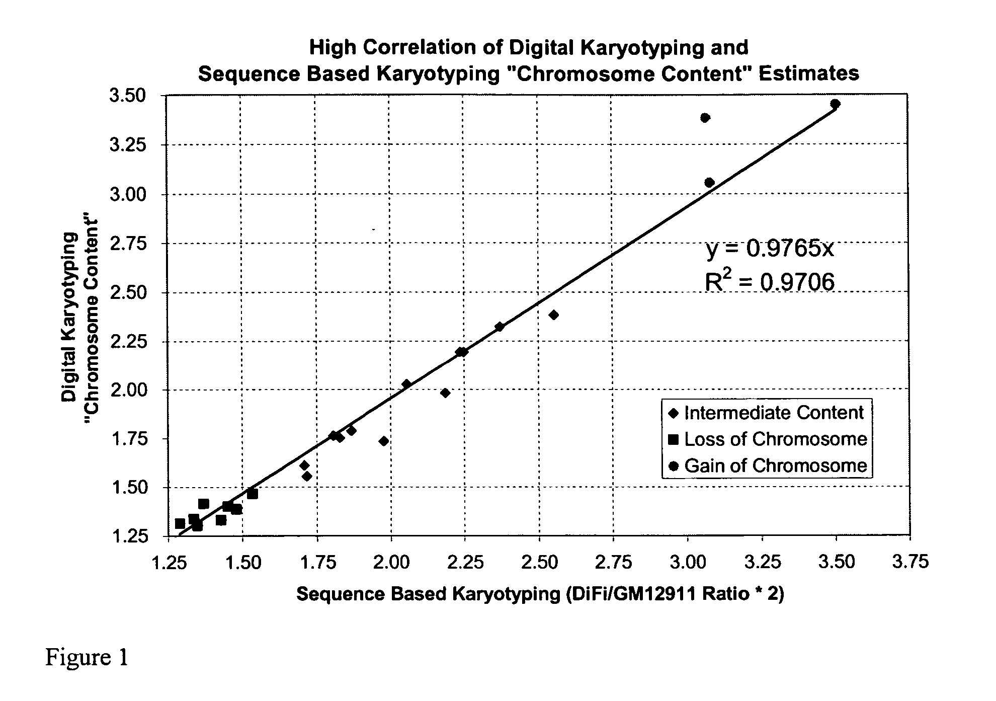
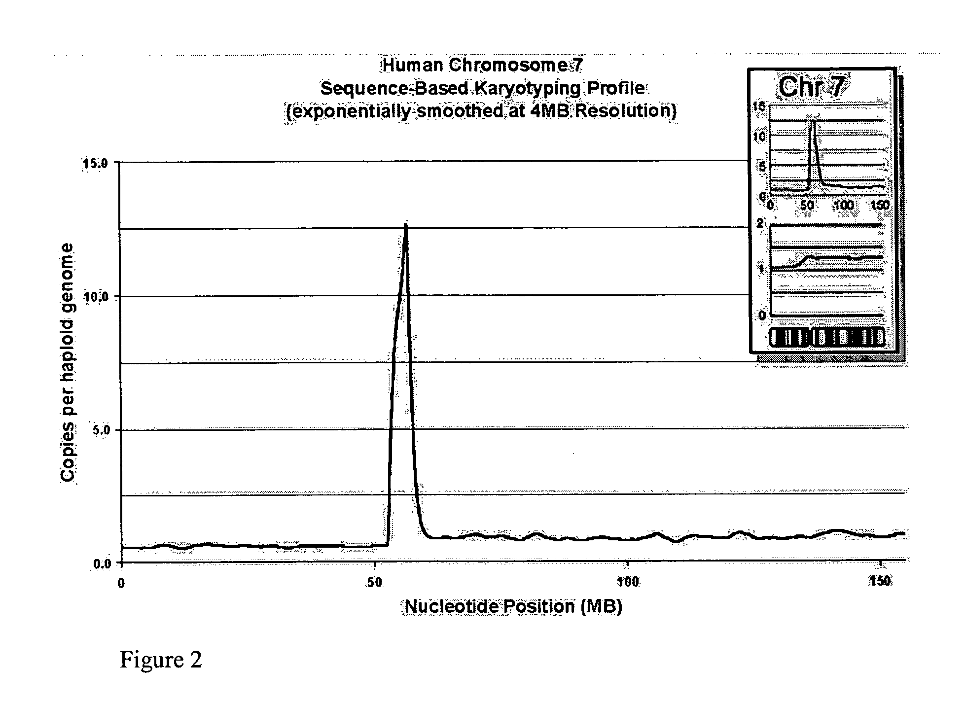
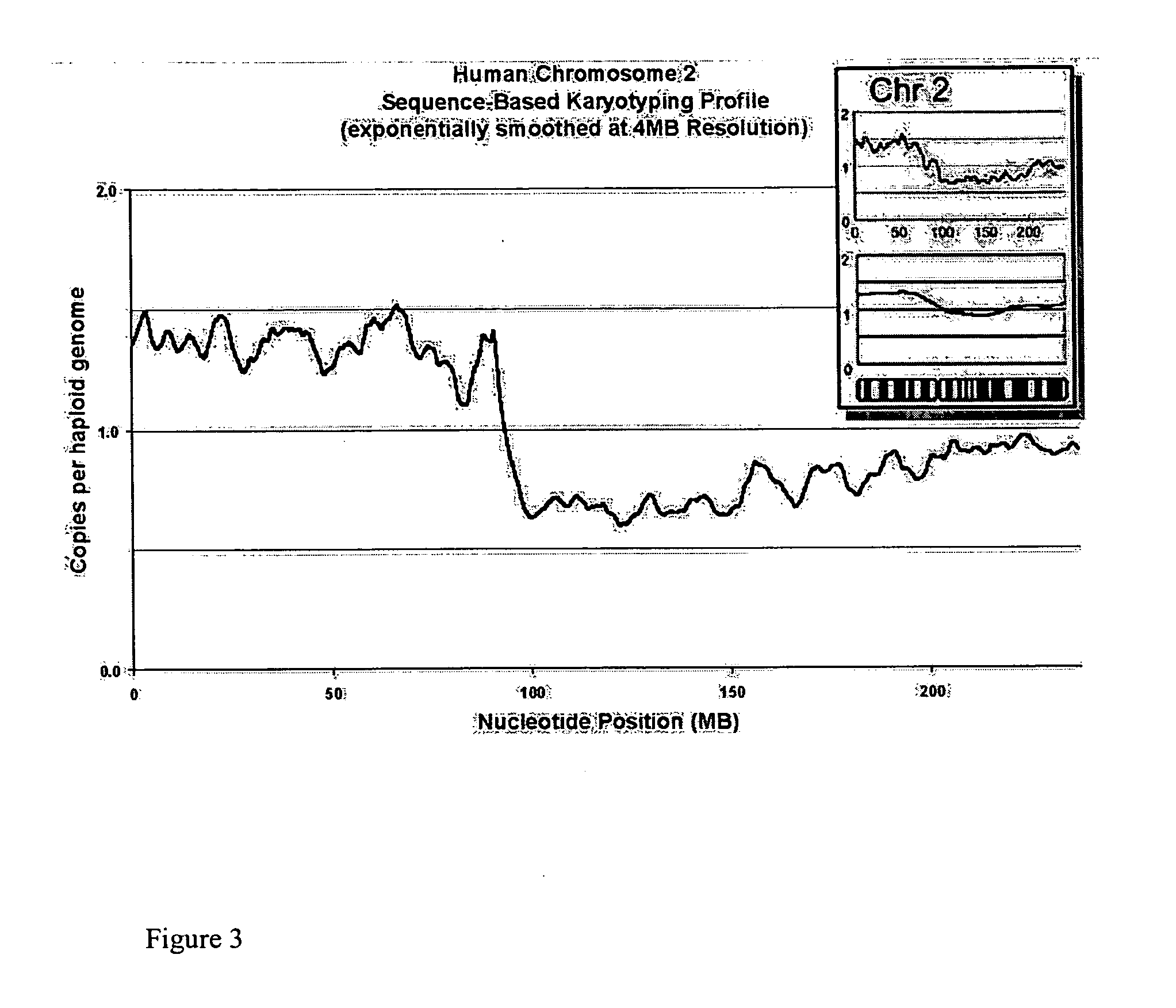
[0246] The sensitivity and specificity of Sequence-Based Karyotyping in detecting genome-wide changes was expected to depend on several factors. The breadth of the region of amplification or deletion and the magnitude of the change in copy number of a given genomic event will directly effect the detection of the change.

[0247] Analysis of Whole Chromosomes

[0248] We attempted to determine whether any loss or gain of chromosomal content was present in DiFi cells that were detectable using Sequence-Based Karyotyping relative to the published findings by digital karyotyping. Briefly, all the DNA sequences obtained were mapped to a genomic scaffold. Sequences that did not map to the genome, either due to incompleteness of the genomic scaffold or issues of sequencing quality, were removed from consideration. Filtering was also performed to remove DNA sequences which mapped to multiple genomic locations (within repeated sequences). Counts of the re...

example 2

Materials and Methods for Sequence-Based Karyotyping

[0257] Sequence-Based Karyotyping was performed on DNA from the DiFi colorectal cancer cell line, and from lymphoblastoid cells of a normal individual (GM1291 1, obtained from Coriell Cell Repositories, NJ). Genomic DNA was isolated using DNeasy or QIAamp DNA blood kits (Qiagen, Chatsworth, Calif.) using the manufacturers' protocols.

[0258] Briefly, DNA is fragmented and size fractionated. Fragments within a several hundred basepair size range are ligated to proprietary adapters to generate templates. These templates are suitable for subsequent PCR and sequencing reactions using the sequencing methods described in this disclosure (454 Life Sciences technology). The adapted templates are amplified using a proprietary oil-water emulsion PCR system. The amplified DNA molecules are then immobilized onto proprietary microscopic beads and collected. The beads containing amplified DNA are subsequently segregated from non-DNA containing b...

example 4

Preparation of DNA Sample For Sequence-Based Karyotyping

[0271] DNA Sample:

[0272] Step 1: DNase I Digestion

[0273] DNA was obtained and prepared to a concentration of 0.3 mg / ml in Tris-HCl (10 mM, pH 7-8). A total of 134 μl of DNA (15 μg) was needed for this preparation. It is recommended to not use DNA preparations diluted with buffers containing EDTA (i.e., TE, Tris / EDTA).

[0274] In a 0.2 ml tube, DNase I Buffer, comprising 50 μl Tris pH 7.5 (1M), 10 μl MnCl2 (1M), 1 μl BSA (100 mg / ml), and 39 μl water was prepared.

[0275] In a separate 0.2 ml tube, 15 μl of DNase I Buffer and 1.5 μl of DNase I (1 U / ml) was added. The reaction tube was placed in a thermal cycler set to 15° C.

[0276] The 134 μl of DNA (0.3 mg / ml) was added to the DNase I reaction tube placed in the thermal cycler set at 15° C. The lid was closed and the sample was incubated for exactly 1 minute. Following incubation, 50 μl of 50 mM EDTA was added to stop the enzyme digestion.

[0277] The digested DNA was purified b...


PUM

PropertyMeasurementUnit
Digital informationaaaaaaaaaa

Abstract

A new method for genomic analysis, termed “Sequence-Based Karyotyping,” is described. Sequence-Based Karyotyping methods for the detection of genomic abnormalities, for diagnosis of hereditary disease, or for diagnosis of spontaneous genomic mutations are also described.

Description

RELATED APPLICATIONS [0001] This application claims the benefit of priority from U.S. Application Nos. 60 / 513,691 and 60 / 513,319, both filed Oct. 22, 2003. All patents and patent applications referenced in this specification are hereby incorporated by reference herein in their entireties.FIELD OF THE INVENTION [0002] The invention relates to the field of genetics. In particular, it relates to the determination of karyotypes of genomes of individuals cells and organisms. BACKGROUND OF THE INVENTION [0003] Structural rearrangements of chromosomes have played a decisive role in the development of abnormalities in animals. It is also known that inversions, translocations, fusions, fissions, heterochromatin variations and other chromosomal changes occur as transient somatic or hereditary mutation events in natural populations. In human cancer, chromosomal changes, including deletion of tumor suppressor genes and amplification of oncogenes, are hallmarks of neoplasia (1). Single copy chan...

Claims


Application Information

Patent Timeline
IPC IPC(8): A61BC12Q1/68G01N33/48G01N33/50G06F19/00G16B20/10G16B20/20G16B30/10G16B40/10
CPCC12Q1/6813C12Q1/6841C12Q1/6876G16B20/00Y02A90/26G16B40/00G16B30/00C12Q2600/156C12Q2545/101Y02A90/10G16B30/10G16B40/10G16B20/20G16B20/10
Inventor SHIMKETS, RICHARDBRAVERMAN, MICHAEL
Owner 454 CORP
Why Eureka
Try Eureka