Methods for high resolution identification of solvent accessible amide hydrogens in polypeptides and for characterization of polypeptide structure

Inactive Publication Date: 2005-10-20
RGT UNIV OF CALIFORNIA
View PDF6 Cites 4 Cited by
  • Summary
  • Abstract
  • Description
  • Claims
  • Application Information

AI Technical Summary

Benefits of technology

[0030] The present invention provides improved methods by which peptide amide hydrogen exchange can be used to characterize protein structure, dynamics, binding interactions and solvent accessibility. The methods of the present invention provide improvements and simplification of the protein degradation and fragmentation methods (performed under amide hydrogen exchange quench-conditions) previously described (U.S. Pat. Nos. 5,658,739; 6,331,400, and 6,291,189) for performing such characterizations, and, in particular, improved methods for calculating the amount of deuterium label at sequence-specific locations within a functionally labeled protein by analysis of deuterium-labeled protein fragmentation data. The elimination of unnecessary steps and improved deconvolution methods substantially increases throughput of the overall technique, without loss of the ability to highly localize deuterium label. The methods of the present invention are illustrated in FIG. 21.
[0031] The simplification omits the progressive degradation steps of the prior art methods (employing carboxypeptidases), which were previously thought critical to the success of high resolution localization of deuterium, and instead employing refined and enhanced methods by which endoproteinases can be made to rapidly produce highly varied and high yield, and highly sequence-overlapping protein fragmentation under optimal slowed exchange conditions. With this improved methodology, exchanged peptide amide hydrogens can be localized at high resolution (within 1-5 amino add residues) within the primary amino add sequence of a polypeptide or protein. The improvement affords simple and efficient methods for studying or mapping polypeptide fine structure, such as binding sites and / or interaction surfaces of a polypeptide or protein.

Problems solved by technology

In general, the techniques that are the easiest to use and which give the quickest answers, result in an inexact and only approximate idea of the nature of the critical structural features.
The complexities of these interactions may confound conventional analytical techniques, as binding function is often lost as soon as one of the 3-dimensional conformations of the several contributing polypeptide sequences is directly or indirectly perturbed.
While these techniques can ideally provide a precise characterization of the relevant structural features, they have major limitations, including inordinate amounts of time required for study, inability to study large proteins, and, for X-ray analysis, the need for protein-binding partner crystals.
These studies do not allow a determination of the identity (location within the protein's primary amino acid sequence) of the exchanging amide hydrogens measured.
Again, determination of the identity of the particular peptide amides experiencing changes in their environment is not possible with these techniques.
However, the techniques described by Rosa and Richards were of marginal utility, primarily due to their failure to optimize certain critical experimental steps.
However, as the fragmentation of hemoglobin proceeds, each fragment's secondary and tertiary structure is lost and the unfolded peptide amides become freely accessible to H2O in the buffer.
Moreover, in Englander's work, there is no appreciation that a suitably adapted exchange technique might be used to identify the peptide amides which reside in the contacting surface of a protein receptor and its binding partner: his disclosures are concerned with the mapping of allosteric changes in hemoglobin
Unfortunately, add proteases are very nonspecific in their sites of cleavage, leading to considerable HPLC separation difficulties.
Even then the fragments were “difficult to separate cleanly”.
Over the succeeding years since this observation was made, no advances have been disclosed which address these critical limitations of the medium resolution hydrogen exchange technique.
Most acid-reactive proteases are in general no more specific in their cleavage patterns than pepsin and efforts to improve the technology by employing other acid reactive proteases other than pepsin have not significantly improved the technique.
However, none of the methods described in the art are capable of localizing the positions of the tritium labels of the labeled proteins at high resolution, the best resolution in the art generally being on the order of ≧14 amino acid residues.
Furthermore, study of proteins by the NMR technique is not possible unless the protein is small (generally less than 30 kDa), large amounts of the protein are available for the study, and computationally intensive resonance assignment work is completed.
The resolution of the deuterium-exchange mass spectrometry work disclosed in these publications therefore remained at the 10-14 amino acid level, with the primary limitation of their art being the ability to generate only a small number of peptides with the endoproteinase pepsin, as they employed it.
Moreover, the prior art teaches that refinement of endopeptidase digestion methods under slowed exchange conditions, including that of pepsin, would be unlikely to prove successful (used alone) in significantly increasing useful fragmentation and therefore produce increased resolution in deuterium label localization.
In particular, the prior art teaches that to produce even the low level of useful fragmentation observed, the enzyme fragmentation times employed with pepsin were already at the maximal duration allowable under quench conditions and that additional time taken for digestion with additional, even slower acid-resistant endoproteinases would produce unacceptable back-exchange deuterium losses.
Thus, using the prior art methods, it would not be thought feasible to produce a dramatic increase in fragmentation within the available time for said digestions under exchange quench conditions, as the prior art regards digestions of more than 10 minutes as producing unacceptable losses of label through back-exchange with solvent.
While progressive proteolysis with the use of carboxypeptidases resulted in a considerable advance in the ability to more precisely localize exchanged deuterium at the time, it was at the cost of approximately doubling the work and time required for analysis.
This means that the use of carboxypeptidases instead of these endoproteinases necessarily incurs a further loss of deuterium signal seen in further analysis, for example using mass spectrometry.
Additionally, carboxypeptidase-P is presently more than fifty times more costly to obtain (on both an activity and weight basis) than the endopeptidases commonly employed for these studies.

Method used


Image

  • Methods for high resolution identification of solvent accessible amide hydrogens in polypeptides and for characterization of polypeptide structure
  • Methods for high resolution identification of solvent accessible amide hydrogens in polypeptides and for characterization of polypeptide structure
  • Methods for high resolution identification of solvent accessible amide hydrogens in polypeptides and for characterization of polypeptide structure

Examples


example 1

Protein Expression and Purification

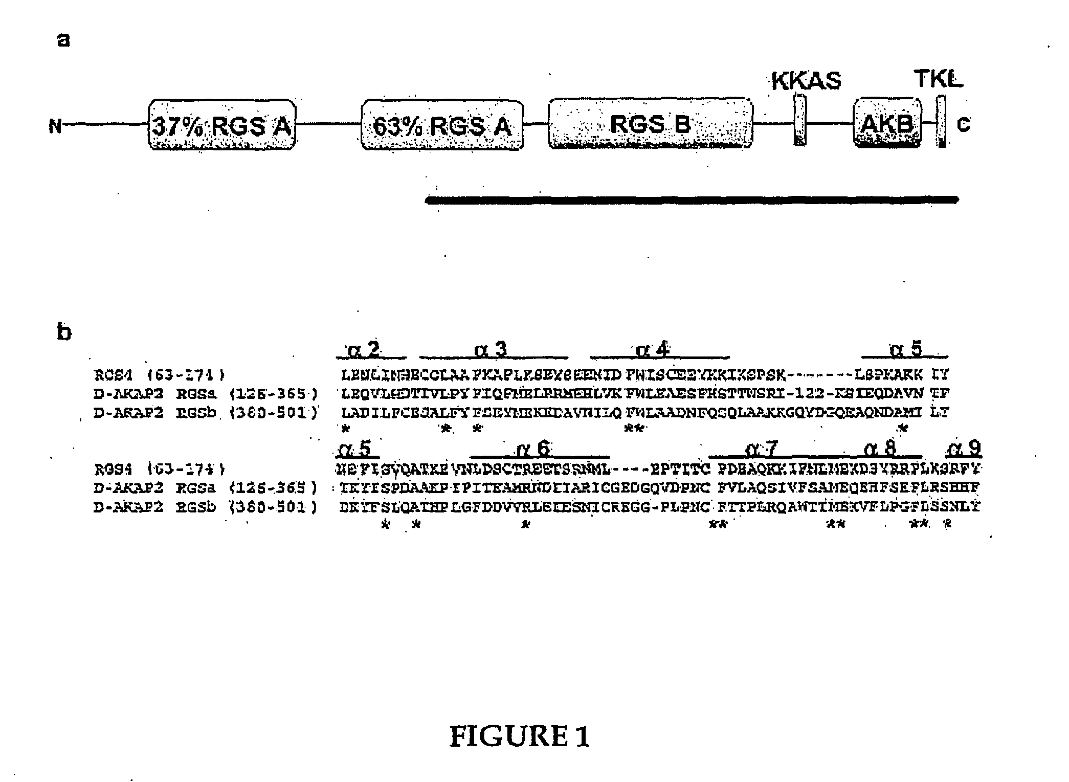
[0218] The sequence of mouse D-AKAP2 (Genbank Accession No. AF021833) was sub-cloned into pET-15b (Invitrogen), using NdeI and XhoI restriction sites after mutating an internal NdeI site. As a result of cloning, three non-native amino acids were attached to the N terminus of the protein after thrombin cleavage. The plasmid was transformed into BL21 (DE3) cells (Novagen) and grown in LB medium with 100 μg / ml of ampicillin at 37° C., 300 rpm. The cells were induced at 0.8 (A600 nm) with 0.5 mM IPTG and the protein expressed for five hours at 24° C. Six liters of culture were pelleted and lysed in 100 ml of lysis buffer (20 mM Tris (pH 8.0), 150 mM NaCl, 5 mM benzamidine) using a French press (1000 psi:1 psi ≈6.9 kPa). The lysate was centrifuged at 17,000 g for 30 minutes at 4° C. The protein was purified from the supernatant using Talon resin (Clontech) and the histidine (His) tag cleaved with three milligrams of thrombin (Sigma). The protein was di...

example 2

Circular Dichroism

[0220] The spectrum was obtained using an AVIV 202 spectropolarimeter in a 0.1 cm rectangular, quartz cuvette (AVIV). The time constant for data collection was 100 ms with a 4 seconds averaging time. Three acquisitions were averaged. The buffer (20 mM NaPO4, 150 mM NaCl, pH 7.0) spectrum was subtracted from the sample spectrum The protein concentration was 2 μM. The mean residue ellipticity (MRE) was calculated according to the following equation:

MRE=θobs(M / 10lcn)

where ζobs is the buffer corrected ellipticity, M is the molecular weight (42,800 Da) of D-AKAP2, 1 is the pathlength, c is the concentration (in molarity) and n is the number of amino acid residues in the protein.

example 3

Limited Proteolysis and Boundary Mapping

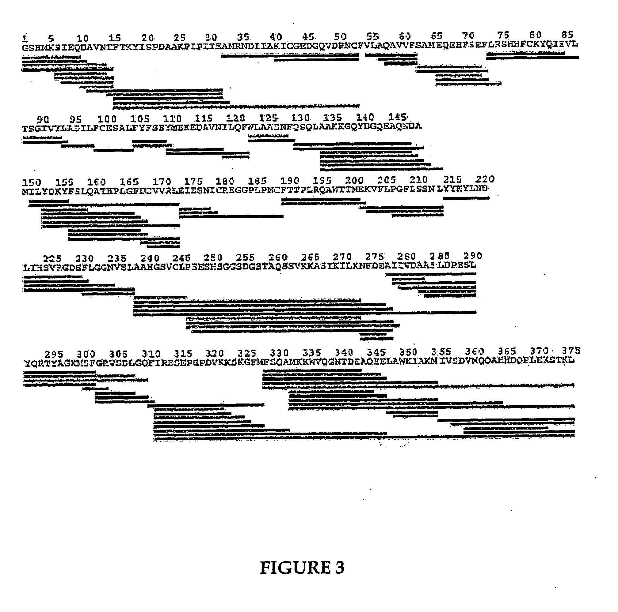
[0221] D-AKAP2 (0.85 mg / ml) was digested separately with both trypsin (Worthington Biochemical Corporation) and endoproteinase Glu-C (Boehringer Mannheim) in a 1:100 (w / w) ratio. At various time-points, aliquots were taken and quenched with 10% (v / v) glacial acetic acid for the trypsin digests or quick-frozen on solid CO2 for the endoproteinase digests. The samples were analyzed by SDS-PAGE and by liquid chromatography-mass spectrometry (LCMS). For each of the various digest time-points, 5.0 μg of total digested protein was loaded onto a Michrom BioResources Magic 2002 microbore HPLC system (Auburn, Calif.) equipped with a 1.0 mm×150 mm Vydac C4 column (5 μm, 300 Å), equilibrated at a flow rate of 50 μl / min and a column temperature of 35° C. A gradient from 10 to 80% solvent B over 60 minutes was then initiated (solvent A, 2% (v / v) acetonitrile, 0.1% (v / v) trifluoroacetic acid (TFA); solvent B, 90% acetonitrile, 0.095% TFA). The eluent from t...


PUM


Abstract

The present invention provides methods of determining, at a resolution of about 1-5 amino acid residues, the position of a peptide amide hydrogen that has been labeled with an isotope of hydrogen other than 1H by determining the quantity of isotope and / or rate of exchange of peptide amide hydrogen(s) with isotope. Invention methods comprise generating a population of sequence-overlapping endopeptidase fragments of the labeled protein under conditions of slowed hydrogen exchange and then deconvoluting fragmentation data. Invention methods allow for the localization of labeled peptide amide positions in an amino acid-specific manner, thereby providing information on residues involved in binding sites, surface conformation and accessibility of residues, and conformational changes at different times or conditions, for example.

Description

RELATED APPLICATIONS [0001] This application claims the benefit of U.S. Provisional Application Ser. No. 60 / 400,614, filed Aug. 1, 2002, and U.S. Provisional Application Ser. No. 60 / 371,366, filed Apr. 10, 2002.FIELD OF THE INVENTION [0002] The present invention relates to methods for characterizing polypeptide structure. In a particular aspect, the invention relates to improved methods for localizing labeled peptide amide hydrogens at high resolution using endoproteinase fragmentation. Invention methods are useful for a variety of applications, for example, determining solvent-accessibility of peptide amide groups, mapping binding interactions, and determining allosteric or conformational changes of a polypeptide, and the like. BACKGROUND OF THE INVENTION [0003] Considerable experimental work and time are required to precisely characterize the structure of a polypeptide of interest. In general, the techniques that are the easiest to use and which give the quickest answers, result i...

Claims


Application Information

Patent Timeline
IPC IPC(8): C12Q1/37G01N33/48G01N33/50G01N33/60G01N33/68G06F19/00
CPCC12Q1/37G01N2458/15G01N33/6848G01N33/60
Inventor WOODS, VIRGIL L. JR.
Owner RGT UNIV OF CALIFORNIA
Why Eureka
Try Eureka