
Chitosan/nanocrystalline hydroxyapatite composite microsphere-based scaffolds

a technology of hydroxyapatite and chitosan, which is applied in the field of bone grafting materials, can solve the problems of pain and risk of infection at the donor site, increased risk of disease transmission and immunological reactions, and severe limitation of autograft quantity and quality, and achieves the effects of increasing the crystallinity of the chitosan molecule, increasing dda, and increasing strength

Inactive Publication Date: 2007-11-01
BUMGARDNER JOEL D +5
View PDF6 Cites 25 Cited by
  • Summary
  • Abstract
  • Description
  • Claims
  • Application Information

AI Technical Summary

Benefits of technology

[0035] Another advantage of the present invention is that the composite material may formulated as microspheres that can be fused together to form complex shapes. This would allow custom grafts to be designed to fit any site.
[0036] In addition, therapeutic agents (e.g., growth factors, drugs, antibiotics, and other medicaments) may be added to the formation solutions to make composite microspheres containing said therapeutic agents. This allows those therapeutic agents to be released at a slower, more controlled rate, and to maintain a particular local concentration of the therapeutic agent for an extended period of time, as desired. Thus, for example, the invention may be used to maintain a high concentration of the therapeutic agent for a longer period of time than using current methods.
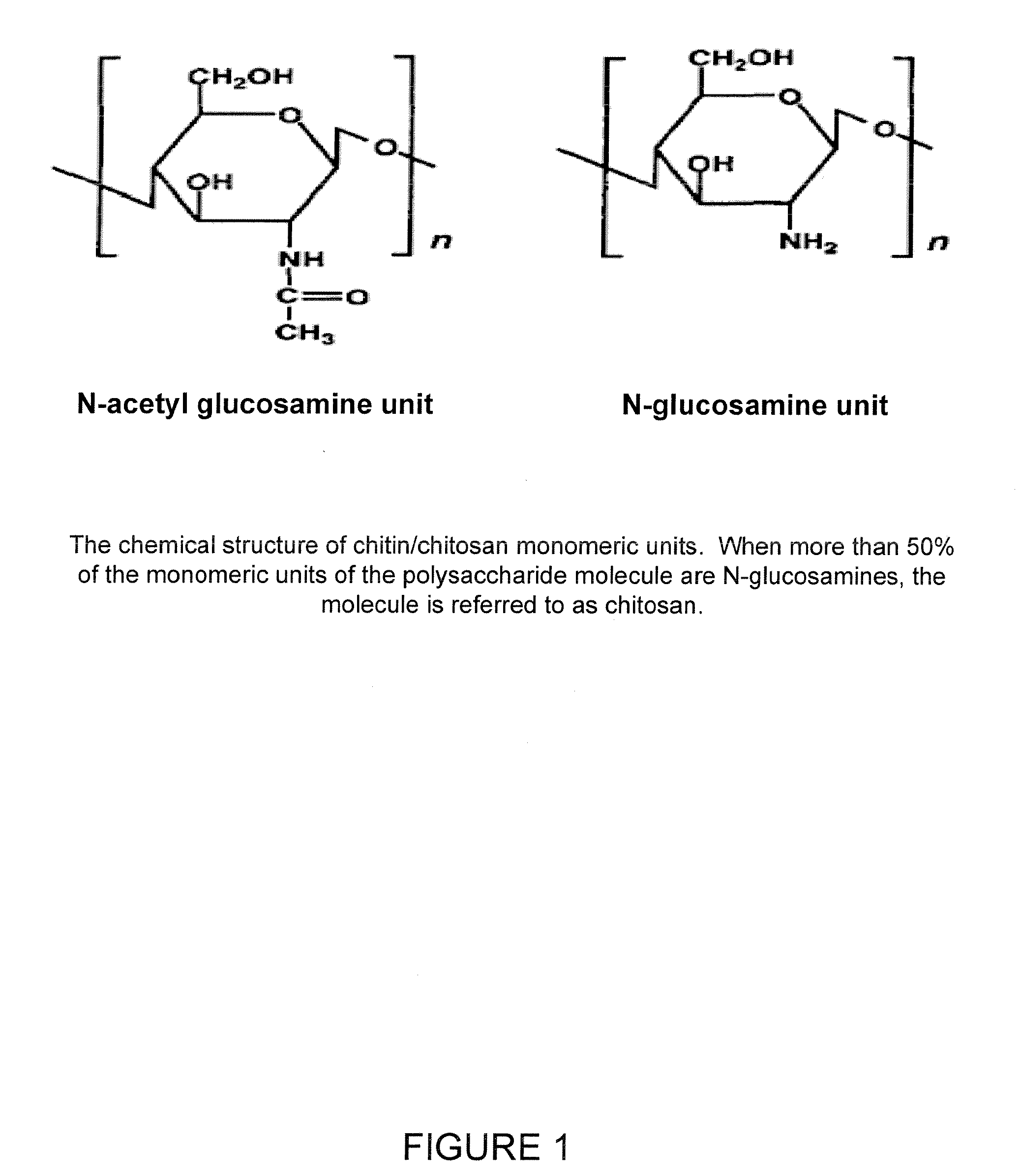
[0037] As seen in FIG. 1, chitosan is a linear polysaccharide co-polymer of N-acetyl-glucosamine and N-glucosamine units. Either an acetamido group (—NH—COCH3) or an amino group (—NH2) is attached to the C-2 carbon of the glucopyran ring. The degree of deacetylation (DDA) represents the percentage of amino groups attached to the polymer glucopyran rings. When more than 50% of the C-2 attachment is an amino group, i.e. >50% DDA, the material is termed chitosan. When more than 50% of the C-2 attachment is the acetomido group, i.e. >50% acetylated, the material is termed chitin.
[0038] The DDA is important to the physiochemical properties of the polysaccharide. Chitosan is soluble in aqueous solutions at pH<6, whereas chitin is not soluble in aqueous solutions. Crystallinity of the chitosan molecule increases with increasing DDA, resulting in polymers with higher strengths, lower moisture content and swelling properties, and lower susceptibility to degradation in physiological environments. Therefore, chitosan-calcium phosphate microspheres can be made to increase or decrease mechanical and swelling properties and degradation rates by selecting chitosans of different DDA to make the composite spheres to meet specific graft substitute and / or drug delivery applications.
[0039] The microspheres in accordance with one exemplary embodiment of the present invention can be used alone, as part of other delivery vessels (such as calcium sulfate or calcium phosphate), or formed into a scaffold of any size or shape to fit exactly into the graft site. The composite of chitosan with nano-sized calcium phosphate presents multiple development opportunities for musculoskeletal treatments. The resulting microspheres may be packed into many forms to make shapes as required for implant applications (e.g., the shape of a palate to treat cleft palate defects), or may be applied in the standard manner of other calcium compounds. The present invention thus has the ability to act both as a degradable bone graft and / or a drug delivery device that can be designed with flexible degradation and elution properties.
[0040] As discussed in greater detail below, testing of samples of various embodiments of scaffolds created in accordance with the present invention have shown an interconnected porous structure with pore sizes that can facilitate bone ingrowth. The resulting scaffolds are composed entirely of biocompatible, biodegradable materials, but does not degrade quickly even in the presence of lysozyme. Osteoblast cells are able to attach and grow well on the composite scaffold, and begin to grow into the interior pores quickly. Furthermore, as shown by dsDNA analysis, cell growth is significantly increased on the composite scaffolds.

Problems solved by technology

However, autografts are severely limited in quantity and sometimes quality, and they lead to pain and risk of infection at the donor site.
However, with allografts the risk of disease transmission and immunological reactions is present.
These materials are osteoconductive, but their degradation rate is difficult to control, and they are very brittle.
However, these materials have been shown to release acidic degradation products that increase inflammation at the implant site and impair healing.
However, these biological compounds do not bind well to many of these materials, and because the degradation rate is difficult to control, the growth factors or other compounds are often released too quickly or not at a biologically driven rate.
Hydroxyapatite (HA) has been widely used as an orthopedic implant coating for more than two decades because of its osteoconductivity, but its use for bone grafting applications is limited to low loading conditions due to its brittle nature.
However, although chitosan is tough and flexible, it lacks sufficient strength to be used alone in load bearing applications.
In general, composites of chitosan and HA or other forms of calcium phosphate can support bone cell growth and differentiation, but many of these scaffolds are produced by lyophilization, a method known to result in small pores, poor interconnected porosity, and weak mechanical properties.
Weak interfacial bonding between chitosan and powdered CaP particles can result in decreased mechanical strength, and poorly integrated CaP particles may be able to migrate out of the chitosan matrix and cause inflammation and tissue damage.
Co-precipitation methods may result in composites with both uniform distribution and strong interfacial bonding of nano-HA crystals in the chitosan matrix, but the pore size of such scaffolds is too small for effective bone ingrowth, and the lyophilization fabrication suffers from the problems described above.

Method used


Image

  • Chitosan/nanocrystalline hydroxyapatite composite microsphere-based scaffolds
  • Chitosan/nanocrystalline hydroxyapatite composite microsphere-based scaffolds
  • Chitosan/nanocrystalline hydroxyapatite composite microsphere-based scaffolds

Examples


example 1

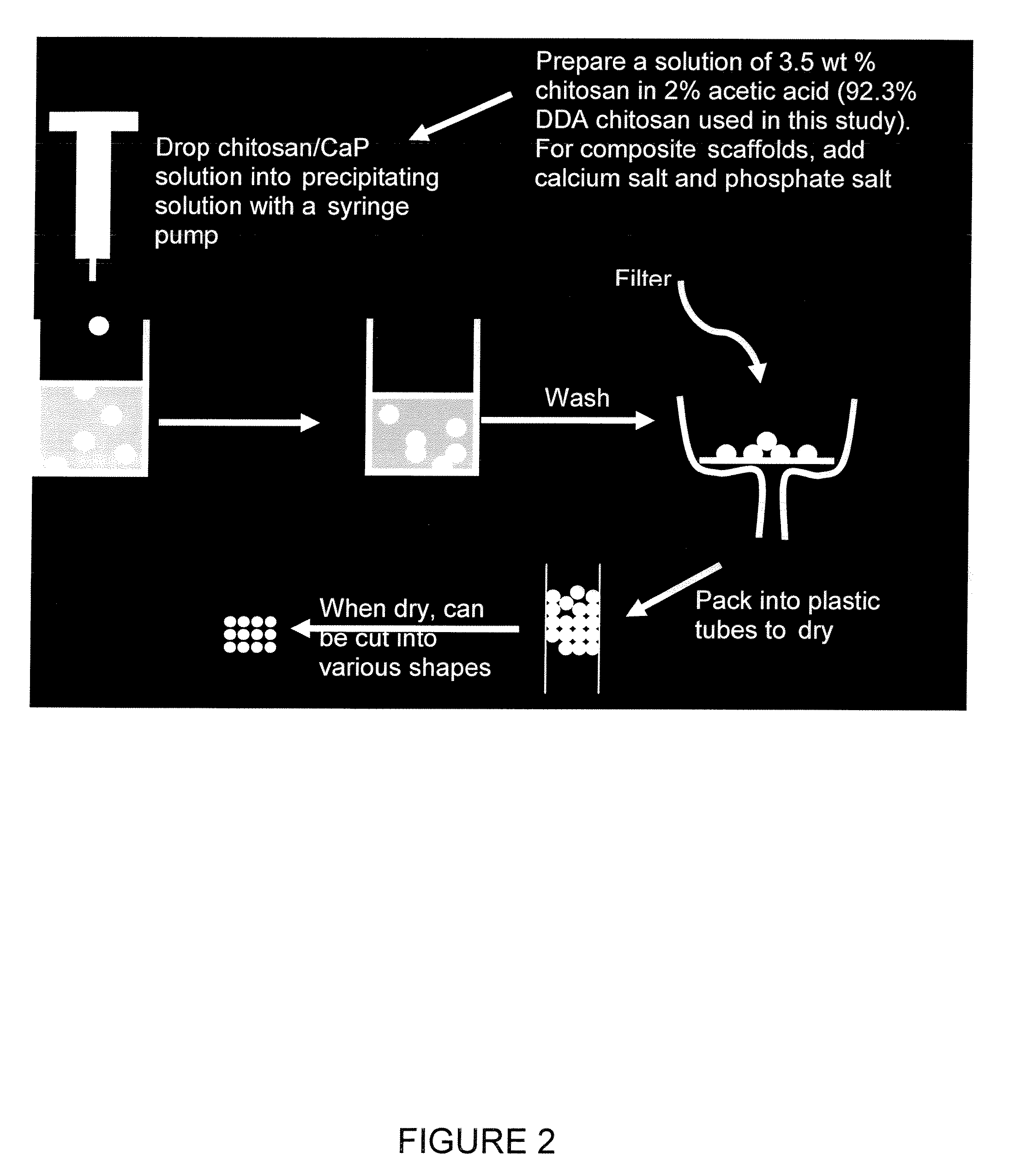
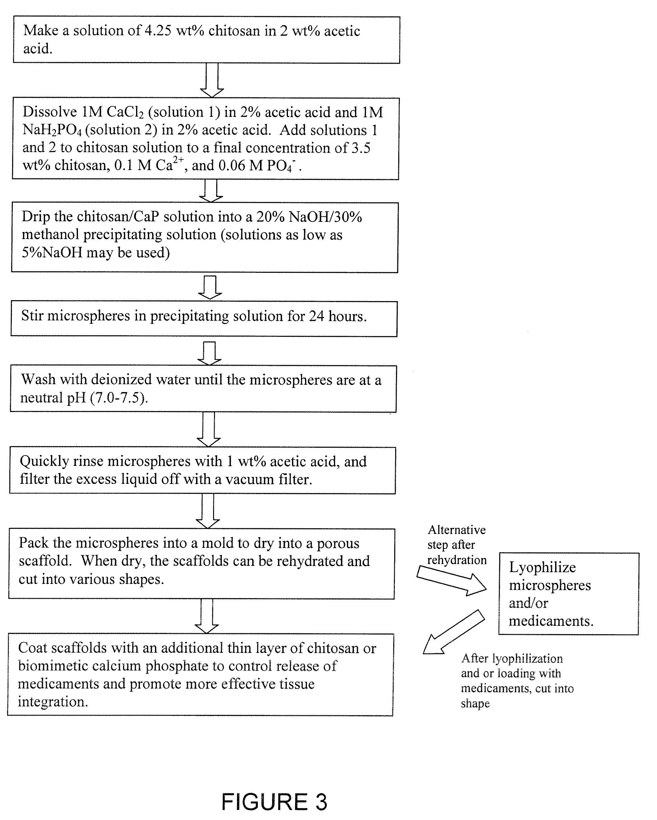
[0043] Chitosan-calcium phosphate composite microspheres are made by dripping a 3.5 wt % chitosan (92.3% DDA) in 2% acetic acid solution containing 100 mM CaCl2, and 60 mM NaH2PO4 into a NaOH / methanol solution. The chitosan-calcium phosphate microspheres are left in the NaOH / methanol solution for 24 hours to allow the initial amorphous CaP to develop into crystalline hydroxyapatite (HA). Then, the microspheres are washed with distilled de-ionized (DI) water until a neutral pH is reached. The microspheres are then quickly washed with 1% acetic acid, packed into 13 mm diameter tubes, and dried at room temperature.

example 2

[0044] Composite chitosan / calcium phosphate scaffolds are formed as follows: 3.57 g of 92.3% DDA chitosan powder was dissolved in 84 mL 2 wt % acetic acid. 10 mL of 1M CaCl2 in 2% acetic and 6 mL of IM NaH2PO4 in 2% acetic acid was added to the chitosan solution to give a final chitosan concentration of 3.44 wt %, a final Ca2+ concentration of 0.1 M, and a final PO4− concentration of 0.06 M (Ca:P ratio=1.67). The chitosan solution was placed in a 10 mL syringe fitted with a 21G needle (BD Medical, Franklin Lakes, N.J.). The syringe was placed in a syringe pump and the chitosan was slowly dripped into a solution composed of 20% NaOH, 30% methanol, and 50% water (pH=13), with constant stirring. This solution caused the chitosan drops to precipitate into solid beads. The beads were left in the basic solution for 24 hours to allow crystalline hydroxyapatite to develop from unstable brushite and amorphous calcium phosphate (ACP), likely according to the following reactions:

10CaHPO4+12OH−...

example 3

[0071]3.57 g of 92.3% degree of deacetylation chitosan is dissolved in 84 mL of 2 wt % acetic acid overnight. A 1 M sodium monobasic phosphate solution was made by dissolving 0.83 g in 6 mL 2 wt % acetic acid, and a 1 M calcium chloride solution was prepared by dissolving 1.47 in 10 mL 2 wt % acetic acid. The solutions are added to the chitosan solution and stirred. The resulting solution is dripped into a 20% NaOH / 30% methanol / 50% H2O precipitating solution using a syringe pump or similar means. When the chitosan solution drips into basic solution, chitosan microspheres instantly precipitate. This process is repeated until all of the chitosan solution has been used. Once all of the microspheres have been made, they are left in the basic solution for 24 hours to allow crystalline hydroxyapatite to form. After 24 hours, the microspheres are washed numerous times with deionized water (DI water) to reduce the pH to neutral.

[0072] After fabrication, the microspheres are rinsed with 1 w...


PUM

PropertyMeasurementUnit
Timeaaaaaaaaaa
Pore sizeaaaaaaaaaa
Pore sizeaaaaaaaaaa

Abstract

A composite chitosan / nano-hydroxyapatite microsphere-based material used for bone-grafting and delivery of therapeutic agents. The composite material may be produced using co-precipitation methods. The composite material may be used to form scaffolds with significantly greater surface area and surface roughness than scaffolds composed of only chitosan. Composite scaffolds exhibit less swelling and greater toughness and flexibility than scaffolds fabricated by other techniques. Composite scaffolds also exhibit greater osteoblast proliferation. Composite scaffolds also may contain therapeutic agents or medicaments, and may be lyophilized.

Description

[0001] This application claims priority to U.S. Provisional Patent Application No. 60 / 794,688, filed Apr. 25, 2006, by Joel D. Bumgardner, et al., and is entitled in whole or in part to that filing date for priority. The complete disclosure, specification and drawings of Provisional Patent Application No. 60 / 794,688 are incorporated herein in their entireties by reference.TECHNICAL FIELD [0002] The present invention relates to a material for bone grafting. More particularly, the present invention relates to a composite material for use as a scaffold for bone engineering and as a vehicle for delivery of medicaments to a graft or wound site. BACKGROUND OF THE INVENTION [0003] More than two million people in the United States each year suffer bone diseases, defects, or traumatic injuries that require orthopedic implants and / or bone grafting materials. The current gold standard for bone grafting is an autograft because there is no risk of disease transmission or immunological rejection....

Claims


Application Information

Patent Timeline
IPC IPC(8): A61K33/42
CPCA61K31/722A61K33/42A61K2300/00
Inventor BUMGARDNER, JOEL D.CHESNUTT, BETSY M.HAGGARD, WARREN O.YUAN, YOULINGUTTURKAR, TANGADHAR MORESHWARREVES, BENJAMIN
Owner BUMGARDNER JOEL D
Learn More