
ADP-ribosyl transferase fusion variant proteins

a technology of ribosyl transferase and variant proteins, which is applied in the direction of peptides, drug compositions, metabolic disorders, etc., can solve the problems of permanent functional impairment, loss of damaged axons, deficits associated with spinal cord injury, etc., and achieve the effect of preventing or inhibiting uncontrolled proliferation

Inactive Publication Date: 2007-11-22
BIOAXONE BIOSCI
View PDF1 Cites 6 Cited by
  • Summary
  • Abstract
  • Description
  • Claims
  • Application Information

AI Technical Summary

Benefits of technology

[0021] The following therapeutic methods are provided: a method of treating macular degeneration in a subject; a method of inhibiting or reducing the rate of subretinal neovascularization and / or proliferation of neovascular tissue associated with macular degeneration in the eye in a subject; a method of protecting retinal photoreceptors from cell death associated with macular degeneration in the eye in a subject; a method of protecting retinal photoreceptors from cell death associated with macular degeneration in the eye in a subject; and / or a method of prevention or inhibition of uncontrolled proliferation or spreading or migration of a metastatic neoplastic cell of a cancer in a subject. In another aspect, the invention relates to a method of prevention or inhibition of uncontrolled proliferation or spreading or migration, within a resection margin of a host tissue proximal to the site of excision of a tumor of a cancer in a subject, or of a metastatic neoplastic cell residing in the resection margin. Administration may be directly on to the surface of the resection margin or below the surface of the resection margin or into the tissue proximal to the resection margin which remains in the subject, and the administration may be in a time interval prior to or subsequent to, or both prior to and subsequent to, excision or removal of the tumor.
[0022] In a further aspect, a method of prevention of growth of a tumor from a malignant cell in a host tissue in a subject is provided. The polypeptide, fusion protein or composition of the invention may simultaneously prevent or inhibit at least two of malignant cell migration, malignant cell proliferation, angiogenesis or tubular structure formation or capillary network growth proximal to the malignant cell, and secretion of an active metalloproteinase from the malignant cell. A method of prevention of growth within a resection margin of a host tissue proximal to a site of excision or removal of a first tumor of a cancer in a subject, of a second tumor comprising a residual tumor cell of the cancer is also provided. Administration may be directly on to the surface of the resection margin or below the surface of the resection margin or into the tissue proximal to the resection margin which remains in the subject, and may be in a time interval prior to or subsequent to, or both prior to and subsequent to, excision or removal of the first tumor, wherein the fusion protein simultaneously prevents or inhibits at least two of residual tumor cell migration, residual tumor cell proliferation, angiogenesis or tubular structure formation or capillary network growth proximal to the residual tumor cell, and secretion of an active metalloproteinase from the residual tumor cell. In an aspect, the cancer is breast, brain, colon, skin, kidney, or hepatic cancer. In another aspect, the cancer is a brain tumor such as, for example, a glial tumor, a neuron tumor, a pineal gland tumor, a menigeal tumor, a tumor of nerve sheath, a lymphoma, a malformative tumor, and a metastatic tumor located in the brain derived from tumors of the lung, breast, melanoma, kidney, or gastrointestinal tract. In a further aspect, the cancer is a brain tumor selected from the group consisting of anaplastic astrocytoma, glioblastoma multiform, pilocytic astrocytoma, oligodendroglioma, ependymoma, myxopapillary ependymoma, subependymoma, choroid plexus papilloma, neuroblastoma, ganglioneuroblastoma, ganglioneuroma, and medulloblastoma, pineoblastoma and pineocytoma, meningioma, meningeal hemangiopericytoma, meningeal sarcoma, Schwannoma (neurolemmoma) and neurofibroma, Hodgkin's lymphoma, non-Hodgkin's lymphoma, primary and secondary subtypes of Hodgkin's lymphoma, primary and secondary subtypes of non-Hodgkin's lymphoma, craniopharyngioma, epidermoid cysts, dermoid cysts and colloid cysts.

Problems solved by technology

Most of the deficits associated with spinal cord injury result from the loss of axons that are damaged in the central nervous system (CNS).
For example, following a white matter stroke, axons are damaged and lost, even though the neuronal cell bodies are alive, and stroke in grey matter kills many neurons and non-neuronal (glial) cells.
The high dose of recombinant C3 needed to promote functional recovery presents a practical constraint or limitation on the use of C3 in vivo to promote regeneration.
If the cells are not triturated, even this dose is ineffective.
The large amount of recombinant C3 protein needed creates significant problems for manufacturing, due to the large-scale protein purification and cost.
It also limits the dose ranges that can be tested because of the large amount of protein needed for minimal effective doses.
Another related limitation with respect to the use of C3 to promote repair in the injured CNS is that it does not easily penetrate the plasma membrane of living cells.
The disadvantage of this method is that the cells needing treatment must express the necessary receptor.
The disadvantage of this system is that much of the C3 in the cell will be restrained within a membrane compartment.
More importantly, two different proteins must be added separately for transport to occur, which makes this system difficult to apply to treatment of disease in vivo.
Ocular ischemic disease, or stroke of the optic nerve results in irreversible death of retinal neurons, leading to permanent visual impairment.
This disease is not expected to respond to current therapies for AMD.

Method used


Image

  • ADP-ribosyl transferase fusion variant proteins
  • ADP-ribosyl transferase fusion variant proteins
  • ADP-ribosyl transferase fusion variant proteins

Examples


example 1

Matrigel Assay Experiment

[0225] Data from a typical Matrigel assay experiment, for example relating to the effect of a pharmaceutical composition comprising a fusion protein designated as SEQ ID NO: 8 on length of an angiogenesis-derived capillary network are summarized in Table 7. These data show that the network formation was inhibited by approximately 13% to about 20% under the dose and formulation conditions used versus the inhibition produced by a control vehicle wherein zero inhibition provides 100% growth. This effect on angiogenesis can be enhanced by using higher doses of fusion protein and by preincubation of the HUVEC cells with SEQ ID NO: 8 prior to addition of the cells to Matrigel. The anti-angiogenesis effect of a composition comprising a polypeptide of this invention comprising an amino acid sequence of a transport agent covalently linked to an amino acid sequence of an active agent, wherein the amino acid sequence of the active agent retains an ADP-ribosyl transfer...

example 2

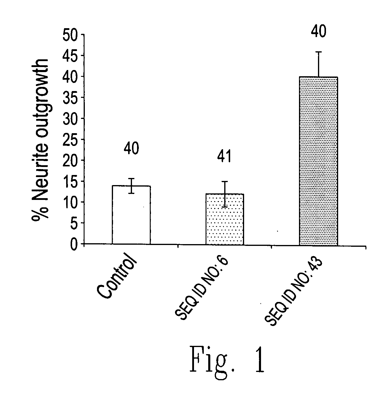
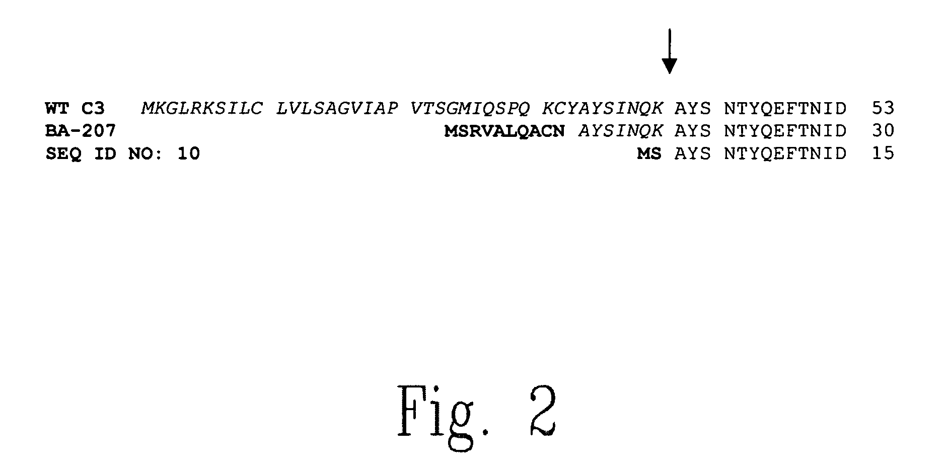
Preparation of SEQ ID NO: 43 and SEQ ID NO: 10

[0226] SEQ ID NO: 43 is prepared by polymerase chain reaction and subcloned into pET9a vector to create SEQ ID NO: 10. Two oligonucleotides are designed to delete amino acids 3-17 by site-directed-mutagenesis using the QuikChange (Stratagene) kit. Polymerase chain reaction is carried out in a thermo cycler using 50 ng of the pET9a-C3-variant, using 42-mer mutant primers: primer 2029F 5′-GGA GAT ATA CAT ATG TC*G GCT TAT TCA AAT ACT TAC CAG GAG-3′ (SEQ ID NO: 11); and primer 2029R 5′-CTC CTG GTA AGT ATT TGA ATA AGC C*GA CAT ATG TAT ATC TCC-3′(SEQ ID NO: SEQ ID NO: 12). The symbol (*) indicates the junction where the 45 nucleotide deletion occurred.

[0227] DpnI digestion is done according to the manufacturer's instructions and 1 μL of this product was used to transform XL1-Blue Super-competent cells (InVitrogen). These plates are then incubated overnight at 37° C. Clones of putative SEQ ID NO: 10 are selected and their plasmid DNA amplifie...

example 3

General Method for Determination of Inhibition of Angiogenesis

[0229] Referring to FIG. 33, the formation of new blood vessels can be studied in a cell culture model by growing endothelial cells in the presence of a matrix of basement membrane (Matigel). Human umbilical vein endothelial cells (HUVEC) are harvested from stock cultures by trypinization, and are resuspended in growth medial consisting of EBM-2 (Clonetics), FBS, hydrocortisone, hFGF, VEGF, R3-IGF-1, ascorbic acid, hEGF, GA-1000, heparin. MATRIGEL® (12.5 mg / mL) is thawed at 4° C., and 50 mL of MATRIGEL® is added to each well of a 96 well plate, and allowed to solidify for 10 min. at 37° C. Cells in growth medium at a concentration of 15,000 cells / well are added to each well, and are allowed to adhere for 6 hours. A fusion protein of this invention, e.g., SEQ ID NO: 8, in phosphate buffered saline (PBS) is added to the well at about 10 mg / ml, and in other wells PBS is added as control. The cultures are allowed to grow for...


PUM

PropertyMeasurementUnit
lengthsaaaaaaaaaa
concentrationsaaaaaaaaaa

Abstract

The present invention relates to novel chimeric C3-like Rho antagonists and their use for promoting repair and neuron survival in injured mammalian central and peripheral nervous system and for treating or preventing cancer.

Description

CROSS-REFERENCE TO RELATED APPLICATIONS [0001] This application is a continuation in part of application Ser. No. 10 / 902,878 filed Aug. 2, 2004, and of application Ser. No. 10 / 902,959 filed Aug. 2, 2004, both applications claiming priority to Canadian Application Nos 2,342,970, 2,362,004, and 2,367,636, filed Apr. 12, 2001, Nov. 13, 2001 and Jan. 15, 2002, respectively, and further claiming priority to U.S. provisional application No. 60 / 506,162 filed Oct. 29, 2003. The entire content of these applications is hereby incorporated by reference.FIELD OF THE INVENTION [0002] The present invention relates to novel chimeric C3-like Rho antagonists and their use for promoting repair and neuron survival in injured mammalian central nervous system, including the retina, and for the inhibition of the proliferation of cancer cells. BACKGROUND [0003] Traumatic injury of the spinal cord results in permanent functional impairment. Most of the deficits associated with spinal cord injury result fro...

Claims


Application Information

Patent Timeline
Patent Type & Authority Applications(United States)
IPC IPC(8): A61K38/00A61P11/00A61P3/00A61P43/00C07K14/00
CPCA61K38/00C12N9/1077C07K2319/03C07K2319/01A61P3/00A61P11/00A61P43/00
Inventor MCKERRACHER, LISAMUNZER, JON SCOTT
Owner BIOAXONE BIOSCI
Learn More