
Nanoparticles Comprising a PDGF Receptor Tyrosine Kinase Inhibitor

a technology of pdgf receptor and nanoparticles, which is applied in the direction of surgery, drug compositions, cardiovascular disorders, etc., can solve the problems of ischemic injury, stroke or myocardial infarction, and no beneficial effects of systemic administration of imatinib against restenosis, and achieve excellent passing ability

Inactive Publication Date: 2009-05-28
KYUSHU UNIV +1
View PDF3 Cites 7 Cited by
  • Summary
  • Abstract
  • Description
  • Claims
  • Application Information

AI Technical Summary

Benefits of technology

[0003]It was now surprisingly found that intracellular delivery of PDGF receptor tyrosine kinase inhibitors by nanoparticle technology represent an advantageous therapeutic strategy for vascular smooth muscle cells growth diseases such as restenosis, atherosclerotic vascular disease and primary pulmonary hypertension.
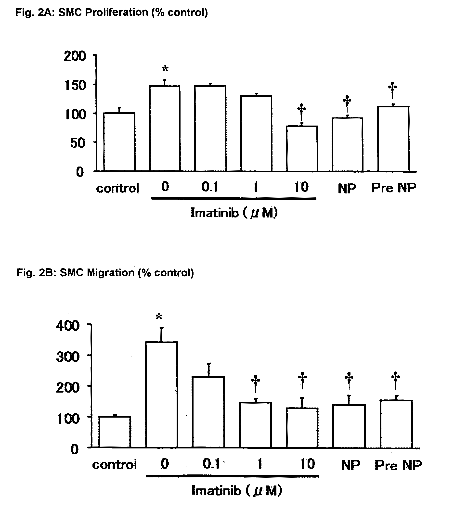
[0060]As shown in the Examples below, intracellular delivery of Imatinib with bio-absorbable polymeric nanoparticle technology effectively suppresses vascular smooth muscle proliferation and migration of vascular smooth muscle cells.
[0066]Complications associated with vascular access devices is a major cause of morbidity in many disease states. For example, vascular access dysfunction in hemodialysis patients is generally caused by outflow stenoses in the venous circulation (Schwam S. J., et al., Kidney Int. 36: 707-711, 1989). Vascular access related morbidity accounts for about 23 percent of all hospital stays for advanced renal disease patients and contributes to as much as half of all hospitalization costs for such patients (Feldman H. I., J. Am. Soc. Nephrol. 7: 523-535, 1996). Additionally, vascular access dysfunction in chemotherapy patients is generally caused by outflow stenoses in the venous circulation and results in a decreased ability to administer medications to cancer patients. Often the outflow stenoses is so severe as to require intervention. Additionally, vascular access dysfunction in total parenteral nutrition (TPN) patients is generally caused by outflow stenoses in the venous circulation and results in reduced ability to care for these patients. Up to the present time, there has not been any effective drug for the prevention or reduction of vascular access dysfunction that accompany the insertion or repair of an indwelling shunt, fistula or catheter, such as a large bore catheter, into a vein in a mammal, particularly a human patient. Survival of patients with chronic renal failure depends on optimal regular performance of dialysis. If this is not possible (for example as a result of vascular access dysfunction or failure), it leads to rapid clinical deterioration and unless the situation is remedied, these patients will die. Hemodialysis requires access to the circulation. The ideal form of hemodialysis vascular access should allow repeated access to the circulation, provide high blood flow rates, and be associated with minimal complications. At present, the three forms of vascular access are native arteriovenous fistulas (AVF), synthetic grafts, and central venous catheters. Grafts are most commonly composed of polytetrafluoroethylene (PTFE, or Gore-Tex). Each type of access has its own advantages and disadvantages.
[0072]The administration may be by one or more of the following routes: via catheter or other intravascular delivery system, intranasally, intrabronchially, interperitoneally or eosophagal. Hollow tubes include circulatory system vessels such as blood vessels (arteries or veins), tissue lumen, lymphatic pathways, digestive tract including alimentary canal, respiratory tract, excretory system tubes, reproductive system tubes and ducts, body cavity tubes, etc. Local administration or application of the PDGF receptor tyrosine kinase inhibitor(s) affords concentrated delivery of said PDGF receptor tyrosine kinase inhibitor(s), achieving tissue levels in target tissues not otherwise obtainable through other administration route. Additionally local administration or application may reduce the risk of remote or systemic toxicity. Preferably the smooth muscle cell proliferation or migration is inhibited or reduced according to the invention immediately proximal or distal to the locally treated or stented area.
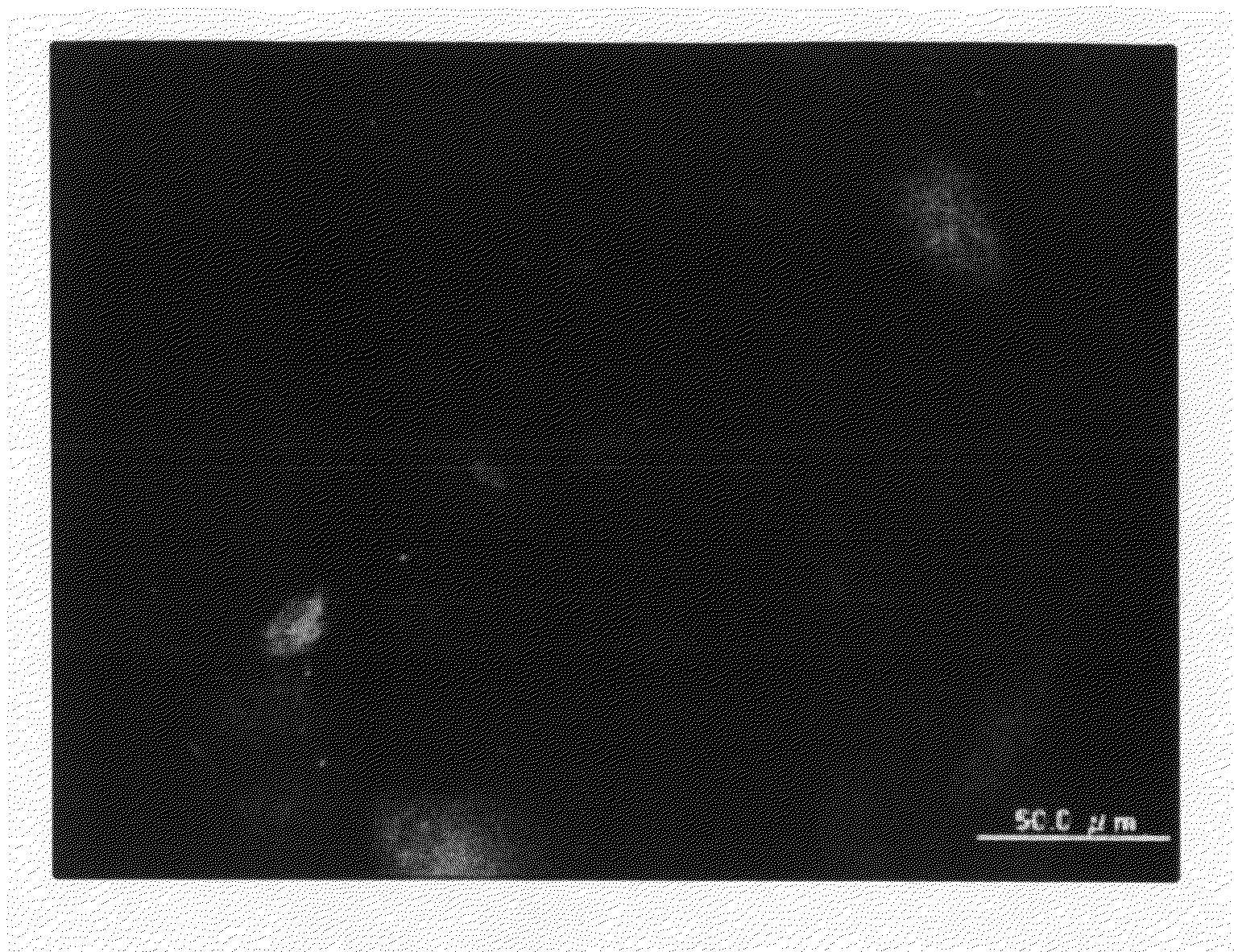
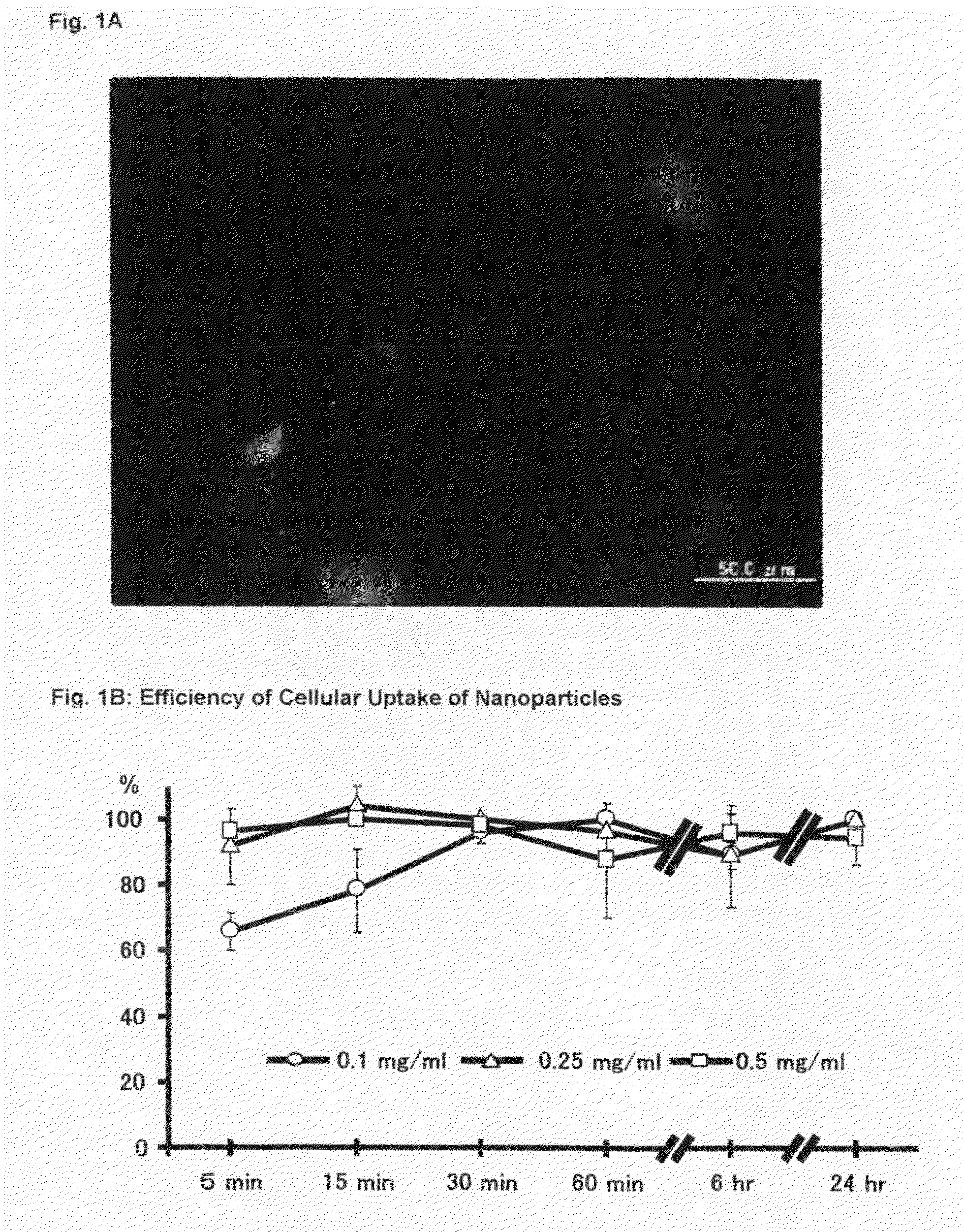
[0081]As shown in the Examples, when incubated with rat aortic and human coronary artery arterial vascular SMCs, nanoparticles loaded with a fluorescence marker instead of a PGDF receptor tyrosine kinase inhibitor enter rapidly into almost all SMCs and reach the peri-nuclear region within 1 hour. In addition, such nanoparticles incorporated into the cells show prolonged retention in the cytoplasm at least for 14 days. As further shown in the Examples, non-encapsulated Imatinib at 0.1, 1.0, and 10 μM inhibit PDGF-induced proliferation / migration of SMCs in a dose-dependent manner: Imatinib at 0.1 μM shows no effect, but Imatinib at 10 μM normalizes the PDGF-induced response. Co- or pre-treatment with nanoparticles containing Imatinib at 0.1 μM completely normalizes PDGF-induced proliferation / migration of SMCs. This demonstrates that the inhibitory potency of nanoparticulated Imatinib is at least 100-times stronger, compared with that of non-encapsulated free Imatinib.

Problems solved by technology

However, no beneficial effects of systemic administration of Imatinib against restenosis was observed in clinical studies reported by D. Zohlnhofer, et al. in J Am Coll Cardiol.
Severe blockage of blood vessels in such humans often leads to ischemic injury, hypertension, stroke or myocardial infarction.
Further lumen narrowing may take place due to constructive remodeling, e.g. vascular remodeling, leading to further intimal thickening or hyperplasia.
Such atherosclerotic lesions or vulnerable plaques are prone to rupture or ulcerate, which results in thrombosis and thus produces unstable angina pectoris, myocardial infarction or sudden death.
Complications associated with vascular access devices is a major cause of morbidity in many disease states.
Additionally, vascular access dysfunction in chemotherapy patients is generally caused by outflow stenoses in the venous circulation and results in a decreased ability to administer medications to cancer patients.
Often the outflow stenoses is so severe as to require intervention.
Additionally, vascular access dysfunction in total parenteral nutrition (TPN) patients is generally caused by outflow stenoses in the venous circulation and results in reduced ability to care for these patients.
Up to the present time, there has not been any effective drug for the prevention or reduction of vascular access dysfunction that accompany the insertion or repair of an indwelling shunt, fistula or catheter, such as a large bore catheter, into a vein in a mammal, particularly a human patient.
If this is not possible (for example as a result of vascular access dysfunction or failure), it leads to rapid clinical deterioration and unless the situation is remedied, these patients will die.
Vascular access dysfunction is the most important cause of morbidity and hospitalization in the hemodialysis population.
Venous neointimal hyperplasia characterized by stenosis and subsequent thrombosis accounts for the overwhelming majority of pathology resulting in dialysis graft failure.

Method used


Image

  • Nanoparticles Comprising a PDGF Receptor Tyrosine Kinase Inhibitor
  • Nanoparticles Comprising a PDGF Receptor Tyrosine Kinase Inhibitor
  • Nanoparticles Comprising a PDGF Receptor Tyrosine Kinase Inhibitor

Examples


example 1

Preparation of Nanoparticles

[0111]Fluorescence marker or Imatinib loaded PEG-PLGA nanoparticles are prepared by the solvent diffusion method. Hydrophobic poly (D, L-lactic-co-glycolic acid) (PLGA) with L:G molar ratio of 75:25 and MW of 20000, polyvinylalcohol (PVA) with MW of 30,000-70,000, fluorescence marker coumarin-6, are dissolved in ethyl acetate. Hydrosoluble polyethylene glycol (PEG with an average molecular weight ranging from 2,000 to 20,000 purchased from Aldrich Chemical Co) is first dissolved in water and then emulsified in the PLGA dissolving organic phase. An oil phase solution of PEG-PLGA is slowly poured into an aqueous solution containing PVA and emulsified using a microtip probe sonicator. The PEG-PLGA copolymer solution also contained 0.05% (w / v) coumarin-6 or 5% (w / v) fluoresceine isothiocyanate (FITC) as fluorescence marker or 15% (w / v) Imatinib, for the preparation of fluorescence marker or Imatinib loaded PEG-PLGA nanoparticles, respectively. The resulted oi...

example 2

Fluorescence Microscopy

[0112]Rat aortic SMCs (Toyobo) are cultured in DMEM (Sigma) supplemented with 10% FBS (Equitech-Bio, Inc.) except where otherwise indicated. Human coronary artery SMCs (Cambrex Bio Science Walkersville, Inc.) are cultured in SmGM-2 (Cambrex Bio Science). Each Cells are used between passages 4 to 8. Rat aortic SMCs are seeded on chambered cover glasses and incubated at 37° C. / 5% CO2 environment until cells are subconfluent. On the day of experiment, the growth medium is replaced with the coumarin-6 loaded PEG-PLGA nanoparticles suspension medium (0.5 mg / ml) and then further incubated for 1 hour. At the end of experiment, the cells are washed three times with PBS to eliminate excess nanoparticles which are not incorporated into the cells. Then, the cells are fixed with 1% formaldehyde / PBS buffer and nuclear is counterstained with propidium iodide (PI). Cellular uptake of coumarin-6 loaded PEG-PLGA nanoparticles is evaluated by fluorescence microscopy.

[0113]Alter...

example 3

Cellular Uptake and Intracellular Distribution of Nanoparticles

[0114]Rat aortic SMCs are seeded on 48-well culture plate to an initial concentration of 1×10⁵ cells per well (n=4 per well). The coumarin-6 loaded PEG-PLGA nanoparticles suspension medium is added to the cells at final concentration ranging from 0.1 to 0.5 mg / ml. To examine the effects of incubation time on intracellular uptake, the duration is varied from 5 minutes to 24 hours. At different time points, the nanoparticle-containing medium is removed, and the cells are washed three times with PBS. The cells are fixed with 1% formaldehyde / PBS buffer. Differential interference contrast (DIC) and fluorescence images are captured with a microscope. The images are digitized and analyzed with Adobe Photoshop and Scion Image Software. The total number of fluorescence positive cells in each field and the number of total cells was counted. Cellular uptake percentage was assessed by the percentage of fluorescence positive cells pe...


PUM

PropertyMeasurementUnit
water-solubilityaaaaaaaaaa
mean diameteraaaaaaaaaa
diameteraaaaaaaaaa

Abstract

The present invention relates to nanoparticles comprising a platelet-derived growth factor (PDGF) receptor tyrosine kinase inhibitor, especially a PDGF receptor tyrosine kinase inhibitor having a water-solubility at 20° C. between about 2.5 g / 100 ml and 250 g / 100 ml, more specifically nanoparticles comprising an N-phenyl-2-pyrimidine-amine derivative of formula I,in which the symbols and substituents have the meanings as given herein above, in free form or in pharmaceutically acceptable salt form; to the intracellular delivery of PDGF receptor tyrosine kinase inhibitors such as Imatinib with bio-absorbable polymeric nanoparticles; the use of such nanoparticles in the manufacture of a pharmaceutical composition for the treatment of vascular smooth muscle cells growth diseases; to a method of treatment of warm-blooded animals suffering from vascular smooth muscle cells growth diseases; to a process to prepare such nanoparticles; to pharmaceutical compositions comprising such nanoparticles; and to drug delivery systems incorporating such nanoparticles for the prevention and treatment of vascular smooth muscle cells growth diseases.

Description

[0001]The present invention relates to nanoparticles comprising a platelet-derived growth factor (PDGF) receptor tyrosine kinase inhibitor, especially nanoparticles comprising a N-phenyl-2-pyrimidine-amine derivative of formula I, in which the symbols and substituents have the meanings as given hereinafter, in free form or in pharmaceutically acceptable salt form; to the intracellular delivery of PDGF receptor tyrosine kinase inhibitors such as Imatinib with bio-absorbable polymeric nanoparticles; the use of such nanoparticles in the manufacture of a pharmaceutical composition for the treatment of vascular smooth muscle cells growth diseases; to a method of treatment of warm-blooded animals, including humans, suffering from vascular smooth muscle cells growth diseases; to a process to prepare such nanoparticles; to pharmaceutical compositions comprising such nanoparticles; and to drug delivery systems incorporating such nanoparticles for the prevention and treatment of vascular smoo...

Claims


Application Information

Patent Timeline
Patent Type & Authority Applications(United States)
IPC IPC(8): A61K9/16A61K31/506
CPCA61K9/5153A61K31/506A61L31/06A61L31/148A61L31/16A61L2400/12A61L2300/434A61L2300/624C08L67/04A61P11/00A61P9/00A61P9/10A61P9/12A61K9/14A61K31/505
Inventor EGASHIRA, KENSUKE
Owner KYUSHU UNIV
Learn More