
Plasma kallikrein fragments as diagnostic biomarkers for lung cancers

a technology of lung cancer and plasma kallikrein, which is applied in the field of plasma kallikrein fragment as a diagnostic biomarker for lung cancer, can solve the problems of difficult detection of low-abonding proteins which can be used as new biomarkers, increase in the incidence of lung cancer and the resulting mortality rate in korea, and achieve the effect of being useful in diagnosing lung cancer

Inactive Publication Date: 2010-11-11
PROTAN BIO INC
View PDF1 Cites 13 Cited by
  • Summary
  • Abstract
  • Description
  • Claims
  • Application Information

AI Technical Summary

Benefits of technology

[0040]Also, the diagnostic composition of the present invention can be immobilized on a suitable carrier or support in order to enhance the rapidness and convenience of diagnosis (Antibodies: A Laboratory Manual, Harlow & Lane; Cold Spring Harbor, 1988). Examples of suitable carriers or supports include agarose, cellulose, nitrocellulose, dextran, Sephadex, Sepharose, liposomes, carboxymethyl cellulose, polyacrylamides, polystyrene, gabbros, filter paper, magnetite, ion-exchange resin, plastic film, plastic tube, glass, polyamine-methyl vinyl-ether-maleic acid copolymer, amino acid copolymer, ethylene-maleic acid copolymer, nylon, cups and flat packs. In addition, other solid substrates include cell culture plates, ELISA plates, tubes and polymeric membranes. The support material may have any possible configuration including spherical (e.g., bead), cylindrical (e.g., inside surface of a test tube or well, or flat (e.g., sheet, test strip).
[0065]The diagnostic markers for lung cancer according to the present invention are highly expressed specifically in the sera of lung cancer patients at high levels, and thus are very useful for diagnosing lung cancer and estimating disease progression and treatment.

Problems solved by technology

Also, the incidence of lung cancer and the resulting mortality rate in Korea are estimated to continue to increase.
Despite the recent development of cancer therapy, the survival rate of lung cancer patients is very low.
In addition, due to abundant serum proteins (such as albumin, IgG and transferrin), it is difficult to detect low abundant proteins which can be used as new biomarkers.
However, the sensitivity and specificity thereof as lung cancer markers are not sufficient (Tarro, G. et al., J Cell Physiol 2005, 203, 1-5).

Method used


Image

  • Plasma kallikrein fragments as diagnostic biomarkers for lung cancers
  • Plasma kallikrein fragments as diagnostic biomarkers for lung cancers
  • Plasma kallikrein fragments as diagnostic biomarkers for lung cancers

Examples


example 1

Isolation and Deglycosylation of Glycoprotein from Human Serum

[0080] Serum Sample

[0081]6 blood samples were collected from 3 lung adenocarcinoma patients and 3 healthy normal persons among 65-year-old smoking men under consent in Kyungpook National University Hospital, Korea (Table 1). The blood samples were centrifuged at 4° C. at 12000 rpm for 10 minutes to collect sera, which were then stored at −70° C. before use in experiments.

TABLE 1Features of lung cancer patients and normal patientsLung cancer groupNormal groupNumber of subjects 3 3Clinical historyLung cancerHealthySexMenMenAge6464SmokingSmokersSmokers

[0082] Isolation and Concentration of Glycoproteins Using Multi-Lectin Affinity Column

[0083]A multi-lectin affinity column was used to isolate and concentrate glycoproteins from the serum samples obtained in Example . The multi-lectin affinity column (Qiagen, USA) was packed with ConA (concanavalinA), LCH (lentil lectin), GNA (snowdrop lectin), WGA (wheat germ agglutinin), SNA ...

example 2

Deglycosylation of Glycoproteins and Preparation of Peptides

[0090] Deglycosylation of Glycoproteins

[0091]The serum glycoproteins concentrated in Example 1 were treated with peptide-N-glycosidase F (PNGase F) to remove the sugar moieties and were subjected to 1D-SDS PAGE.

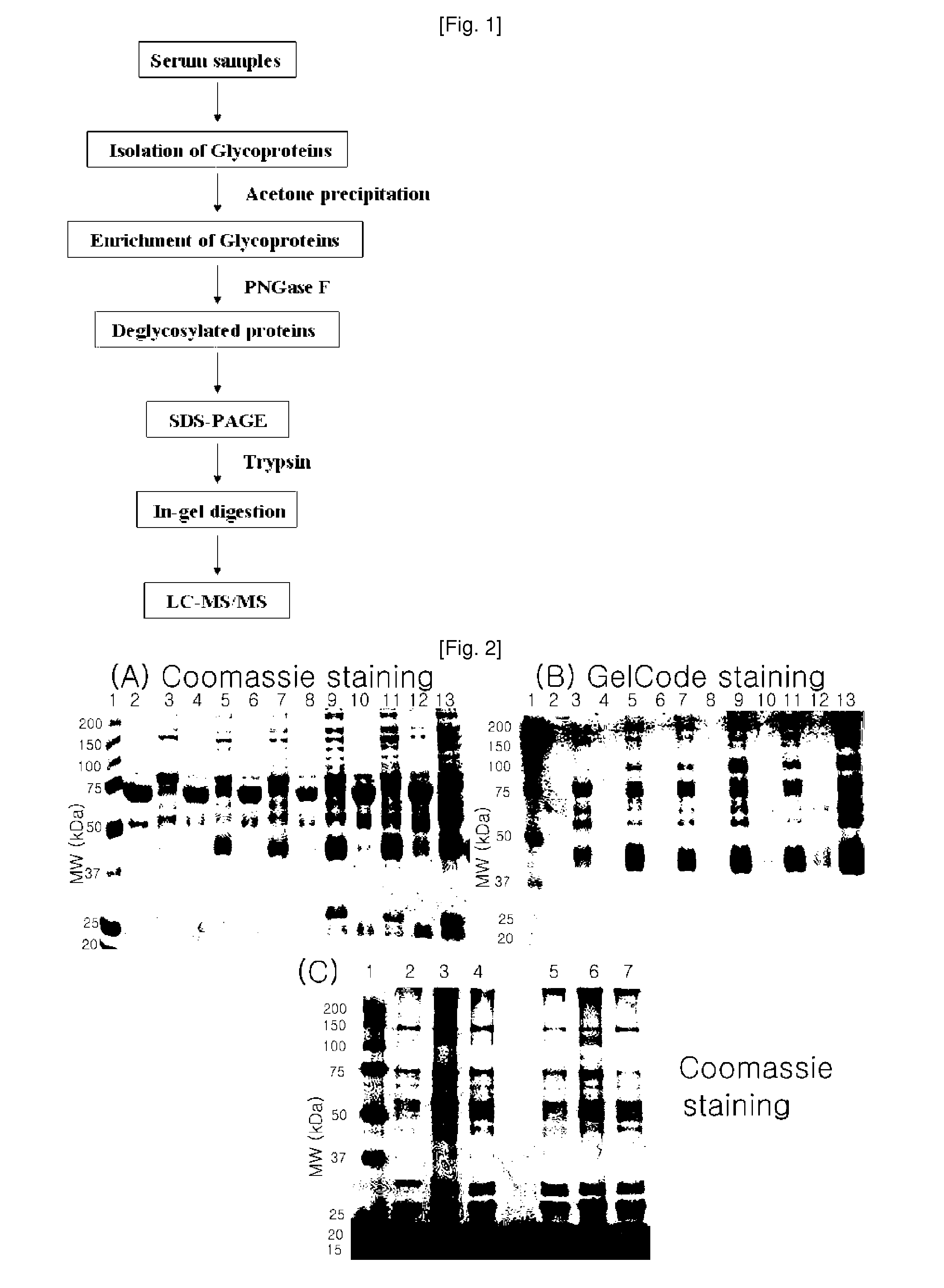
[0092]15 □ of the concentrated glycoproteins were denatured in a buffer, containing 100 mM mercaptoethanol and 2% octyl beta-D-glucopyranoside, at 100° C. for 10 minutes, and were cooled at room temperature. Then, the proteins were incubated in a reaction buffer, containing 5 □ of PNGase F (Sigma, Germany), at 37° C. for 3 hours. The deglycosylated proteins were loaded onto SDS-PAGE to separate the samples and were subjected to Coomassie brilliant straining. The SDS-PAGE results are shown in FIG. 2C.

[0093] In-Gel Digestion

[0094]The gel Coomassie-stained in Example was cleaved, and the resulting protein bands were desalted by treatment with 75 mM ammonium bicarbonate / 40% ethanol (1:1). After desalting, a sufficient a...

example 3

LC-ESI-MS / MS Analysis

[0095] LC-ESI-MS / MS

[0096]The peptide mixture produced in Example 2 was analyzed in the following manner using LC-MS / MS. The LC-MS / MS was performed using a Thermo Finnigan's ProteomeX workstation LTQ linear ion trap MS (Thermo Electron, San Jose, Calif., USA) equipped with nonospray ionization sources (NSI sources, San Jose, Calif.). 12 □ of the peptide mixture was injected and loaded into a peptide trap cartridge (Agilent, Palo Alto, Calif.). The trapped peptide was eluted using a 10-cm reversed-phase PicoFrit column (5 □, 300 Å diameter C18) packed in a housing and was separated by gradient elution in a reverse phase column (RP column). As the mobile phase, each of solutions A (H2O) and B (acetonitrile, ACN) was used, and the solutions all contained 0.1% (v / v) formic acid. The flow rate was maintained at 200 nL / min.

[0097]The gradient elution started with 2% mobile phase, and linear gradient elution was performed such that the mobile phase reached 60% within 50 ...


PUM

PropertyMeasurementUnit
Currentaaaaaaaaaa
Atomic weightaaaaaaaaaa
Fractionaaaaaaaaaa

Abstract

Disclosed herein are diagnostic markers for lung cancer, isolated from serum glycoproteins. The disclosed diagnostic markers for lung cancer are specifically expressed only in the sera of lung cancer patients at high levels, and thus will be very useful for diagnosing lung cancer and estimating disease progression and treatment.

Description

TECHNICAL FIELD[0001]The present invention relates to the use of a plasma kallikrein fragment as a diagnostic biomarker for lung cancer.BACKGROUND ART[0002]Lung cancer is the most common form of cancer in the world, which is estimated to account for 12.3% of all cancers and 17.8% of cancer-related deaths (Parkin, D. M., Lancet Oncol 2001, 2, 533-43). Also, the incidence of lung cancer and the resulting mortality rate in Korea are estimated to continue to increase. Despite the recent development of cancer therapy, the survival rate of lung cancer patients is very low. This is because the cancer is diagnosed at a late stage in most cases. Thus, there is an urgent need to develop markers which can diagnose lung cancer at an early state to increase the survival rate of lung cancer patients.[0003]Meanwhile, human body fluids, such as blood and urine, are useful for recognizing the pathological conditions (conditions associated with tumors, immune responses and vascular diseases) of the b...

Claims


Application Information

Patent Timeline
IPC IPC(8): G01N33/53C12N9/16C12N9/48C12N9/50G01N33/566C07K14/00C07K16/00C07K16/42C07H21/04C07K16/18C07K16/40
CPCG01N33/57423C12Q1/6837C12Q1/6886G01N33/57488G01N2800/52G01N2800/50G01N2333/4704
Inventor CHO, JE-YEOLPARK, JAE-YONGLEE, SEUNG-JIN
Owner PROTAN BIO INC
Learn More