
Diagnostic reagent, containing bioparticles, method for production thereof and use thereof as internal standard in nucleic acid preparation and nucleic acid detection methods

Inactive Publication Date: 2011-08-04
QIAGEN GMBH
View PDF35 Cites 6 Cited by
  • Summary
  • Abstract
  • Description
  • Claims
  • Application Information

AI Technical Summary

Benefits of technology

[0040]A major problem in the steps for nucleic acid preparation and nucleic acid amplification, both on a laboratory scale and with microfluidic devices, is incorporating reliable checking of the method in order to avoid, more particularly, false-negative results. False negative means that the target nucleic acid is present in the sample because, for example, a certain pathogen is present, but is not detected owing to errors in the test. The lysis of the cells, the isolation of the nucleic acids and also the amplification may be prone to error. Especially PCR is sensitive to disadvantageous effects of inhibitors, many of which are often present in biological samples, such as heme and its metabolic products, acidic polysaccharides, detergents, and chaotropic salts for example. Likewise, the lysis of the cells does not always proceed reliably, more particularly in the case of difficult-to-lyse cells, as described above.
[0041]It has therefore already been proposed (WO 02 / 052041, WO 2005 / 04762) to add internal control substances to the PCR, which are designed such that there is confirmation of the amplification being carried out correctly. The internal standard may be added to the sample before the first processing step, for example before the purification or the lysis, or even before the PCR. Inter alia, there is produced a synthetic oligonucleotide construct which is different from the target region of the test detection system (internal control target). Appropriate selection of the internal control is very complex and difficult. Internal control standards known in the prior art are purified nucleic acids, protein-complexed nucleic acids (armored RNA from Ambion), with capsid for example, or inactive virus standards.
[0042]A problem with the purified nucleic acids is their low stability. Especially RNA is degraded in blood or other biological sample materials within a very short period of time. In addition, the use of nucleic acids does not make it possible to check whether the lysis, i.e., the sample disruption including nucleic acid release, has proceeded correctly; their use only makes it possible to check the amplification.
[0043]Armored RNA is distinctly more stable than “naked” nucleic acids, but is distinctly easier to lyse than intact cells and therefore not comparable with these cells and not well suited as a lysis control.
[0044]The inactive virus standards are essentially vaccines, and the chemicals present, more particularly formaldehyde, change the surface structure of the viruses, with respect to active forms, by chemical crosslinking. Therefore, the lysis behavior is not comparable with that of living viruses, and the inactive virus standards are not really suitable as a lysis control.
[0045]WO 03 / 02959 relates to products obtained by freeze-drying, comprising certain defined amounts of bioparticles. The application covers, according to information from the applicant in the last submission in the EP procedure of Aug. 22, 2006, the product BioBall®, which is freeze-dried beads which are used for quantitative, microbiological quality control. They are, to date, used in the food, pharmaceutical, water, and sewage industries, but can also be used for cosmetics, personal care, clinical laboratories, and research and academic institutions. Further constituents of the beads or use as internal controls in nucleic acid preparations is not disclosed. BioBalls® are available with different bacteria, for example, enterococci, E. coli, salmonellae. There is disclosed, inter alia, a set of substantially solid products which comprise a defined number of from 1 to 1000 microorganisms, selected from viruses, bacteria, yeasts, fungi, parasites, protozoa, cells, and mixtures thereof, with the solid products being able to be transferred between containers without loss of microorganisms and the microorganisms being able to be released in a liquid, and with the deviation of the number of the microorganisms from the defined number being not more than 10%. The purpose of these products is to provide an exactly defined, very low number of microorganisms for comparative samples in quality control.

Problems solved by technology

Often, valuable time is lost as a result.
Cells can be disrupted with different levels of difficulty.
The agents required for the lysis of such cells not only break open the cells but also fragment much of the genetic information.
However, the genetic information is not fragmented to such an extent that the desired analyses would be compromised as a result.
The greater the extent of fragmentation of the nucleic acids, the more problematic it is to carry out the desired analyses.
However, ultrasonication leads to particularly extensive fragmentation of genetic information.
However, in all three cases, the genetic information is fragmented to different extents.
As a result, it is possible to detect the presence or absence of a target DNA; however, the method gives no information about the starting concentration of the target DNA.
A major problem in the steps for nucleic acid preparation and nucleic acid amplification, both on a laboratory scale and with microfluidic devices, is incorporating reliable checking of the method in order to avoid, more particularly, false-negative results.
False negative means that the target nucleic acid is present in the sample because, for example, a certain pathogen is present, but is not detected owing to errors in the test.
The lysis of the cells, the isolation of the nucleic acids and also the amplification may be prone to error.
Especially PCR is sensitive to disadvantageous effects of inhibitors, many of which are often present in biological samples, such as heme and its metabolic products, acidic polysaccharides, detergents, and chaotropic salts for example.
Likewise, the lysis of the cells does not always proceed reliably, more particularly in the case of difficult-to-lyse cells, as described above.
Appropriate selection of the internal control is very complex and difficult.
A problem with the purified nucleic acids is their low stability.
In addition, the use of nucleic acids does not make it possible to check whether the lysis, i.e., the sample disruption including nucleic acid release, has proceeded correctly; their use only makes it possible to check the amplification.
Armored RNA is distinctly more stable than “naked” nucleic acids, but is distinctly easier to lyse than intact cells and therefore not comparable with these cells and not well suited as a lysis control.
Therefore, the lysis behavior is not comparable with that of living viruses, and the inactive virus standards are not really suitable as a lysis control.
The internal standards mentioned are all still not satisfactory.
Firstly, most of them can be used only as a PCR control but not as a lysis control, since they are not intact microorganisms having a structure comparable to that of the microorganisms to be detected.
Secondly, they are, in some cases, instable products which are not storable under standard conditions.
For a simple procedure, the suspensions or solutions, some of which are known, are additionally disadvantageous because they are more laborious and inexact to dose and to add than, for example, solid specimens.
With microfluidic methods, in contrast, the internal standard has to be added at the beginning of the method and be suitable for checking all steps, since adding is no longer possible in subsequent steps owing to the method being carried out in a closed system.
As explained above, there are to date still no satisfactory internal standards which make it possible to reliably check all steps of a detection method for bioparticles by analysis of nucleic acids thereof (i.e., preparation / lysis of the cells, extraction, amplification, and detection).

Method used


Image

  • Diagnostic reagent, containing bioparticles, method for production thereof and use thereof as internal standard in nucleic acid preparation and nucleic acid detection methods
  • Diagnostic reagent, containing bioparticles, method for production thereof and use thereof as internal standard in nucleic acid preparation and nucleic acid detection methods
  • Diagnostic reagent, containing bioparticles, method for production thereof and use thereof as internal standard in nucleic acid preparation and nucleic acid detection methods

Examples


example 1

Production of Tablets

[0129]a) Tablet A (Corynebacterium glutamicum)

[0130]3 g of hydroxyethyl starch, 0.5 ml of 20% PEG 8000, 1.5 ml of TE (10 mM Tris-Cl, pH 8.0, 1 mN EDTA), 1.5 ml of an overnight culture of Corynebacterium glutamicum (obtained as described above) in 2YT medium (yeast extract tryptone medium), and a few crumbs of fuchsine (Fluka) are added to a single-use weighing dish and mixed to a homogeneous mass with a spatula. The mass is transferred to a syringe with the spatula and portioned onto Parafilm film as small beads (diameter of 1-2 mm) via a blunt cannula. Subsequently, the tablets obtained are dried overnight in the fume cupboard. There are obtained in this way, depending on the aliquots used, 50 to 200 tablets.

b) Tablet B (Corynebacterium glutamicum)

[0131]1 g of hydroxyethyl starch, 500 of 20% PEG 8000, 500 of an overnight culture of Corynebacterium glutamicum in 2YT medium (obtained as described above), 1 small spatula tip of methylene blue, 750 of Buffer P (1.4...

example 2

Nucleic Acid Preparation and Nucleic Acid Amplification

a) Lysis / preparation

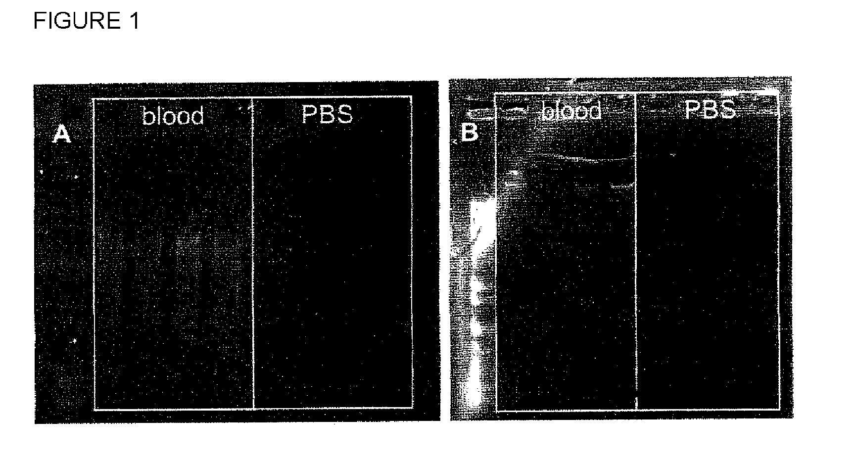
[0136]One each of Tablet A was added to a 2 ml tube, and 200 μl of blood (example 2a) or 200 μl of PBS buffer (example 2b) were added. To each tube, 20 μl of QIAGEN proteinase K solution and 40 μl of lysozyme solution (20 mg / ml in water in each case) were added in each case. The incubation was carried out over 10 minutes at 56° C. on an Eppendorf Thermomixer at a shaking speed of 1400 rpm. Subsequently, 200 μl of Buffer G (3 M GITC (guanidinium thiocyanate), 20% Nonidet® P40) were added, and reincubation was carried out over 10 minutes at 56° C. on an Eppendorf Thermomixer at a shaking speed of 1400 rpm. Then, 200 μl of ethanol were added and mixed well with the tube content.

b) Purification

[0137]The following steps were then carried out according to the QIAamp DNA Micro Handbook with each of the two samples:[0138]Binding: the entire mixture was added to a QIAamp MinElute spin column and centrifuged for 1 minu...

example 3

Quantitative PCR (Specific for C. glutamicum)

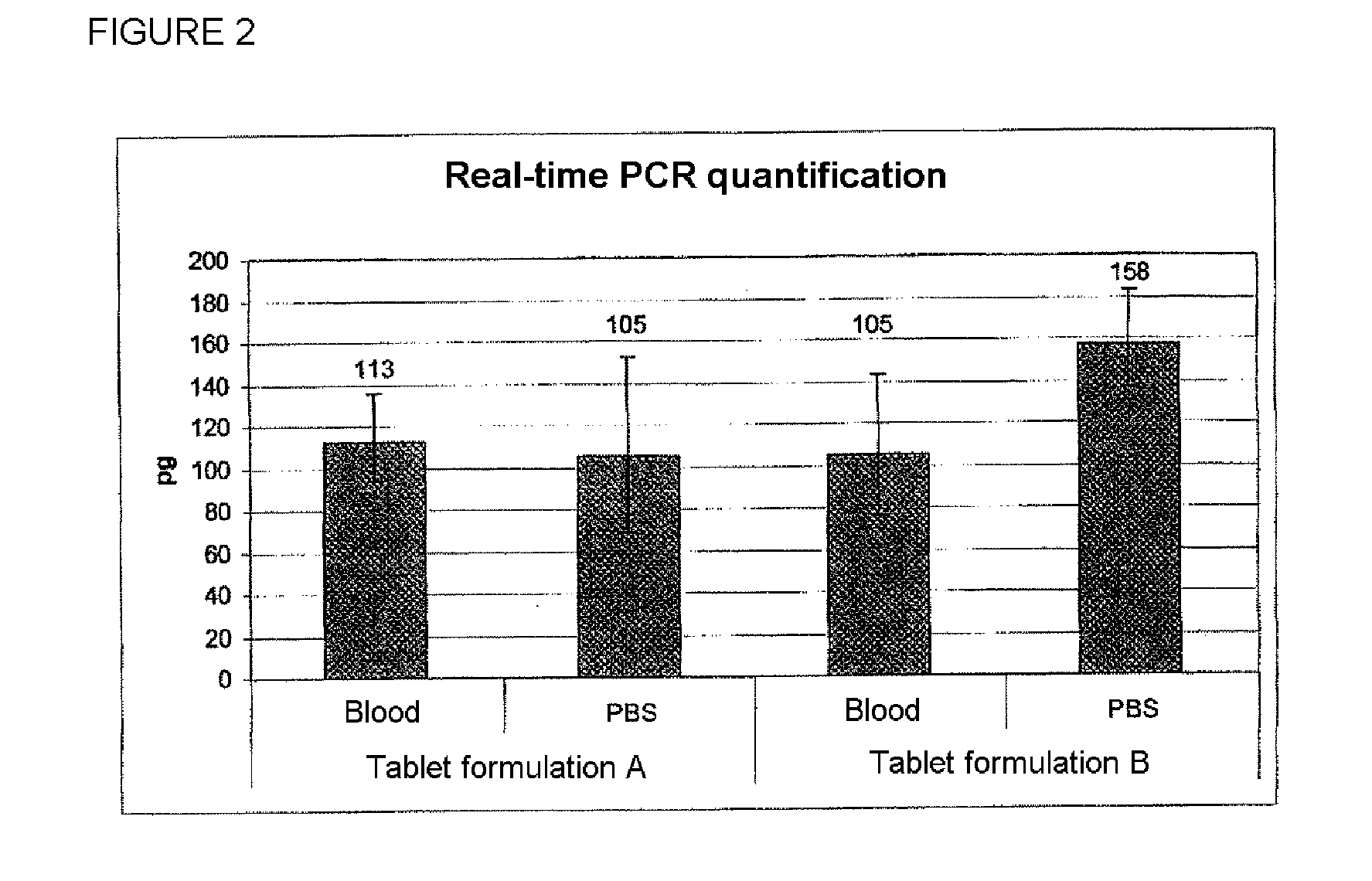
[0149]The real-time PCR specific for C. glutamicum was carried out with 10.0 μl of QIAGEN QuantiTect Probe PCR Master Mix, 0.1 μl of CG Forward (SEQ ID NO: 1) (100 μM), 0.10 μl of CG Reverse (SEQ ID NO: 2) (100 μM), 0.05 μl of CG Probe (SEQ ID NO: 3) (100 μM), 7.75 μl of double-distilled water, and 2.0 μl of template aliquots of the eluates obtained in example 2 with blood and PBS samples with, in each case, tablets A and B. The temperature cycles were 15 minutes at 95° C. and 40×(15 seconds at 95° C., 1 minute at 60° C.). The amplification was carried out in each case with eluates of 4 individual nucleic acid preparations.

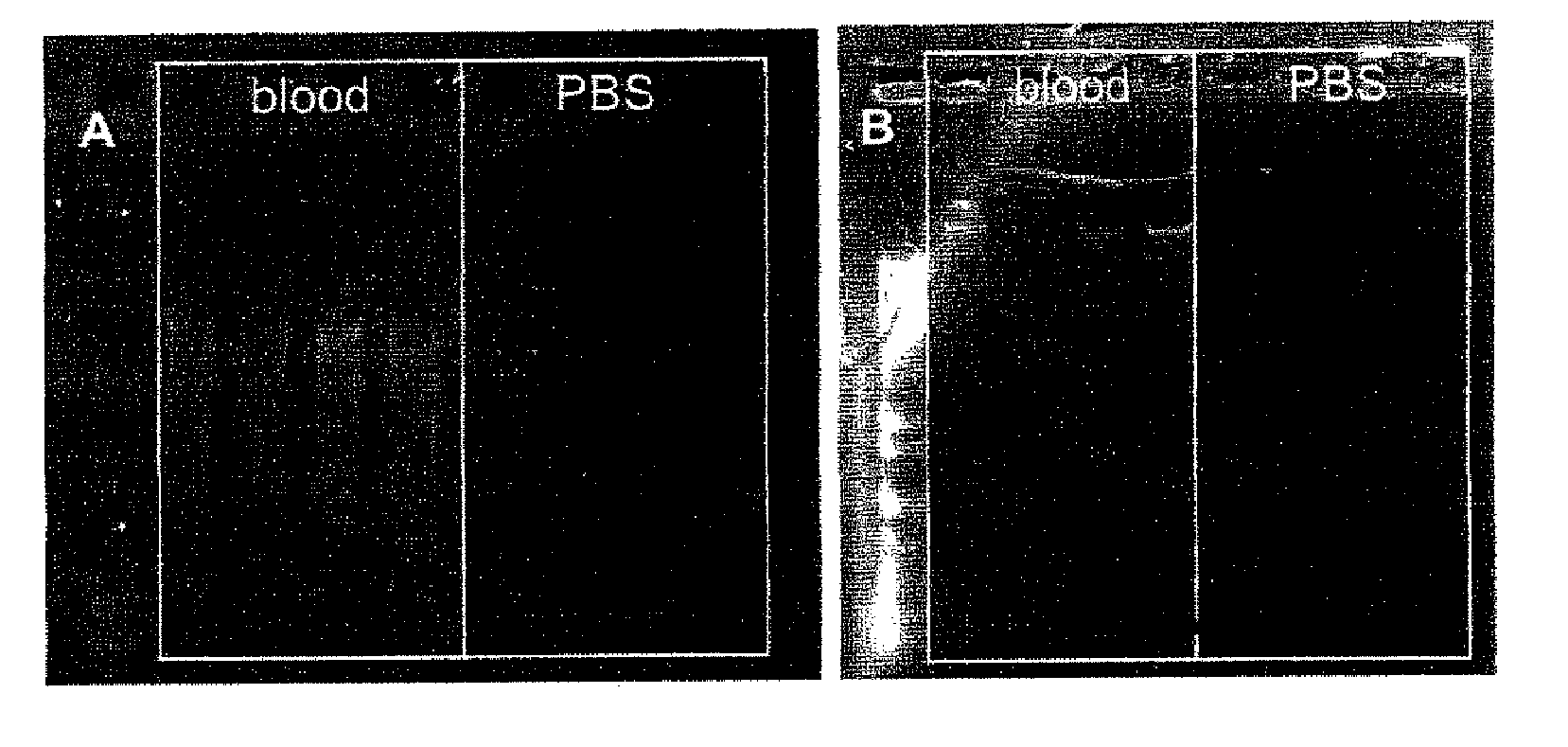
[0150]The results are shown in FIG. 2. The histogram shows the amount of C. glutamicum DNA detected by means of the PCR (in pg) per reaction with a sample size of 4. In each case, the Ct values in the RT-PCR were determined by means of fluorescence spectroscopy. With the aid of a calibration curve, the respective starti...


PUM

PropertyMeasurementUnit
Massaaaaaaaaaa
Massaaaaaaaaaa
Massaaaaaaaaaa

Abstract

A diagnostic reagent in the form of a composition dimensionally stable under standard conditions, comprising bioparticles and also customary pharmaceutical excipients, wherein the bioparticles are selected from the group consisting of bacteria, viruses, fungi, protozoa, bacteriophages, yeasts, spores, parasites, plant cells, animal or human cells, gametes, plasmids, and viroids.

Description

[0001]The present invention relates to a diagnostic reagent in the form of a composition which is dimensionally stable under standard conditions and which comprises at least one type of bioparticle selected from the group consisting of bacteria, viruses, fungi, protozoa, bacteriophages, yeasts, spores, parasites, plant parts, animal or human cells, gametes, plasmids, and viroids and also customary pharmaceutical excipients, to a method for production thereof, and to use thereof as an internal standard in methods for nucleic acid preparation and nucleic acid detection, more particularly in microfluidic devices. The invention further relates to a kit-of-parts, comprising the diagnostic reagent, and also to a method for detecting the bioparticles mentioned using the diagnostic reagent.BACKGROUND OF THE INVENTION[0002]Detection of bioparticles, such as bacteria, viruses, fungi, protozoa, or the like, in samples of different origin is increasingly of great importance. For example, when s...

Claims


Application Information

Patent Timeline
IPC IPC(8): C12Q1/68C12Q1/70C12Q1/02C12M1/34
CPCA61K9/2009A61K9/2027A61K9/2068A61K9/2054A61K9/2059A61K9/2031
Inventor HIMMELREICH, RALF
Owner QIAGEN GMBH
Learn More