System and method for identification of biological tissues

a biological tissue and system technology, applied in the field of biological tissue identification systems and methods, can solve the problems of insufficient information provided by ct, mri, and relatively poor resolution of nuclear imaging techniques, and achieve the effects of reducing the mass of healthy tissues, increasing the detection of tumour spots, and increasing the precision of tissue removal

Active Publication Date: 2012-06-21
MICROMASS UK LTD +1
View PDF7 Cites 105 Cited by
  • Summary
  • Abstract
  • Description
  • Claims
  • Application Information

AI Technical Summary

Benefits of technology

[0037]Advantages of the methods, systems and devices at surgery application include:
[0038]Application of the method, systems and devices of the present invention allows surgeons to detect, how different types of tissues are distributed over surgical area. This feature increases detection of tumour spots, increases the precision of tissue removal in case of tumour removal surgeries and removal of necrotized / ischemic tissues, and also minimizes the mass of healthy tissues removed.
[0039]The methods, devices and systems of the present invention enable the real-time, in-situ identification of tissue parts during surgery.
[0040]The methods, devices and systems of the present invention allow for automatic alerting of surgeons when malignant tissues are dissected during surgery.
[0041]Overall benefits of use of method, device and system of the present invention include:
[0042](a) Decreased invasiveness of tissue removal surgeries, hence faster recovery rate; less surgical preparation; automatic or semi-automatic objective tissue identification;

Problems solved by technology

Certain imaging methods (CT, MRI) do not provide sufficient information for the identification of malignant proliferations, but give high resolution imaging.
Other methods, especially nuclear imaging techniques provide relatively poor resolution, however, easily identify proliferating tissue parts, including various types of cancer.
However, in contrast to the amount of information available before and after surgery, there is only little information available for the surgeon about the actual position of malignant tissue relative to visible features on surgical site.
Although the procedure is used widely, it has number of disadvantages, including about 20 minutes time demand, while the patient is in the operating room with open an surgical wound, and low reliability of the results caused by sub-optimal processing of samples.
Sonography and X-ray fluoroscopy has long been used to follow surgical procedure, though their application generally causes interruption of the surgical intervention.
Although these systems were proven to be extremely useful in certain applications, e.g. spinal surgeries, they are not capable of identifying minor amounts of tumour tissue on surgical area or minor proximal metastases.
Weakness of these methods lies in their selective nature to certain tumours, their incompatibility with surgical techniques and the undesired side effects of labels.
It has to be noted, that melanoma can be detected by means of near-infrared two photon laser induced fluorescence without labeling, however this technique can only be used for detection of primary melanoma on skin surface.
One disadvantage of these ionization methods is that they lack the capability of analysis of non-volatile compounds.
In spite of the excellent spatial resolution, widespread application of SIMS is strongly hindered by limited molecular weight range of molecules, which undergo SIMS ionization.
One disadvantage of LSIMS is that samples have to be dissolved in glycerol or nitrobenzyl-alcohol prior to analysis.
This step often involves solubility problems, and dissolution of solid samples obviously excludes any kind of spatially resolved analysis.
Further disadvantages include the milder, but still existing limitation on molecular weight of species ionized this way.
This latter technique was termed ‘fast atom bombardment’ (FAB) and had incremental advantages compared to LSIMS [Williams, D H et al, JACS, 103 (19): 5700-5704 1981], however the method kept practically all disadvantages of the original method, including strong limitations on molecular weight and loss of capability of spatially resolved analysis.
MCI still carried the disadvantage of limited molecular weight range, complicated instrumentation and cross contamination between samples due to sputtering effect of impacting glycerol droplets.
Although the method is theoretically capable of spatially resolved analysis, known prior art attempts to develop this capability have all failed.
A common disadvantage of the described methods is that they generally work strictly under high vacuum conditions.
Simple laser desorption ionization, similarly to SIMS, gives poor ionization efficiencies and they can only be used for the investigation of a relatively limited number of molecules.
Since the matrix compound is used in excessive amounts, the resulting sample consists of matrix compound crystals with analyte molecules embedded into its crystal lattice.
Main disadvantages of MALDI include the low ion yield, production of predominantly singly charged ions and the fact that natural surfaces can only be investigated after deposition of matrix compounds.
Desorption ionization methods utilizing collimated beam of atoms, ions, molecules, or molecular clusters cannot be used under atmospheric pressure conditions, since particles cannot be accelerated to suitable velocities at high pressure due to consecutive collisions with gas molecules.
Same phenomenon is also responsible for the extreme divergence of particle beams at higher pressures, which also hinders the formation of practical analytical beams.
(2000), Anal. Chem., 72, pages 652-657; however the technique did not gain popularity due to low ion yield which problem is further increased by the 99% ion loss in atmospheric interface, and workplace safety issues generally associated with the use of laser in open experimental setups.
Although these methods cannot be used for fast identification of tissues, they provide invaluable information on marker molecules characteristic to one or another type of tissue.
SIMS analysis of tissues gives characteristic spectra showing mainly phospholipid fragments, however, the technique works exclusively under high vacuum conditions, and hence it cannot be applied for in vivo analysis of tissues.
Although both types of spectra are characteristic, and show unique features in case of malignant tumours, the method still cannot applied for in vivo analysis, since deposition of matrix compounds is incompatible with living organisms.
This method is fully compatible with in vivo analysis (these infrared lasers are widely used in surgery), however tissue identification in this case has not been demonstrated until now.
DESI analysis of living tissues, however, does not yield conclusive data, due to interference from blood and interstitial fluid leaking from surface being investigated.
Further disadvantage of DESI analysis is the safety concern associated with the use of 4-5 kV DC in proximity of living organisms.
From the above analysis of the state of the art in MS it can be concluded that both abundant proteins and phospholipids give characteristic distribution in DI mass spectra of various tissues, however for in vivo MS analysis there is no appropriate ionization method developed yet.
Experimental implementation of these experiments was limited to contact heating of pure, crystalline analyte compounds.

Method used


Image

  • System and method for identification of biological tissues
  • System and method for identification of biological tissues
  • System and method for identification of biological tissues

Examples


example 1

Analysis and Identification of Tissue Parts During Surgery

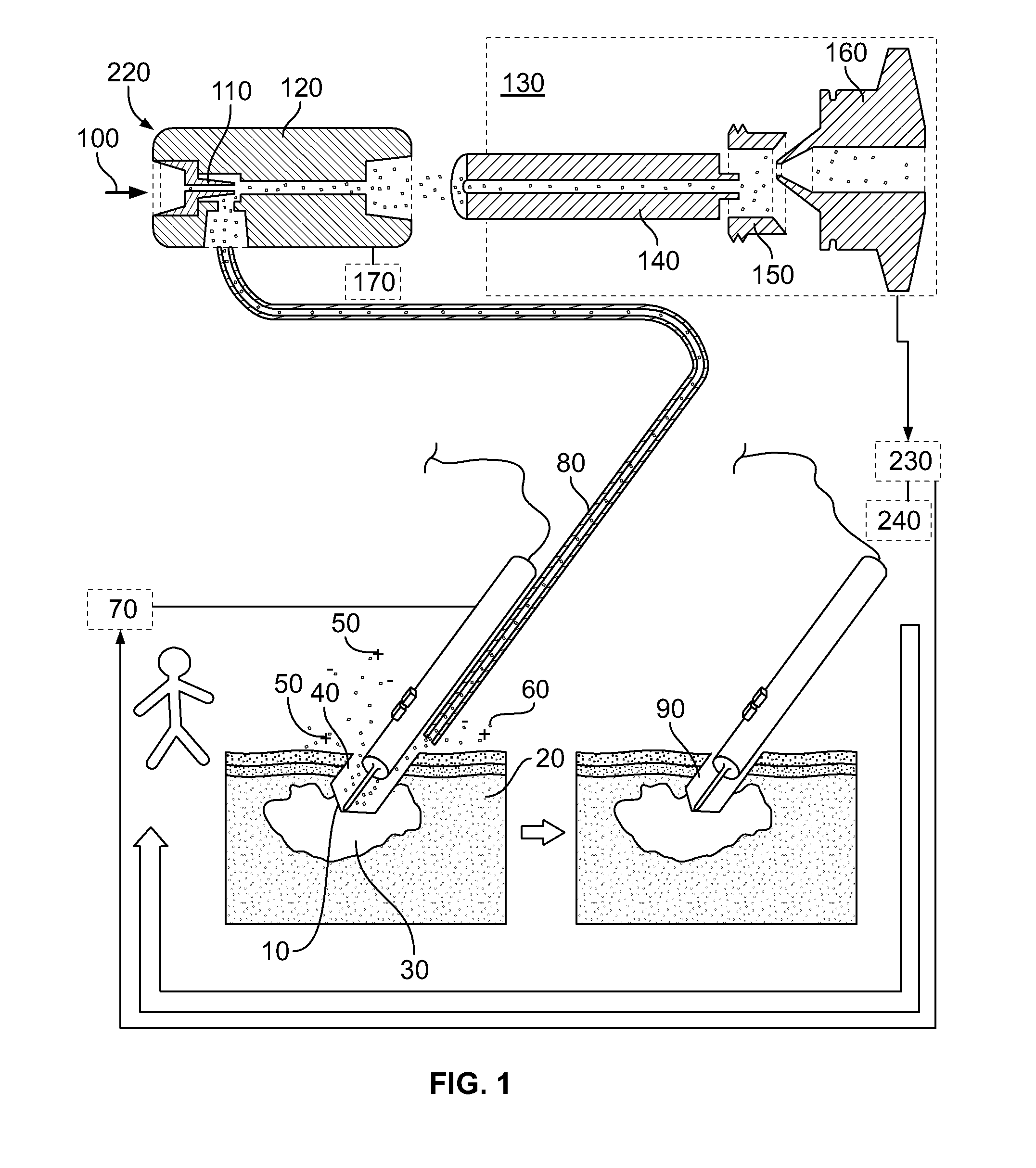
[0114]By reference for example to FIG. 1, an electrosurgical unit (ICC 350, Erbe Elektromedizin GmbH) is used in combination with quadrupole ion trap mass spectrometer (LCQ Duo, ThermoFinnigan). Electrosurgical cutting electrode 10 was equipped with commercially available smoke removal unit 80 (Erbe), which was connected to fluid pump 220 (VAC 100, Veriflo) through 8⅛″ OD 2 mm ID PTFE tubing. Fluid pump 220 was mounted on LCQ instrument using heavily modified DESI ion source (OmniSpray, Prosolia) platform. Mass spectrometer 130 was operated in negative ion mode. Ions in the range of 700-800 were isolated in ion trap, and were activated using collisional activation with neutral helium atoms. Spectra were acquired in range of m / z 700-800.

[0115]Canine in vivo and ex vivo data was acquired from dogs with spontaneous tumours from veterinary oncology praxis.

[0116]Electrosurgical electrode 10 was used to remove malignant melanoma tu...

example 2

Determination of Drugs in Tissues for Localization of Tumour Cells

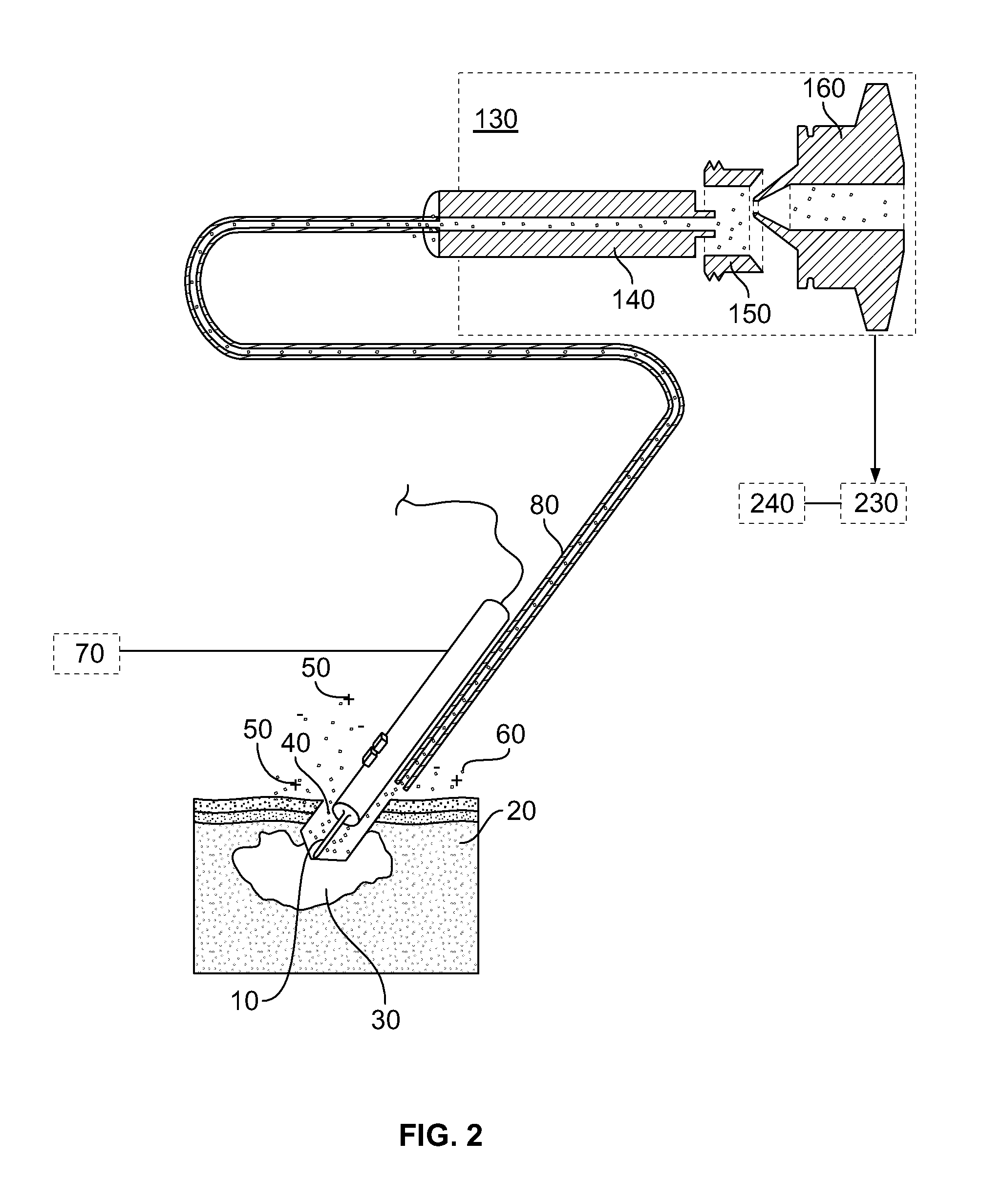
[0118]An electrosurgical unit (ICC 350, Erbe Elektromedizin GmbH) is used in combination with quadrupole ion trap mass spectrometer (LCQ Duo, ThermoFinnigan). Electrosurgical cutting electrode 10 was equipped with commercially available smoke removal unit 80 (Erbe), which was connected to fluid-pump 220 (VAC 100, Veriflo) through 8⅛″ OD 2 mm ID PTFE tubing. Fluid pump 220 was mounted on LCQ instrument using heavily modified DESI ion source (OmniSpray, Prosolia) platform. Fluid pump 220 was equipped with secondary electrospray post-ionization unit, comprising capillary 180 and high voltage power supply 170. Electrospray 260 and mass spectrometer 130 were operated in positive ion mode. Ions at m / z 447 and 449 were monitored with m / z 446 as background signal.

[0119]Nude mice carrying NCI-H460 human non-small cell lung cancer xenograft were house in a temperature- and light-controlled room, feed and water were supplied ad ...

example 3

Localization and Identification of Bacterial Infections on Mucous Membranes

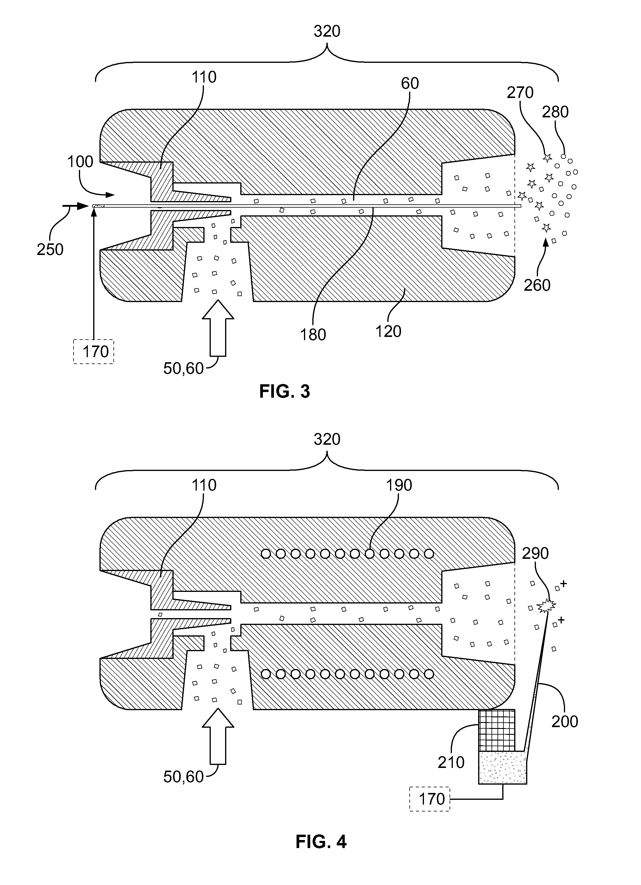
[0123]Home built thermal tissue disintegration device comprising DC power supply 70 and metal electrodes 10 is used in combination with quadrupole ion trap mass spectrometer (LCQ Duo, ThermoFinnigan). Metal electrodes 10 were connected to fluid pump 220 (VAC 100, Veriflo) through 8⅛″ OD 2 mm ID PTFE tubing. Fluid pump 220 was mounted on LCQ instrument using heavily modified DESI ion source (OmniSpray, Prosolia) platform. Mass spectrometer 130 was operated in negative ion mode. Ions in the range of 640-840 were isolated in ion trap, and were activated using collisional activation with neutral helium atoms. Spectra were acquired in range of m / z 640-840.

[0124]Electrodes 10 were used to sample upper epithelial layer of mucous membranes infected by various bacteria. Since present application of method and device is aimed at minimally invasive analysis of mucous membranes, about 0.1-0.4 mg of total material compris...


PUM

PropertyMeasurementUnit
thickaaaaaaaaaa
molecular weightaaaaaaaaaa
energyaaaaaaaaaa

Abstract

The present invention provides for a system, method, and device for analyzing, localizing and/or identifying tissue types. The method includes analyzing, localizing and/or identifying one or more tissue samples, characterized in that the method comprises: (a) generating gaseous tissue particles from a site in the one or more tissue samples, (b) transporting the gaseous tissue particles from the site to an analyser, (c) using the analyser for generating tissue-related data based on the gaseous tissue particles, and (d) analyzing, localizing and/or identifying the one or more tissue samples based on the tissue-related data. The invention can either be used in close conjunction with a surgical procedure, when one or more surgical tools are an integrated part of ionization, or as a separate mass spectrometric probe for the analysis of one or more tissue parts.

Description

RELATED PRIORITY APPLICATION[0001]This application claims the benefit of the filing date of U.S. Provisional Application No. 61 / 181,421, filed May 27, 2009, the content of which is specifically incorporated herein by reference.FIELD OF THE INVENTION[0002]The present invention relates to devices, systems and methods for analyzing, localizing and / or identifying tissues. More specifically, the present invention relates to devices, systems and methods for analyzing, localizing or identifying tissues in real time and in situ by combining disintegration of tissues and an analytical method such as mass spectrometry.BACKGROUND OF THE INVENTION[0003]Throughout this application, various references are cited in square brackets to describe more fully the state of the art to which this invention pertains. The disclosure of these references is hereby incorporated by reference into the present disclosure.[0004]Identification of pathological or abnormal tissues has crucial importance during the dia...

Claims


Application Information

Patent Timeline
Patent Type & Authority Applications(United States)
IPC IPC(8): G01N27/62C12M1/34C12Q1/02
CPCG01N1/02G01N2001/028G01N27/62G01N2001/2886H01J49/04G01N2001/045A61B10/02H01J49/165H01J49/168A61B2010/0208G01N27/623
Inventor TAKATS, ZOLTAN
Owner MICROMASS UK LTD
Why Eureka
Try Eureka