

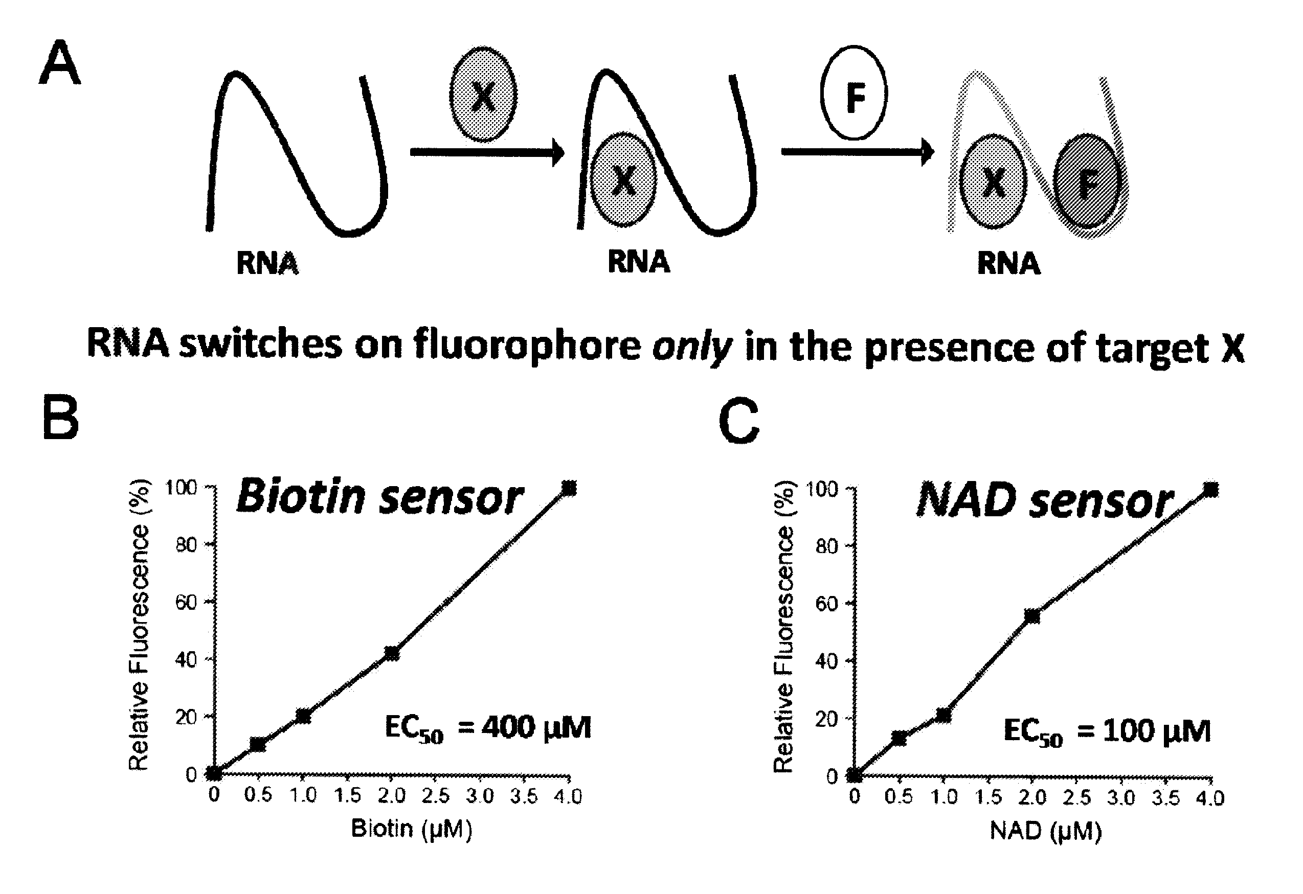
Coupled recognition/detection system for in vivo and in vitro use
a recognition/detection system and in vitro technology, applied in the field of in vitro and in vitro use of coupled recognition/detection systems, can solve the problems of inability to achieve homogeneous assays, limited rna visualization technologies, and inability to provide similar simple and straightforward technologies for rna and small molecule visualization, etc., to achieve rapid, simple and general approach, and improve the number of molecules
- Summary
- Abstract
- Description
- Claims
- Application Information
AI Technical Summary
Benefits of technology
Problems solved by technology
Method used
Image

Examples
example 1
Synthesis GFP-Mimetic Fluorophore
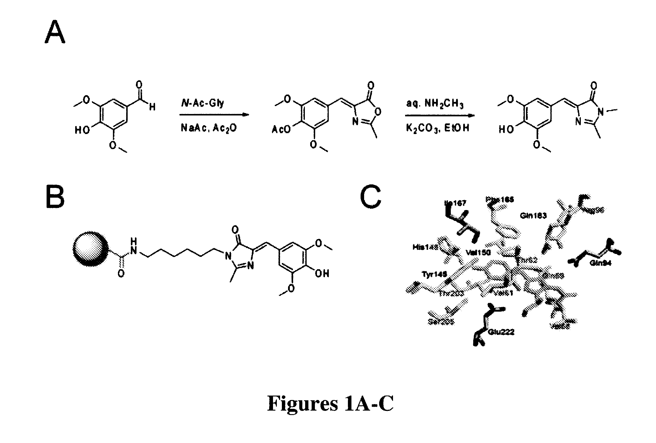
[0269]The most widely used approach for the synthesis of 4-arylidene-5-imidazolinones like those found in green fluorescent protein was first developed by Kojima et al. (“Fluorescent Properties of Model Chromophores of Tyrosine-66 Substituted Mutants of Aequorea Green Fluorescent Protein (GFP),”Tet. Lett. 39:5239-5242 (1998), which is hereby incorporated by reference in its entirety) and involves an Erlenmeyer azlactone formation between N-acetyl glycine and a 4-hydroxybenzaldehyde. The resulting azalactone is then subjected to aminolysis and subsequent cyclization under alkaline conditions to form the final 4-hydroxybenzylidene-5-imidazolinone (HBI).
[0270]Fundamentally the same synthesis was used here, except that a different substituted benzaldehyde was used to generate the novel fluorophore 3,5-dimethoxy-4-hydroxybenzylidene-5-imidazolinone (DMHBI). It was expected that the addition of these substituents would provide additional points of interact...
example 2
Synthesis of Resin-Immobilized Fluorophore
[0273]To perform SELEX and pull out RNAs that could bind to the designed fluorophores of the present invention, a strategy was needed to immobilize the designed fluorophores on resin. Thus, a general synthesis mechanism for generating fluorophores containing an aminoalkyl linker was developed. The aminoalkyl linker could be linked to a commercially available NHS-activated resin (FIG. 1B).
[0274]The azalactone intermediate (prepared in Example 1) was first reacted with N-t-butyloxycarbonyl (BOC)-1,6-hexanediamine for aminolysis and subsequent cyclization. After silica gel purification of the BOC-protected product, the BOC moiety was removed with trifluoroacetic acid (TFA) treatment in dichloromethane. Silica gel purification yielded a pure fluorophore with a hexyl linker and a free amine to couple to an amine-reactive resin.
[0275]Linker-modified DMHBI was then coupled to NHS-activated agarose resin. The fluorophore was first dissolved in DMSO ...
example 3
Fluorescence Characterization of Novel GFP-Mimetic Fluorophores
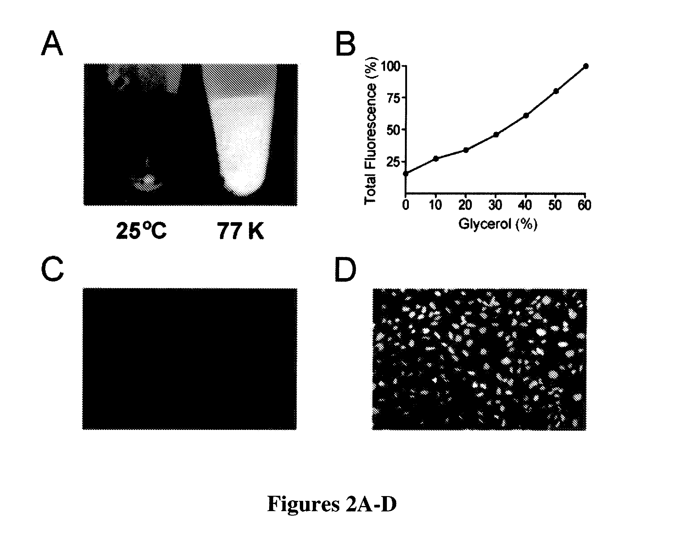
[0276]In GFP, the chromophore is held inside the protein β-barrel structure and rigidified by direct interactions with over a dozen amino acid residues (FIG. 1C). This inflexible positioning of the chromophore by the protein is the reason for its large fluorescent quantum yield. Similarly, the synthetic GFP chromophores 4-hydroxybenzilidene-1,2-dimethyl-imidazol-5-one (“HBDI”) is capable of bright fluorescence only when rigidified by solidified solvents (Niwa et al., “Chemical Nature of the Light Emitter of the Aequorea Green Fluorescent Protein,”Proc. Nat'l Acad. Sci. USA 93:13617-13622 (1996), which is hereby incorporated by reference in its entirety). Therefore, immobilization of DMHBI was performed to confirm whether DMHBI also exhibits this immobilization-dependent fluorescence that is seen in the native fluorophore. As an initial experiment DMHBI was first dissolved in ethanol and frozen in liquid nitrogen to creat...
PUM
| Property | Measurement | Unit |
|---|---|---|
| dissociation constant | aaaaa | aaaaa |
| dissociation constant | aaaaa | aaaaa |
| pH | aaaaa | aaaaa |
Abstract
Description
Claims
Application Information